The ubiquitin-proteasome pathway in cell cycle control
Bạn đang xem bản rút gọn của tài liệu. Xem và tải ngay bản đầy đủ của tài liệu tại đây (721.52 KB, 35 trang )
Results Probl Cell Differ (42)
P. Kaldis: Cell Cycle Regulation
DOI 10.1007/b136681/Published online: 6 July 2005
© Springer-Verlag Berlin Heidelberg 2005
The ubiquitin-proteasome pathway in cell cycle control
Steven I. Reed
Department of Molecular Biology, MB-7, The Scripps Research Institute, 10550 North
Torrey Pines Road, La Jolla, CA 92037, USA
Abstract Ubiquitin-mediated proteolysis is one of the key mechanisms underlying cell
cycle control. The removal of barriers posed by accumulation of negative regulators,
as well as the clearance of proteins when they are no longer needed or deleterious,
are carried out via the ubiquitin-proteasome system. Ubiquitin conjugating enzymes
and protein-ubiquitin ligases collaborate to mark proteins destined for degradation by
the proteasome by covalent attachment of multi-ubiquitin chains. Most regulated pro-
teolysis during the cell cycle can be attributed to two families of protein-ubiquitin
ligases. The anaphase promoting complex/cyclosome (APC/C) is activated during mitosis
and G1 where it is responsible for eliminating proteins that impede mitotic progres-
sion and that would have deleterious consequences if allowed to accumulate during G1.
SCF (Skp1/Culin/F-box protein) protein-ubiquitin ligases ubiquitylate proteins that are
marked by phosphorylation at specific sequences known as phosphodegrons. Targeting
of proteins for destruction by phosphorylation provides a mechanism for linking cell
cycle regulation to internal and external signaling pathways via regulated protein kinase
activities.
1
Introduction
The importance of ubiquitin-mediated proteolysis for cell cycle progression
and control is now well beyond dispute and can be illustrated in a variety of
ways. One of the most telling comes from the assignment of function to the
initial collection of cell division cycle (cdc) genes identified by Hartwell and
colleagues (Hartwell et al. 1974). Of the original 35 genes described in the
Hartwell screen based on a division-defective phenotypic endpoint, seven, or
fully 20%, either encode components of protein-ubiquitin ligases or ubiquitin
conjugating enzymes. For comparison, only five genes in this set encode pro-
tein kinases or phosphatases. Even though this exercise cannot be construed
as being of high quantitative significance, it does underscore the importance
of ubiquitin-mediated proteolysis and at least suggests its rough equivalence
with regulatory phosphorylation as an underlying mechanism in cell cycle
progression. We now know that ubiquitin-mediated protein turnover and
protein phosphorylation are not separable processes, but are indeed highly
interconnected, collaborating to form a network of pathways and regulatory
148 S.I. Reed
loops that control the cell cycle. The purpose of this review is to provide
a current view the role of ubiquitylation, ubiquitin-mediated proteolysis, and
proteasomes in cell cycle control in both yeasts and metazoans.
2
The ubiquitin-proteasome pathway
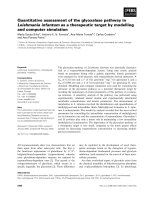
The process of ubiquitin-mediated proteolysis begins with the covalent
attachment of multi-ubiquitin chains to targeted proteins (Fig. 1) (Her-
shko 1983). Ubiquitin is a small (76 amino acid) highly conserved protein
(Hershko 1983). A cascade of enzyme-catalyzed reactions first activates
monomeric ubiquitin and then effects the processive attachment of ubiqui-
tin monomers first to lysines on the targeted protein and then to lysines of
previously attached ubiquitins. The activation of ubiquitin by the formation
of a high energy thioester bond with the C-terminal carboxylate of ubiquitin
is carried out by a ubiquitin-activating enzyme or E1. The transfer of acti-
vated ubiquitin to the lysines of target proteins to form an isopeptide bond
between the C-terminal carboxylate of ubiquitin and a lysine epsilon amino
group is carried out through a collaboration between ubiquitin conjugating
enzymes (E2) and protein-ubiquitin ligases (E3). The processive addition of
ubiquitin monomers to the lysines of already attached ubiquitins leads to the
decoration of proteins with long ubiquitin chains. Although several differ-
ent lysines on ubiquitin can serve as acceptor sites for ubiquitin addition,
the predominant linkage for protein turnover is through lysine 48 (Chen
and Pickart 1990). Once chains of significant length have been produced, the
Fig. 1 The ubiquitin proteasome pathway. Ubiquitin is initially activated by formation
of a thioester bond with E1 at the expense of a molecule of ATP. Activated ubiqutin is
then transferred via a complex of E2 (ubiquitin conjugating enzymer) and E3 (protein–
ubiquitin ligase) to the target molecule (T), Processive addition of ubiquitin to previously
conjugated ubiquitins forms multi-ubiquitylated chains that are recognized by the 26S
proteasome, leading to degradation of T
The ubiquitin-proteasome pathway in cell cycle control 149
protein is recognized by a complex protease known as the proteasome, ul-
timately leading to its processing into small peptides and the recycling of
ubiquitin.Theactive(26S)proteasomeiscomposedofabarrelshapedcata-
lytic core containing multiple protease activities on the inside surface (the
20S particle) and a regulatory (19S) particle at either end (Pickart and Cohen
2004). The regulatory particle is responsible for recognizing ubiquitylated
targets, removing the polyubiquitin chains for recycling of ubiquitin, unfold-
ing the protein to be degraded and opening up a pore in the 20S particle
so that unfolded substrate proteins can enter and contact the protease active
sites. The 19S cap contains a receptor for polyubiquitin chains. However, effi-
cient substrate recognition of at least some polyubiquitinated targets requires
“adapter” proteins that themselves bind ubiquitylated targets and dock to the
19S regulatory cap (Elsasser et al. 2004; Verma et al. 2004). The best charac-
terized of these is Rad23, which contains two ubiquitin-binding Uba domains
and a ubiquitin-like (Ubl) domain that interacts with the proteasome. One
final level of regulation concerns activities known as E4, which lengthen ubiq-
uitin chains on already polyubiquitylated proteasome substrates, presumably
to prevent substrate escape due to deubiquitylating activities (Koegl et al.
1999; Hatakeyama et al. 2001).
3
Protein-ubiquitin ligases in the cell cycle core machinery
One of the earliest observations relevant to the molecular basis for cell cycle
regulation was periodic synthesis and destruction of major proteins in sea
urchin cleavage embryos (Evans et al. 1983). Although the function of these
proteins, termed cyclins, was not known at the time, their accumulation dur-
ing interphase and turnover during mitosis was suggestive of a critical cell
cycle role. We now know that the cyclins observed in these sea urchin embryo
studies are positive regulatory subunits of the cyclin-dependent kinase (Cdk),
Cdk1 (Draetta et al. 1989; Meijer et al. 1989), which controls the mitotic state
for all eukaryotes: activation of Cdk1 establishes the mitotic state, whereas in-
activation determines mitotic exit into interphase. The inactivation of Cdk1,
potentiating mitotic exit is mediated for the most part by the ubiquitin-
mediated proteolysis of mitotic cyclins (Murray et al. 1989; Ghiara et al. 1991;
Surana et al. 1993). Investigation into the basis for mitotic cyclin degrada-
tion has led to the discovery and characterization of a complex protein-
ubiquitin ligase known as the anaphase promoting complex/cyclosome or
APC/C (King et al. 1995; Sudakin et al. 1995). Composed of at least 13 core
subunits (Zachariae et al. 1998b) and two alternative regulatory subunits
(Visintin et al. 1997) (Fig. 2), the APC/C targets not only mititotic cyclins
(A and B) but many other proteins that need to be degraded during mitosis
150 S.I. Reed
Fig. 2 The anaphase promoting complex/cyclosome (APC/C)
and/or the subsequent G1 interval (Table 1). Typically, the APC/Crecognizes
targets whose destruction at the population level is mandated at the times
when the APC/C is active, from the metaphase-anaphase transition to the
G1–S phase transition.
While the initial observations leading to the discovery of mitosis-specific
protein degradation came from studies on the early embryonic cell cycles of
Table 1 Cell cycle targets of the anaphase promoting complex
Substrate Organism Specificity factor Cell cycle function
Securin S. cerevisiae Cdc20 Anaphase inhibitor
(Pds1) (metazoan)
Clb2 S. cerevisiae Cdc20/Cdh1 B-type cyclin (mitosis)
Clb5 S. cerevisiae Cdc20 B-type cyclin (S phase)
Cyclin B Metazoan Cdc20/Cdh1 Mitosis
Cyclin A Metazoan Cdc20/Cdh1 S phase, mitosis
Cdc20 S. cerevisiae, Cdh1 Mitosis
metazoan
Cdc5/Plk S. cerevisiae, Cdh1 Mitosis
metazoan
Aurora A vertebrate Cdh1 Mitosis
Dbf4 S. cerevisiae Cdc20 Replication
Ase1 S. cerevisiae Cdh1 Mitosis
Nek2A Vertebrate Cdh1 Centrosome development
Cdc6 Vertebrate Cdh1 Replication
Geminin Metazoan Cdh1 Replication licensing
Cin8/Kip1 S. cerevisiae Cdh1 Mitotic spindle motor
Xkid Vertebrate Cdh1 Mitotic spindle motor
Hsl1 S. cerevisiae Cdc20/Cdh1 Mitosis
Cdc25A Vertebrates Cdh1 S phase, mitosis
Skp2 Vertebrates Cdh1 SCF cofactor
The ubiquitin-proteasome pathway in cell cycle control 151
Fig. 3 SCF protein-ubiqutin ligases
marine invertebrates (Evans et al. 1983; Swenson et al. 1986), first insights into
the role of proteolysis at the G1–S phase transition were a byproduct of ge-
netic analysis of the yeast cell cycle. As mentioned above, a number of the first
cell division cycle (cdc) mutants to be characterized were ultimately found
to define components of protein-ubiquitin ligases and associated proteins. Of
these, cdc4 and cdc34 conferred arrest at the G1/S boundary (Hartwell et al.
1974). In a subsequent round of cdc mutant isolation, mutations in a third
gene, cdc53, were found to confer a similar phenotype. We now know that
Cdc53 defines part of the catalytic core of a class of protein-ubiquitin lig-
ases known as SCF (Skp1-Cullin-F-box protein) (Willems et al. 1996) (Fig. 3),
whereas Cdc4 constitutes one of at least several SCF substrate specificity
factors (Feldman et al. 1997; Skowyra et al. 1997). Cdc34 is the associated
ubiquitin conjugating enzyme that works in conjunction with SCF to transfer
ubiquitin to target proteins (Goebl et al. 1988). The cell cycle arrest phe-
notype conferred by mutations in the genes encoding these proteins results
from an inability to degrade a Cdk inhibitor, Sic1 (Nugroho and Menden-
hall 1994; Schwob et al. 1994). Since Cdk1 activity is required for initiation
of DNA replication in yeast, failure to degrade Sic1 leads to arrest at the G1-
S phase boundary. Subsequently, numerous other cell cycle targets of SCF
ubiquitin ligases have been identified both in yeasts and metazoans. However,
unlike the APC/C, SCF activities are also simultaneously targeted to non-
cell-cycle-related proteins (although roles for the APC/C have recently been
described in post-mitotic cells). This is possible because the SCF system is
largely activated at the substrate level by substrate phosphorylation, allowing
simultaneous targeting of individual marked proteins within diverse popula-
tions and with distinct functions. Nevertheless, as will be described below,
SCF ligases constitute a core component of the cell cycle machinery. It is also
interesting to note that the APC/C and SCF systems do not operate in isola-
tion from each other. Recent evidence suggests that they are mutually toggled
presumably to enforce coordination of cell cycle events (see below).
152 S.I. Reed
3.1
APC/C protein-ubiquitin ligases
AswithSCF,anumberofcomponentsoftheAPC/C were represented in the
original collection of yeast cdc mutants assembled by Hartwell and colleagues
(Hartwell et al. 1974). Of the genes defined, four (CDC16, CDC23, CDC26 and
CDC27) encode subunits of the ligase itself (Zachariae and Nasmyth 1996;
Zachariae et al. 1996, 1998b), whereas one (CDC20) (Visintin et al. 1997)
encodes an essential positive regulatory factor of the APC/C. Conditional
mutations in all of these essential genes confer a mitotic arrest phenotype
characterized by unseparated sister chromatids. However, the functions of
these genes and their products was revealed only when it was discovered
that cyclin B was degraded via the ubiquitin-proteasome pathway (Glotzer
et al. 1991) and the activity responsible for ubiquitylating cyclin B was pu-
rified from mitotic clam and frog oocytes, respectively (King et al. 1995;
Sudakin et al. 1995). The large (20S) ubiquitin ligase from frog oocytes was
found to contain homologs of the yeast Cdc16 and Cdc27 proteins providing
a rationale for mitotic arrest phenotypes associated with cdc16 and cdc27 mu-
tants and leading a direct demonstration that cdc16, cdc23 and cdc27 mutants
were defective in mitotic cyclin degradation in yeast (Zachariae and Nasmyth
1996). Analysis of purified complexes from both metazoans and yeast has
revealed that the APC/C consists of 13 core polypeptides (Zachariae et al.
1998b; Grossberger et al. 1999) (Fig. 2). Three of these subunits (Cdc16, Cdc23
and Cdc27) contain a repeating motif known as TPR (for tetratricopeptide
repeat) that is involved in protein-protein interactions important for assem-
bling the APC/C macromolecular complex (Sikorski et al. 1990; Lamb et al.
1994). It is not clear why so many subunits are required for activity, since
most protein-ubiquitin ligases are much smaller. Cryoelectron microscopy
indicates that the APC/C has a hollow asymmetric structure (Gieffers et al.
2001). However, until the substrate binding and catalytic sites have been
identified within this structure, its significance remains obscure. This should
be possible, since two the APC/C subunits, Apc2 and Apc11, share signifi-
cant homology with subunits of the catalytic core of SCF, Cul1/Cdc53 and
Roc1/Rbx1, respectively (Ohta et al. 1999; Seol et al. 1999). In this context,
Apc11 and Roc1/Rbx1 contain “ring finger” motifs that are a characteristic
of the catalytic site of a major class of protein-ubiquitin ligases (Lorick et al.
1999).
The APC/C core is inactive without one of two structurally related pos-
itive regulatory cofactors, known as Cdc20 and Cdh1, respectively (Schwab
et al. 1997; Visintin et al. 1997). One function of these regulatory factors
is to recruit substrates (Hilioti et al. 2001; Pfleger et al. 2001; Schwab et al.
2001). It has been shown that Cdc20 preferentially recognizes a sequence
known as the D-box with a consensus R-X-X-L-X-X-X-X-N/D/E(Glotzer
et al. 1991; King et al. 1996), whereas Cdh1 recognizes both the D-box se-
The ubiquitin-proteasome pathway in cell cycle control 153
quence as well as a second sequence known as the KEN-box (Pfleger and
Kirschner 2000) and a third known as an A-box (Littlepage and Ruderman
2002), found specifically in the mitotic kinase Aurora A. However, the mech-
anisms of substrate recognition and targeting by different forms of APC/C
have yet to be completely elucidated. For some targets, e.g. cyclin B, a sin-
gle D-box is sufficient to mediate ubiquitylation and destruction, whereas for
others, e.g. cyclin A, both a D-box and a KEN-box are required (Geley et al.
2001). Furthermore, a recent report suggests that the APC/C itself contributes
to substrate recognition and binding, independent of Cdc20 and Cdh1, in
that a D-box-containing affinity matrix retained APC/CwithoutCdc20(Ya-
mano et al. 2004). This result suggests that Cdc20 and Cdh1 may provide an
activation function in addition to substrate recruitment.
Whereas Cdc20 and Cdh1 share a high degree of structural homology and
presumably provide analogous positive regulatory functions at the enzymatic
level, their biological functions are quite distinct. APC
Cdc20
is primarily re-
sponsible for mediating the metaphase-anaphase transition and early phases
of mitotic exit (Schwab et al. 1997; Visintin et al. 1997; Fang et al. 1999).
APC
Cdh1
completes mitotic exit and restricts mitotic proteins to low levels
during the subsequent G1 phase (Schwab et al. 1997; Visintin et al. 1997; Fang
et al. 1999). This division of labor is orchestrated in part by a complex reg-
ulatory relationship between Cdc20 and Cdh1. Whereas Cdc20 accumulates
based on periodic transcription late in the cell cycle, largely accounting for
theactivewindowofAPC
Cdc20
(Prinz et al. 1998), APC
Cdh1
is expressed con-
stitutively throughout the cell cycle (Weinstein 1997; Prinz et al. 1998; Zhu
et al. 2000). However, APC
Cdh1
is negatively regulated by Cdk phosphoryla-
tion of Cdh1 (Zachariae et al. 1998a; Lukas et al. 1999; Sorensen et al. 2001).
It is the APC
Cdc20
-mediated ubiquitylation and degradation of S-phase and
mitotic cyclins that allows dephosphorylation and activation of Cdh1 during
mitotic exit. On the other hand, the inhibition of Cdh1 at the G1–S phase
transition allows the accumulation of S phase cyclins, which in turn pro-
motes the biosynthesis of Cdc20 via transcription. Cdh1 inhibition at the
G1–S phase transition is initially mediated in mammalian cells by E2F-driven
accumulation of the Cdh1/Cdc20 inhibitor, Emi1 (Hsu et al. 2002) and auto-
ubiquitylation and degradation of a ubiquitin conjugating enzyme, Ubc10,
that serves as a cofactor with APC
Cdh1
(Rape and Kirschner 2004). The re-
sultant accumulation of cyclin A and activation of Cdk2 provides additional
inhibition of Cdh1 via phosphorylation. Accumulation of Cdc20 per se is
not sufficient for activation of APC
Cdc20
,asCdc20isalsoregulatedpost-
translationally. Like Cdh1, Emi1 also inhibits Cdc20 (Reimann et al. 2001a,b).
Phosphorylation-dependent degradation of Emi1 upon mitotic entrance (see
below) potentiates Cdc20 activation (Margottin-Goguet et al. 2003). In add-
ition, the spindle assembly checkpoint, which will be discussed in greater
detail below, maintains Cdc20 in an inactive state until bipolar attachment of
154 S.I. Reed
replicated chromosomes to a functional mitotic spindle is accomplished, thus
triggering anaphase (Lew and Burke 2003). Although phosphorylation of the
APC/C by cyclin B-Cdk1 has been suggested to be critical for mitotic activa-
tion, the precise targets and mechanism(s) have remained elusive (Sudakin
et al. 1995; Kraft et al. 2003). Finally, genetic experiments in budding yeast
have revealed that CDC20 is essential, whereas CDH1 is dispensable (Visintin
et al. 1997). This is because of a redundant pathway for downregulating Cdk
activity in late mitosis and G1, allowing mitotic exit in the absence of com-
plete cyclin proteolysis (Visintin et al. 1998; Shirayama et al. 1999). Indeed,
it has been possible to dispense with the APC/C entirely in yeast if the pri-
mary target of APC
Cdc20
, the anaphase inhibitor Pds1, is eliminated and Cdk
activity is down regulated by non-proteolytic mechanisms (Thornton and
Toczyski 2003).
3.2
APC/C substrates and biology
An increasing number of proteins has been shown to be targeted by the
APC/C (Table 1). However, only for a few has this targeting been demon-
strated to be absolutely essential. Yeast Pds1 (known generically as securin in
other organisms) is perhaps the most critical target of the APC/C(Cohen-Fix
et al. 1996; Yamamoto et al. 1996b; Zou et al. 1999; Zur and Brandeis 2001)
(Fig. 4). Prior to mitotic activation of the APC/C, Pds1/securin is bound to
the protease known as separase (Esp1 in yeast) (Ciosk et al. 1998). Although
Pds1 has positive regulatory roles with regard to Esp1 (e.g. nuclear localiza-
tion in yeast and chaparonin functions in mammalian cells) (Jensen et al.
2001; Hornig et al. 2002; Waizenegger et al. 2002), its primary function is to
inhibit Esp1 protease activity (Ciosk et al. 1998; Waizenegger et al. 2002). Esp1
is the substrate level trigger of anaphase, mediated via the endoproteolytic
cleavage of the Scc1 component of cohesin, a protein complex that binds sis-
ter chromatids together subsequent to DNA replication (Uhlmann et al. 1999;
Waizenegger et al. 2002). Release from cohesion allows spindle-generated
forces to separate sister chromatids and initiate anaphase. Esp1/separase also
then targets other proteins that regulate anaphase spindle functions (Jensen
et al. 2001; Stegmeier et al. 2002; Sullivan et al. 2001). Interestingly, dele-
tion of PDS1 in yeast is not lethal at moderate temperatures, although pds1
nullizygous cells grow poorly (Yamamoto et al. 1996a). The explanation is
most likely due to the balanced loss of both positive and negative Esp1 reg-
ulatory functions, as well as parallel secondary pathways that can restrict
proteolytic targeting of Scc1 to an appropriate time frame (Alexandru et al.
2001). The other critical targets of the APC/C are cyclins (King et al. 1995; Su-
dakin et al. 1995; Zachariae and Nasmyth 1996). As mentioned above, in order
to exit from mitosis, Cdk activities need to be down-regulated. The primary
mechanism whereby this requirement is met is via the APC/C-dependent
The ubiquitin-proteasome pathway in cell cycle control 155
Fig. 4 The spindle assembly checkpoint. Unattached kinetochores establish an inhibitory
complex consisting of Bub3, Mad3/BubR1 and Mad2, that binds to the APC/Ccofactor
Cdc20. Bipolar attachment of chromosomes with adequate tension leads to loss of the
inhibitory complex. Free Cdc20 activates the APC/C, which leads to ubiquitylation and
degradation of the separase inhibitor securin. Active separase cleaves cohesin, leading to
loss of cohesion, sister chromatid separation and anaphase
degradation of mitotic cyclins. Initially, these cyclins are targeted by APC
Cdc20
but they are also recognized by APC
Cdh1
, which presumably is responsible for
completing mitotic exit and restricting mitotic cyclin expression during G1
(Yeong et al. 2000; Wasch and Cross 2002). One key issue that remains un-
resolved with regard to the targeting of mitotic cyclins by the APC/Cisthe
differential kinetics of ubiquitylation of cyclin A relative to cyclin B (Pines
and Hunter 1990; Hunt et al. 1992; den Elzen and Pines 2001). Although both
are targeted by APC
Cdc20
, cyclin A is always ubiquitylated and degraded ear-
lier in mitosis than cyclin B. This relationship is intrinsic to mitotic cyclins
and the APC/C system, since it is observed in amphibian oocytes and early
embryonic cell cycles as well as in vertebrate somatic cells. In addition to se-
curin and cyclins, the APC/C has been shown to target many proteins for
destruction as cells exit mitosis (Table 1). Although the turnover of these pro-
156 S.I. Reed
teins is not necessary for mitotic exit or survival, it is presumed that their
clearance is required for optimal cellular function.
3.3
APC/C and meiosis
Although meiosis constitutes a modified cell cycle, its unusual chromosome
segregation characteristics would suggest that degradation of securin only be
required at the second meiotic division, where sister chromatid segregation
occurs. This is consistent with what has been reported for meiosis in Xeno-
pus oocytes, where depletion of APC/C does not appear to affect progress
through the first division (Peter et al. 2001; Taieb et al. 2001). However, in
mouse oocytes, worms, and yeast, APC function is required for the first mei-
otic division (Salah and Nasmyth 2000; Davis et al. 2002; Terret et al. 2003).
This is rationalized if one assumes that loss of sister chromatid cohesion
is required to resolve meiotic recombination-generated cross-overs between
chromosome arms prior to the first (reductional) division. Presumably, APC
function is also required to allow gametes to exit from meiosis after the sec-
ond (equational) division has been completed.
3.4
SCF protein-ubiquitin ligases
SCF protein-ubiquitin ligases constitute the second class of ubiquitinat-
ing enzymes that are central to cell cycle regulation in both lower and
higher eukaryotes. The core of the SCF ligase consists of three polypep-
tides: Cul1/Cdc53, Rbx1/Roc1 and (Feldman et al. 1997; Lisztwan et al. 1998;
Skowyra et al. 1999). Catalytic activity of the complex resides in a dimer com-
posed of Cul1/Cdc53 and Rbx1/Roc1 (Ohta et al. 1999; Seol et al. 1999), the
latter being a ring-finger protein, characteristic of many protein-ubiquitin
ligases (Lorick et al. 1999). Structural studies also suggest that Cul1/Cdc53
serves as a scaffold for binding the substrate-specificity component of the
SCF complex (Zheng et al. 2002). This consists of Skp1, an adapter protein
that binds directly to Cul1/Cdc53, and one of several F-box-containing pro-
teins. The 42-48 amino acid F-box motif constitutes a Skp1-binding domain
(Bai et al. 1996). Although genomic analysis has revealed the existence of
a large number of F-box proteins in both lower and higher eukaryotes, to
date only a few have been confirmed as components of SCF protein ubiq-
uitin ligases, although the number is likely to increase significantly. On the
other hand, Skp1 has been shown to participate in complexes other than SCF,
presumably recruiting some F-box proteins for roles distinct from ubiquitin
ligation (Connelly and Hieter 1996; Russell et al. 1999). The F-box proteins
involved in SCF function generally contain an F-box motif near their amino
termini and one of several protein-protein interaction motifs carboxy ter-
The ubiquitin-proteasome pathway in cell cycle control 157
minal to the F-box. In addition, some members of the F-box protein family
contain dimerization motifs amino terminal to the F-box, although the mech-
anistic implications of dimerization remain to be elucidated (Kominami et al.
1998; Suzuki et al. 2000). Interestingly, there are three classes of related F-
box proteins in both yeast and mammalian cells that are involved in cell cycle
control. Yeast Cdc4, originally identified via the Hartwell cdc mutant screen
(Hartwell et al. 1974), has mammalian and Drosophila homologs, known as
hCdc4/Fbw7 (Koepp et al. 2001; Strohmaier et al. 2001) and Archipelago
(Ago) (Moberg et al. 2001), respectively. Cdc4 and its homologs contain eight
tandem WD40 repeats that form a beta-propeller structure (Orlicky et al.
2003). This constitutes the substrate recruitment domain. hCdc4 also contains
a dimerization domain upstream of the F-box (O. Sangfelt, F. van Drogen and
S.I. Reed, unpublished data). In fission yeast (S. pombe), Cdc4 is expressed
as two separate genes that encode closely related proteins, Pop1 and Pop2
(Kominami et al. 1998; Wolf et al. 1999). The active form has been shown to
consist of a heterodimer, although the reason for this is not clear, since each
monomer contains an F-box and a substrate interaction domain. It is conceiv-
able that dimerization is required to configure a substrate binding domain
properly with respect to the catalytic site of the SCF core. The essentiality
of dimerization in the function of other Cdc4/Fbw7 homologs remains to be
determined.
The second class of SCF cofactor F-box proteins involved in cell-cyclin con-
trol is defined by vertebrate β-TrCP (Fuchs et al. 1999; Kroll et al. 1999; Latres
et al. 1999; Shirane et al. 1999; Suzuki et al. 1999; Tan et al. 1999; Winston et al.
1999), Drosophila Slimb (Bocca et al. 2001), and yeast Met30 (Kaiser et al.
1998; Patton et al. 1998). These F-box proteins share a similar topology with
Cdc4/Fbw7 in that they contain WD40 repeats (seven for β-TrCP and five for
Met30), an F-box and an amino terminal dimerization domain (Suzuki et al.
2000). As with Cdc4/Fbw7, the role and importance of dimerization has not
been established.
The third class of cell-cycle relevant F-box protein is Skp2 (Lisztwan et al.
1998; Lyapina et al. 1998) in vertebrates and Grr1 (Li and Johnston 1997;
Skowyra et al. 1997; Kishi et al. 1998) in yeast. Structurally, these SCF speci-
ficity factors are different from Cdc4/Fbw7 and β-TrCP in that the substrate
interacting domain contains a motif known as a leucine-rich repeat (Kobe
and Deisenhofer 1994, 1995) instead of WD40 repeats. Structural determin-
ation of Skp2 reveals that the 12 leucine-rich repeats of this molecule form
aconcavesurfacewheresubstratebindingislikelytooccur(Schulmanetal.
2000). Although yeast Grr1 can be modeled to the Skp2 structure, there
is little primary structure homology between the proteins, and it not clear
whether they are functionally homologous in any sense.
158 S.I. Reed
3.5
SCF substrates and biology
Whereas APC/C activity is regulated primarily at the level of its cofactors
Cdc20 and Cdh1, the primary mode of regulation of SCF activity appears to
be substrate activation via phosphorylation (Skowyra et al. 1997), although
regulation of F-box protein levels has also been reported (see below). SCF
ubiquitin ligases mediate ubiquitylation and turnover of a large number of
proteins involved in cell cycle control (Table 2). For both Cdc4/Fbw7 and
β-TrCP, a specific phosphorylated consensus sequence on the substrate, des-
ignated a phosphodegron, has been described. The optimal phosphodegron
sequence for Cdc4/Fbw7 is I/L-I/L/P-pT-P where basic residues are disfa-
vored at the next four positions carboxy terminal to the proline at position
+1 from the phosphothreonine (Nash et al. 2001). The crystal structure of
yeast Cdc4 bound to a peptide corresponding to an ideal phosphodegron has
revealed interactions between the negatively charged phosphate and several
arginine residues of the surface created by the WD40-generated β-propeller
structure (Orlicky et al. 2003). Mutation of any of the key arginines is suffi-
cient to functionally inactivate Cdc4 (Koepp et al. 2001; Orlicky et al. 2003).
It has been shown that substrates of SCF
Cdc4/Fbw7
can either contain a sin-
Table 2 Cell cycle targets of SCF ubiquitin ligases
Substrate Organism F-box protein Cell cycle function
Sic1/Rum1 S. cerevisiae,Cdc4/ Pop1/2 G1–S transition inhibitor
S. pombe
Far1 S. cerevisiae Cdc4 G1–S transition inhibitor
Cdc6/Cdc18 S. cerevisiae,Cdc4/ Pop1/2 DNA replication
S. pombe
Cln1,2 S. cerevisiae Grr1 G1 cyclin
Gic1,2 S. cerevisiae Grr1 Budding
Swe1 S. cerevisiae Met30 Mitosis inhibitor
Met4 S. cerevisiae Met30 G1-S transition inhibitor
Wee1 Vertebrates β –TrCP Mitosis inhibitor
Cyclin E Metazoans Cdc4/Fbw7/Ago G1-S cyclin
p27
Kip1
Mammals Skp2 G1-S transition inhibitor
p21
Cip1
Mammals Skp2 G1-S transition inhibitor
p130 Mammals Skp2 G1-S transition inhibitor
Ctd1 Mammals Skp2 DNA replication
Orc1 Mammals Skp2 DNA replication
Emi1 Vertebrates β –TrCP APC/Cinhibitor
Cdc25A Mammals β –TrCP S phase, mitosis
c-Myc Mammals Cdc4/Fbw7/Ago S phase
The ubiquitin-proteasome pathway in cell cycle control 159
gle high efficiency phosphodegron that closely matches the derived consensus
or multiple low-efficiency sites, most of which are poor matches (Nash et al.
2001). It has been proposed that having a requirement for phosphorylation
of multiple inefficient phosphodegron sequences within a protein constitutes
a means of delaying ubiquitylation and turnover of a regulatory protein until
kinase levels are sufficiently high to assure appropriate timing for a cell cycle
transition (Nash et al. 2001). This model is largely based on the yeast Cdk in-
hibitor Sic1, which requires minimally six phosphorylation events creating six
poor phosphodegron sequences in order to interact effectively with SCF
Cdc4
(Nash et al. 2001). Achieving this level of phosphorylation requires robust ac-
cumulation of the G1 cyclins Cln1 and Cln2 and concomitant full activation
of Cdk1, indicating that cells are ready to enter S phase. Ubiquitylation and
degradation of Sic1 then leads to activation of S phase Cdk activities, allowing
DNA replication to proceed. A similar inhibitor, Rum1, is targeted by the ho-
mologous ligase, SCF
Pop1/Pop2
in fission yeast (Kominami and Toda 1997; Wolf
et al. 1999). Although Sic1 appears to be the most critical target for cell cycle
progression in budding yeast, deletion of SIC1 has revealed another key cell
cycle target of SCF
Cdc4
,ascdc4 sic1 double mutants arrest in mitosis (Goh and
Surana 1999). However, the critical M phase target remains to be identified.
Other yeast cell cycle targets of SCF
Cdc4
are listed in Table 2.
In mammalian and other metazoan cells, the best known target of
SCF
Cdc4/Fbw7
is the G1 cyclin, cyclin E (Koepp et al. 2001; Moberg et al. 2001;
Strohmaier et al. 2001). Unlike Sic1, cyclin E contains a phosphodegron se-
quence that conforms precisely to the optimized consensus (Nash et al. 2001),
although a second less efficient phosphodegron can interact with SCF
Cdc4/Fbw7
when the primary phosphodegron is mutated (Strohmaier et al. 2001). Acti-
vation of the primary cyclin E phosphodegron requires autophosphorylation
by Cdk2 at a site carboxyerminal to the phosphodegron, which then primes
phosphorylation of the phosphodegron itself by the kinase GSK3β (Welcker
et al. 2003). Since, unlike Sic1, cyclin E is a positive regulator of cell cycle
progression, the inability to degrade cyclin E does not block cell cycle pro-
gression. However inability to degrade cyclin E on schedule is associated
with chromosome instability and polyploidy (Spruck et al. 1999). It appears
that persistence cyclin E during mitosis causes defects in mitosis itself (Ra-
jagopalan et al. 2004) as well as pre-replication complex assembly during
mitotic exit (Ekholm-Reed et al. 2004), possibly accounting for these pheno-
types. Since genomic instability is a driving force behind human malignancy,
it is not surprising that CDC4/FBW7 has been found to be mutated in a broad
spectrum of cancers (Moberg et al. 2001; Strohmaier et al. 2001; Spruck et al.
2002; Calhoun et al. 2003; Mao et al. 2004; Rajagopalan et al. 2004). However,
although loss of Cdc4/Fbw7 function confers phonotypes very analogous to
those associated with mutational stabilization/deregulation of cyclin E, it is
difficult to attribute the tumorigenicity of Cdc4 loss solely to defects in cy-
160 S.I. Reed
clin E turnover, since several other targets of SCF
Cdc4/Fbw7
are associated with
genomic instability and malignancy. Of these, c-Myc is this most noteworthy
(Welcker et al. 2004a,b; Yada et al. 2004). c-Myc is a transcription factor asso-
ciated with entry into S phase and apoptosis, although the critical targets for
these responses have not yet been clearly established (Nilsson and Cleveland
2003). Overexpression of c-Myc has been associated with genomic instabil-
ity (Mai and Mushinski 2003). Loss of Cdc4/Fbw7 function has been shown
to result in overexpression and deregulation of c-Myc (Welcker et al. 2004b;
Yada et al. 2004), most likely contributing to the overall associated genomic
instability. Other possible targets of SCF
Cdc4/Fbw7
associated with malignancy
are cytoplasmic signaling domains of Notch proteins (Gupta-Rossi et al. 2001;
Oberg et al. 2001; Wu et al. 2001; Tetzlaff et al. 2004; Tsunematsu et al. 2004).
Interestingly, complete loss of Cdc4/Fbw7 may not be necessary to promote
tumorigenesis. In a mouse model, haploinsufficiency of Cdc4/Fbw7 was ob-
served in a high percentage of tumors isolated from p53 heterozygous mice
(Mao et al. 2004). These data suggest that in some cells, Cdc4/Fbw7 is likely
to be rate-limiting for turnover of important SCF targets.
Although β-TrCP has a similar substrate binding motif composed of WD40
repeats, its preferred phosphodegron is quite different. β-TrCP recognizes
the sequence D-pS-G-(X)
n
-pS, where (X)
n
can be two or several amino acids
and both serines need to phosphorylated (Yaron et al. 1997, 1998; Hattori
et al. 1999; Orian et al. 2000). As with, Cdc4/Fbw7 and its phosphodegron,
the phosphates of the β-TrCP phosphodegron interact with arginines on the
WD40-repeat surface of β-TrCP (Wu et al. 2003). Although several proteins
have been shown to be substrates of SCF
β–TrCP
, those most relevant to cell
cycle control are Emi1 (Guardavaccaro et al. 2003; Margottin-Goguet et al.
2003) and Wee1 (Watanabe et al. 2004). Emi1 is an inhibitor of the APC/C
(Reimann et al. 2001a,b; Hsu et al. 2002). It binds to both Cdh1 and Cdc20,
preventing them from interacting with the APC/C core. Emi1 accomplishes
two important cell cycle functions. Its E2F-driven accumulation at the G1/S
boundary downregulates Cdh1 activity, allowing levels of APC/Ctargetsre-
quired for S phase and mitosis to begin to rise (Hsu et al. 2002). Notably,
stabilization of cyclin A allows further inhibition of Cdh1 via phosphoryla-
tion and progression through S phase. Emi1 then couples APC/Cactivation
to mitotic kinase activation. The primary kinase responsible for phosphode-
gron phosphorylation of Emi1 is Plk (polo-like kinase) (Hansen et al. 2004;
Moshe et al. 2004). However, cyclin B–Cdk1 strongly stimulates this reaction,
directly linking Emi1 destruction to mitotic entry (Hansen et al. 2004; Moshe
et al. 2004). Loss of Emi1 then potentiates activation of APC
Cdc20
and ini-
tiation of anaphase. A second mitotic inhibitor, Wee1, has also been shown
to be targeted for turnover by SCF
β–TrCP
(Watanabe et al. 2004). Wee1, a ki-
nase, prevents activation cyclin B–Cdk1 by phosphorylating Cdk1 on tyrosine
15, thereby preventing premature entrance into mitosis. Phosphorylation by