Senescence and Cell Cycle Control
Bạn đang xem bản rút gọn của tài liệu. Xem và tải ngay bản đầy đủ của tài liệu tại đây (261.56 KB, 14 trang )
Results Probl Cell Differ (42)
P. Kaldis: Cell Cycle Regulation
DOI 10.1007/001/Published online: 23 November 2005
© Springer-Verlag Berlin Heidelberg 2005
Senescence and Cell Cycle Control
Hiroaki Kiyokawa
Department of Molecular Pharmacology and Biological Chemistry,
Feinberg School of Medicine, Northwestern University, 303 E. Chicago Avenue,
Chicago, IL 60611, USA
Abstract In response to various stresses, such as telomere shortening during continuous
proliferation, oxidative stress, DNA damage and aberrant oncogene activation, normal
cells undergo cellular senescence, which is a stable postmitotic state with particular
morphology and metabolism. Signaling that induces senescence involves two major tu-
mor suppressor cascades, i.e., the INK4a-Rb pathway and the ARF-p53 pathway. Diverse
stimuli upregulate these interacting pathways, which orchestrate exit from the cell cycle.
Recent studies have provided insights into substantial differences in senescence-inducing
signals in primary cells of human and rodent origins. This review is focused on recent
advances in understanding the roles of the tumor-suppressive pathways in senescence.
1
Senescence
Senescence was originally defined as an “irreversible” state of cell cycle arrest
that reflects consumed proliferative capacity of the cell (Hayflick and Moor-
head 1961). In eukaryotic cells each chromosome shortens from telomeres
during every round of DNA replication (Smogorzewska and de Lange 2004;
Campisi 2001). The structure of telomeres with repetitive sequences func-
tions as a cap to prevent chromosome end fusions and genomic instability (de
Lange 1998; Sharpless and DePinho 2004). While germ cells express telom-
erase, which resynthesizes the telomeric repeats to maintain the chromoso-
mal length, most human somatic cells do not express telomerase. In somatic
cells proliferating continuously, attrition of telomeres beyond a threshold
triggers a response leading to “replicative” senescence. Recent studies have
indicated that telomere attrition provokes DNA damage-responsive signaling
pathways (d’Adda et al. 2003; Gire et al. 2004; Herbig et al. 2004). In addition,
senescent cells exhibit a particular flat morphology with enlarged cytoplasm,
and also express particular biochemical markers, such as senescence associ-
ated β-galactosidase activity (Dimri et al. 1995). “Premature” senescence or
stasis could be triggered in cells without telomere attrition by ectopic onco-
gene activation, DNA damage, oxidative stress and other stressful conditions
(Serrano et al. 1997; Chang et al. 2002; Chen and Ames 1994). These two
forms of senescence are morphologically indistinguishable, and are likely to
258 H. Kiyokawa
depend on common signaling pathways leading to cell cycle arrest. The path-
ways that play key roles in senescence induction, i.e., the ARF-p53-p21
Cip1
pathway and the p16
INK4a
-Rb pathway, are major tumor-suppressor cascades
(Sherr and DePinho 2000). Thus, it has been postulated that senescence is
a potent tumor-suppressive mechanism, like programmed cell death or apop-
tosis (Lowe and Sherr 2003). While there have been interesting discussions
on whether cellular senescence is involved in organismal aging (Pelicci 2004),
this review focuses on the tumor-suppressor pathways and cell cycle control
during cellular senescence.
2
Role of the p53 Pathway in Senescence
The tumor-suppressor p53 plays a key role in induction of senescence as
evidenced by studies using mouse embryonic fibroblasts (MEFs) from p53-
deficient (knockout) mice (Livingstone et al. 1992; Lowe et al. 1994). Under
standard culture conditions, MEFs from wild-type embryos proliferate up to
15–25 population doublings, followed by induction of senescence. In con-
trast, p53-null MEFs continue to proliferate without obvious cell cycle arrest
or senescence-like morphology. The same immortal phenotype is seen in
wild-type MEFs expressing dominant-negative p53 mutants, short interfer-
ing RNA (siRNA) against p53, or the papilloma virus oncoprotein E6, which
facilitates p53 degradation (Munger and Howley 2002). Thus, loss of p53 func-
tion is sufficient to abrogate the senescence checkpoint in MEFs (Sharpless
and DePinho 2002). The immortalization step dependent on p53 perturbation
renders mouse cells susceptible for activated Ras-induced malignant trans-
formation. In contrast, Ras activation in wild-type MEFs induces premature
senescence, as already described (Serrano et al. 1997). Although the role of
Ras in senescece induction remains to be fully understood, Ras activates the
external transcribed spacer transcription factors (ets) through the mitogen-
activated protein (MAP) kinase pathway, which may upregulate p16
INK4a
(see
Sect. 4 for details). Furthermore, Ras activation leads to accumulation of in-
tracellular reactive oxygen species [ROS] (see Sect. 5 for details). These cellu-
lar changes are apparently involved in Ras-induced senescence response. Hu-
man diploid fibroblasts (HDFs) taken from p53-heterozygous patients with
Li–Fraumenisyndromeshowprolongedreplicativelifespansinculture,as-
sociated with loss of heterozygosity (Boyle et al. 1998). Studies using HDFs,
keratinocytes, and mammary epithelial cells suggest that loss of p53 is not
sufficient for cooperating with Ras activation to transform human cells (Dray-
ton and Peters 2002). It has been described that a combination of SV40 large
T-antigen (T-Ag), activated Ras and the telomerase catalytic subunit (hTERT)
can transform HDFs and human keratinocytes (Hahn et al. 1999). T-Ag inac-
tivates the p53- and Rb-dependent senescence-inducing pathways and hTERT
Senescence and Cell Cycle Control 259
eliminates telomere-mediated signaling, allowing cells to undergo transform-
ation in response to Ras activation. Interestingly, human mammary epithelial
cells seem to have higher requirements for transformation, being insensitive
to the combinatory treatment (Hahn et al. 1999). These data suggest substan-
tial diversity of senescence control in different types of cells and in different
species.
Activation of p53 could result in one of the two cell fates, senescence
or apoptosis. Loss of p53 function could contribute to immortalization and
enhanced survival, both of which are hallmarks of cancer cells. Thus, p53
is a multifuctional tumor suppressor, playing a central role in preventing
malignant transformation. While the proapoptotic function of p53 involves
a number of genes, the only known mediator of its prosenescent function is
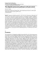
Fig. 1 The p16
INK4a
-Rb and ARF-p53 tumor-suppressor pathways control senescence. The
INK4a/ARF locus on human chromosome 9q21 encodes two tumor-suppressor proteins,
p16
INK4a
and p14
Arf
(p19
ARF
in mice). While p16
INK4a
directly inhibits Cdk4, p14
Arf
in-
duces p21
Cip1
, which inhibits Cdk2. Cdk inhibition results in repression of the E2F target
genes via reduced phosphorylation of the retinoblastoma (Rb) family proteins
260 H. Kiyokawa
the Cdk inhibitor p21
Cip1
(Fig. 1) (Sharpless and DePinho 2002; Vousden and
Prives 2005; Xiong et al. 1993; Harper et al. 1993). In an early study, p21
Cip1
was isolated as a senescence-related gene (Noda et al. 1994). During induc-
tion of senescence, p53 transactivates the p21
Cip1
gene (el Deiry et al. 1993),
and p21
Cip1
protein binds to and inhibits G1-regulatory cyclin-dependent ki-
nases (Cdk), especially Cdk2 in complex with cyclins E and A (Sherr and
Roberts 1999). In addition to Cdk inhibition, p21
Cip1
appears to affect expres-
sion and functions of various proteins, which may lead cells to senescence
in an orchestrated manner (Roninson 2002). Importantly, forced expression
of p21
Cip1
results in cellular accumulation of ROS with undefined mechan-
isms (Macip et al. 2002). ROS could cause DNA damage, which then activates
p53-dependent pathways, possibly forming a positive feedback. Disruption
of p21
Cip1
in HDFs prolongs the replicative life span in culture (Brown et al.
1997). These observations suggest a central role for p21
Cip1
in senescence.
However, studies using knockout mice provide a conflicting view. p21
Cip1
-
null MEFs undergo senescence normally (Pantoja and Serrano 1999), and
p21
Cip1
-null mice display only limited susceptibility to spontaneous tumori-
genesis (Martin-Caballero et al. 2001). Thus, p21
Cip1
may not be essential for
senescence of mouse cells. An alternative possibility is that p21
Cip1
-null mice
undergo developmental adaptation to the absence of p21
Cip1/Waf1
,forwhich
other Cdk inhibitors and possibly p130 (Coats et al. 1999) may compensate.
Studies using acute disruption of p21
Cip1
by conditional gene targeting will
be informative for better understanding of the role of p21
Cip1
in senescence
of mouse cells.
3
Role of the Rb Pathway in Senescence
The Rb-family pocket binding proteins, i.e., Rb, p107 and p130, also play
critical roles in cell fate determination between senescence and immortaliza-
tion. These proteins bind to the E2F family transcription factors and main-
tain the repressor function of E2F (Hatakeyama and Weinberg 1995; Stevaux
and Dyson 2002). Phosphorylation of the pocket binding proteins by Cdk
complexes, such as cyclin D/Cdk4 (or Cdk6) and cyclin E/Cdk2, results in
dissociation of the proteins from E2F complexes, and is thought to medi-
ate derepression or transactivation of E2F target genes (Fig. 1). Senescence of
MEFs induced by p53 overexpression depends on the repressor activity of E2F
(Rowland et al. 2002), suggesting that the senescence-inducing signal from
p53 converges to the Rb/E2F pathway. This signaling crosstalk could result
from p21
Cip1
inhibition of Cdk2 and possibly Cdk4/6. Inactivation of these
pocket binding proteins by the papillomavirus E7 oncoprotein, together with
telomerase activation by hTERT expression, has been shown to immortalize
primary human epithelial cells (Kiyono et al. 1998), although immortaliza-
Senescence and Cell Cycle Control 261
tion of this system may involve additional mutations. In MEFs, disruption
of Rb, p107 and p130 results in increased proliferation with a shortened G1
phase and immortalization (Dannenberg et al. 2000), whereas MEFs with
any one of the three proteins still exhibit senescence in culture (Sage et al.
2000). These observations suggest that the pocket binding proteins have over-
lapping functions in controlling senescence. However, acute disruption of
Rb by the Cre-Loxp recombination system has been demonstrated to im-
mortalize MEFs (Sage et al. 2003). This apparent discrepancy suggests that
germline disruption of one or two pocket binding proteins leads to devel-
opmental adaptation, which helps cells retain the senescence checkpoint by
a compensatory mechanism. For instance, p107 expression is upregulated
in Rb-null MEFs. Interestingly, the same study showed that Rb disruption
induced cell cycle reentry in a small fraction of apparently senescent cells,
suggesting that Rb plays a key role in maintenance of the postmitotic status in
senescent cells. Indeed, Rb, but not p107 or p130, is found in the senescence-
associated heterochromatic foci (SAHF) (Narita et al. 2003), which may play
a critical role in long-term transcriptional repression specific in senescent
cells. Interestingly, similar cell cycle reentry from senescence has been de-
scribed in MEFs infected with lentivirus for anti-p53 short hairpin RNA
(Dirac and Bernards 2003). While these studies intriguingly suggest that
senescence may not necessarily be an irreversible process, this notion awaits
further investigations on regulation of SAHF and other characteristics of
senescence.
Immortalization requires aberrant activation of the cell cycle machin-
ery. Cdk4 activation plays a key role when cells overcome the senescence
checkpoint, presumably via phosphorylation of the pocket binding proteins.
MEFs with targeted Cdk4
R24C
mutation, which express a constitutively ac-
tive Cdk4 protein insensitive to the INK4 inhibitors, exhibit an immortal
phenotype in culture (Rane et al. 2002). Activated Ras is sufficient to trans-
form Cdk4
R24C
MEFs, and mice with the Cdk4
R24C
mutation spontaneously
develop various tumors, such as endocrine and skin tumors (Sotillo et al.
2001; Rane et al. 2002). In contrast, MEFs from Cdk4-null mice are resis-
tant to immortalization induced by a dominant negative p53 mutant (DNp53)
or disruption of the INK4a/ARF locus (Zou et al. 2002). Cdk4-null MEFs
undergo transformation poorly in response to Ras plus DNp53 or Myc.
Consistent with the resistance to transformation, Cdk4-null mice are refrac-
tory to skin carcinogenesis in response to the keratin-5-Myc transgene or
the tumor initiator 7,12-dimethylbenz[4a4]anthracene plus the tumor pro-
moter 12-O-tetradecanoylphorbol-13-acetate (Rodriguez-Puebla et al. 2002;
Miliani de Marval et al. 2004). Genetic alterations that activate Cdk4, such
as overexpression of Cdk4 or D-type cyclins and deletion of the Cdk4 in-
hibitor p16
INK4a
, are observed in the majority of human cancers (Ortega
et al. 2002). Thus, derepression of E2F target genes as a consequence of
Cdk4 activation seems to be required for cellular immortalization. While