Đôi mắt
Bạn đang xem bản rút gọn của tài liệu. Xem và tải ngay bản đầy đủ của tài liệu tại đây (18.85 MB, 94 trang )
<span class='text_page_counter'>(1)</span><div class='page_container' data-page=1>
<b>DEPARTMENT OF ANATOMY</b>
<b>DEPARTMENT OF ANATOMY</b>
THE EYES
THE EYES
THE EYES
</div>
<span class='text_page_counter'>(2)</span><div class='page_container' data-page=2></div>
<span class='text_page_counter'>(3)</span><div class='page_container' data-page=3></div>
<span class='text_page_counter'>(4)</span><div class='page_container' data-page=4></div>
<span class='text_page_counter'>(5)</span><div class='page_container' data-page=5></div>
<span class='text_page_counter'>(6)</span><div class='page_container' data-page=6></div>
<span class='text_page_counter'>(7)</span><div class='page_container' data-page=7></div>
<span class='text_page_counter'>(8)</span><div class='page_container' data-page=8>
CORNEA
CORNEA
CORNEA
CORNEA
The cornea represents the strongest part of the refracting power of the eye, providing about 80% of
the power of the system. The index of refraction of the cornea is about 1.376. Rays pass from the cornea
into the watery fluid known as the aqueous humor which has an index of refraction of about 1.336,
so most of the refraction is at the cornea-air interface
</div>
<span class='text_page_counter'>(9)</span><div class='page_container' data-page=9>
CORNEA
CORNEA
CORNEA
CORNEA
1. THE REFRACTING POWER: +45D (2/3)
2. THICK: 0.5mm-0.74/1mm
3. 11.6mm-10.6mm
1. THE REFRACTING POWER: +45D (2/3)
2. THICK: 0.5mm-0.74/1mm
</div>
<span class='text_page_counter'>(10)</span><div class='page_container' data-page=10>
1
1
1(8ws)
1(8ws)
2
2
2 (thin scar)
2 (thin scar)
3
3
3 (thick scar)
3 (thick scar)
4
4
4 (very elastic)
4 (very elastic)
5
5
5
5
-3500/4000cell de 2500cell adult
-Na/ATPase bump-aqueous humour supp: glucose, miner, vitC..
-Dehydration / transparent cornea
-3500/4000cell de 2500cell adult
-Na/ATPasebump-aqueous humour supp: glucose, miner, vitC..
-Dehydration / transparent cornea
</div>
<span class='text_page_counter'>(11)</span><div class='page_container' data-page=11>
SCLERA
SCLERA
SCLERA
SCLERA
-thick: 1mm
-68%H<sub>2</sub>O
-thick: 1mm
-68%H2O
</div>
<span class='text_page_counter'>(12)</span><div class='page_container' data-page=12></div>
<span class='text_page_counter'>(13)</span><div class='page_container' data-page=13>
CARCINOMA IN SITU
CARCINOMA IN SITU
</div>
<span class='text_page_counter'>(14)</span><div class='page_container' data-page=14></div>
<span class='text_page_counter'>(15)</span><div class='page_container' data-page=15></div>
<span class='text_page_counter'>(16)</span><div class='page_container' data-page=16></div>
<span class='text_page_counter'>(17)</span><div class='page_container' data-page=17></div>
<span class='text_page_counter'>(18)</span><div class='page_container' data-page=18>
CHOROID
CHOROID
CHOROID
CHOROID
1
1
2
2
3
3
4
4 55
6
</div>
<span class='text_page_counter'>(19)</span><div class='page_container' data-page=19></div>
<span class='text_page_counter'>(20)</span><div class='page_container' data-page=20></div>
<span class='text_page_counter'>(21)</span><div class='page_container' data-page=21></div>
<span class='text_page_counter'>(22)</span><div class='page_container' data-page=22></div>
<span class='text_page_counter'>(23)</span><div class='page_container' data-page=23>
<b>Σ</b>
<b>Σ</b>
<b>pΣ</b>
<b>pΣ</b>
</div>
<span class='text_page_counter'>(24)</span><div class='page_container' data-page=24></div>
<span class='text_page_counter'>(25)</span><div class='page_container' data-page=25></div>
<span class='text_page_counter'>(26)</span><div class='page_container' data-page=26></div>
<span class='text_page_counter'>(27)</span><div class='page_container' data-page=27></div>
<span class='text_page_counter'>(28)</span><div class='page_container' data-page=28></div>
<span class='text_page_counter'>(29)</span><div class='page_container' data-page=29>
prod aque. humour
prod aque. humour
<b>cir. mus. pΣ</b>
<b>cir. mus. pΣ</b>
rad. mus. pΣ
(aque.hum. escape)
rad. mus. pΣ
(aque.hum. escape)
<b>obli. mus. pΣ</b>
<b>obli. mus. pΣ</b>
Zinn lig.
Zinn lig.
ACCOMMDATION
ACCOMMDATION
PRO. VITREOUS BODY
PRO. VITREOUS BODY
</div>
<span class='text_page_counter'>(30)</span><div class='page_container' data-page=30></div>
<span class='text_page_counter'>(31)</span><div class='page_container' data-page=31></div>
<span class='text_page_counter'>(32)</span><div class='page_container' data-page=32></div>
<span class='text_page_counter'>(33)</span><div class='page_container' data-page=33></div>
<span class='text_page_counter'>(34)</span><div class='page_container' data-page=34></div>
<span class='text_page_counter'>(35)</span><div class='page_container' data-page=35></div>
<span class='text_page_counter'>(36)</span><div class='page_container' data-page=36></div>
<span class='text_page_counter'>(37)</span><div class='page_container' data-page=37></div>
<span class='text_page_counter'>(38)</span><div class='page_container' data-page=38>
RETINA
RETINA
RETINA
RETINA
0.35mm
0.35mm
1.5mm
</div>
<span class='text_page_counter'>(39)</span><div class='page_container' data-page=39></div>
<span class='text_page_counter'>(40)</span><div class='page_container' data-page=40></div>
<span class='text_page_counter'>(41)</span><div class='page_container' data-page=41></div>
<span class='text_page_counter'>(42)</span><div class='page_container' data-page=42>
1/3
1/3
2/3
</div>
<span class='text_page_counter'>(43)</span><div class='page_container' data-page=43></div>
<span class='text_page_counter'>(44)</span><div class='page_container' data-page=44></div>
<span class='text_page_counter'>(45)</span><div class='page_container' data-page=45>
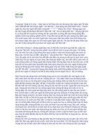
HYPERTENSIVE RETINOPATHY IS TRADITIONALLY DIVIDED INTO FOUR GRADES.
A, Grade 1 shows very early and minor changes in a young patient: increased tortuosity of a retinal
vessel and increased reflectiveness (silver wiring) of a retinal artery are seen at 1 o’clock in this view.
Otherwise, the fundus is completely normal.
B, Grade 2 also shows increased tortuosity and silver wiring (arrowheads). In addition, there is “nipping”
of the venules at arteriovenous crossings (arrow).
C, Grade 3 shows the same changes as grade 2 plus flame-shaped retinal hemorrhages and soft
“cotton wool” exudates.
D, In grade 4, there is swelling of the optic disc (papilledema), retinal edema is present, and hard
exudates may collect around the fovea, producing a typical “macular star.”
HYPERTENSIVE RETINOPATHY IS TRADITIONALLY DIVIDED INTO FOUR GRADES.
A, Grade 1 shows very early and minor changes in a young patient: increased tortuosity of a retinal
vessel and increased reflectiveness (silver wiring) of a retinal artery are seen at 1 o’clock in this view.
Otherwise, the fundus is completely normal.
B, Grade 2 also shows increased tortuosity and silver wiring (arrowheads). In addition, there is “nipping”
of the venules at arteriovenous crossings (arrow).
C, Grade 3 shows the same changes as grade 2 plus flame-shaped retinal hemorrhages and soft
“cotton wool” exudates.
</div>
<span class='text_page_counter'>(46)</span><div class='page_container' data-page=46></div>
<span class='text_page_counter'>(47)</span><div class='page_container' data-page=47></div>
<span class='text_page_counter'>(48)</span><div class='page_container' data-page=48></div>
<span class='text_page_counter'>(49)</span><div class='page_container' data-page=49>
AQUEOUS HUMOUR
AQUEOUS HUMOUR
AQUEOUS HUMOUR
AQUEOUS HUMOUR
0.25ml
0.25ml
0.05ml
0.05ml
1. maintains-regu intraocular pressure
2. nutrients, metabolic exchange for cornea-lens
3. constancy of the ocular dimensions of the eyeball
1. maintains-regu intraocular pressure
2. nutrients, metabolic exchange for cornea-lens
</div>
<span class='text_page_counter'>(50)</span><div class='page_container' data-page=50>
GLAUCOMA
GLAUCOMA
GLAUCOMA
GLAUCOMA
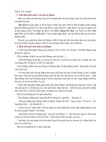
- Aqueous is formed by the nonpigmented cells of the
ciliary body by ultrafiltration of the blood, diffusion,
and active enzymatic secretion.
It flows into the posterior chamber, around the lens,
through the pupil into the anterior chamber and exits
the eye through the trabecular meshwork and the
Canal of Schlemm which leads to aqueous collector
veins which lead back to the systemic circulation.
The lower panel shows what happens in Primary Open
Angle Glaucoma, when the drainage ability of the
trabecular meshwork is impaired, aqueous cannot
drain, and intraocular pressure builds up.
Pilocarpin2%
Pilocarpin2%
Stress-catechole
</div>
<span class='text_page_counter'>(51)</span><div class='page_container' data-page=51>
In glaucoma, the visual field becomes constricted, leading to loss of peripheral
vision first and ultimately to blindness.
In glaucoma, the visual field becomes constricted, leading to loss of peripheral
vision first and ultimately to blindness.
</div>
<span class='text_page_counter'>(52)</span><div class='page_container' data-page=52>
LENS
LENS
LENS
LENS
1
1
About 9mm in diameter and 4 mm thick, the crystalline lens provides perhaps 20% of the refracting
power of the eye. Hecht likens it to a tiny transparent onion with some 22,000 fine layers. The index
ranges from about 1.406 at the center to about 1.386 in outer layers, making it a gradient index lens.
It is pliable, and changes shape to accomplish accommodation for close focusing
2
2
3
</div>
<span class='text_page_counter'>(53)</span><div class='page_container' data-page=53>
LENS
LENS
LENS
LENS
-65%H2O, 35%protein, K+, ascorbic a., glutathione
- Oxygen de, H2O, Na, Ca incre, Protein de / UV B= CATARACT
-65%H<sub>2</sub>O, 35%protein, K+, ascorbic a., glutathione
</div>
<span class='text_page_counter'>(54)</span><div class='page_container' data-page=54>
ACCOMMODATION
ACCOMMODATION
ACCOMMODATION
ACCOMMODATION
When the eye is relaxed and the interior lens is the least rounded, the lens has its maximum focal length
for distant viewing . As the muscle tension around the ring of muscle is increased and the supporting
fibers are thereby loosened, the interior lens rounds out to its minimum focal length.
</div>
<span class='text_page_counter'>(55)</span><div class='page_container' data-page=55>
ACCOMMODATION
ACCOMMODATION
ACCOMMODATION
ACCOMMODATION
To model the accommodation of the eye, the scale model eye was used with the cornea through the front
surface of the lens held constant at the model values. The matrix for that part of the eye was calculated
and then the thickness d and back surface power P were varied.
</div>
<span class='text_page_counter'>(56)</span><div class='page_container' data-page=56>
ACCOMMODATION
ACCOMMODATION
ACCOMMODATION
ACCOMMODATION
When the ciliary muscles contract, they loosen the ciliary fibers which are attached to the envelope
of the crystalline lens. Because the lens is pliable, it relaxes into a more curves shape, increasing it's
refractive power to accommodate for closer viewing. The iris serves as the aperture stop for the eye,
closing to about 2mm in diameter in bright light and opening to a maximum of about 8mm in dim light.
</div>
<span class='text_page_counter'>(57)</span><div class='page_container' data-page=57>
CATARACT
CATARACT
CATARACT
CATARACT
This slide shows a nuclear cataract; the lens seen within the pupil has a yellow-orange colour to it,
rather than being clear as in younger life. Nuclear cataracts dull the light path into the eye, affect
colour vision and acuity. They are most common in older eyes.
This slide shows a nuclear cataract; the lens seen within the pupil has a yellow-orange colour to it,
rather than being clear as in younger life. Nuclear cataracts dull the light path into the eye, affect
colour vision and acuity. They are most common in older eyes.
This slide shows a posterior subcapsular cataract. There is a plaque of whitish appearing material that
covers the back surface of the lens. A posterior subcapsular cataract scatters the light, producing glare,
especially at night with oncoming lights. It also significantly affects visual acuity. Posterior subcapsular
cataracts occur for many reasons, the most common being diabetes, intraocular inflammation, topical or
systemic steroid use, and trauma.
This slide shows complete opacity of the anterior cortex, a condition that used to be called a "ripe" or
"mature" lens. This will completely obscure the vision, allowing only light to pass into the eye.
</div>
<span class='text_page_counter'>(58)</span><div class='page_container' data-page=58>
VITREOUS BODY
VITREOUS BODY
VITREOUS BODY
VITREOUS BODY
-99%H
2O, gel-like, (hyalocytes)Na-hyaluronate- glycosaminoglycan frame
-Absent at birth, 4-5
thy and incre.
-
Hyaloid canal – disappear 6ws bef birth
Hyaloid canal – disappear 6ws bef birth
-99%H
<sub>2</sub>O, gel-like, (hyalocytes)Na-hyaluronate- glycosaminoglycan frame
-Absent at birth, 4-5
thy and incre.
</div>
<span class='text_page_counter'>(59)</span><div class='page_container' data-page=59></div>
<span class='text_page_counter'>(60)</span><div class='page_container' data-page=60></div>
<span class='text_page_counter'>(61)</span><div class='page_container' data-page=61>
Nearsightedness or myopia, occurs when light entering the eye focuses in front of the retina instead of
directly on it. This is caused by a cornea that is steeper, or an eye that is longer, than a normal eye.
Nearsighted people typically see well up close, but have difficulty seeing far away.
Nearsightedness or myopia, occurs when light entering the eye focuses in front of the retina instead of
directly on it. This is caused by a cornea that is steeper, or an eye that is longer, than a normal eye.
Nearsighted people typically see well up close, but have difficulty seeing far away.
Farsightedness or hyperopia, occurs when light entering the eye focuses behind the retina, instead of
directly on it. This is caused by a cornea that is flatter, or an eye that is shorter, than a normal eye.
Farsighted people usually have trouble seeing up close, but may also have difficulty seeing far away
as well.
</div>
<span class='text_page_counter'>(62)</span><div class='page_container' data-page=62>
LASIK
LASIK
LASIK
LASIK
LASIK (laser in situ keratomilieusis) is a laser vision correction technique that uses an excimer laser
to reshape the cornea without disturbing adjacent cell layers.
1. The eye is numbed using anesthetic drops.
2. An eyelid holder is used to prevent blinking.
3. A small suction ring is placed on the cornea.
4. Using a microkeratome, a flap is created.
5. Using an excimer laser, a predetermined amount of corneal tissue is removed to re-sculpt the cornea.
6. The flap is placed back into position where it naturally re-adheres.
LASIK (laser in situ keratomilieusis) is a laser vision correction technique that uses an excimer laser
to reshape the cornea without disturbing adjacent cell layers.
1. The eye is numbed using anesthetic drops.
2. An eyelid holder is used to prevent blinking.
3. A small suction ring is placed on the cornea.
4. Using a microkeratome, a flap is created.
</div>
<span class='text_page_counter'>(63)</span><div class='page_container' data-page=63></div>
<span class='text_page_counter'>(64)</span><div class='page_container' data-page=64>
2mm
2mm
</div>
<span class='text_page_counter'>(65)</span><div class='page_container' data-page=65>
CONJUNCTIVA
CONJUNCTIVA
CONJUNCTIVA
CONJUNCTIVA
-to regenerate epithe-cornea
-immune system
-mucin
-to regenerate epithe-cornea
-immune system
</div>
<span class='text_page_counter'>(66)</span><div class='page_container' data-page=66></div>
<span class='text_page_counter'>(67)</span><div class='page_container' data-page=67></div>
<span class='text_page_counter'>(68)</span><div class='page_container' data-page=68></div>
<span class='text_page_counter'>(69)</span><div class='page_container' data-page=69></div>
<span class='text_page_counter'>(70)</span><div class='page_container' data-page=70></div>
<span class='text_page_counter'>(71)</span><div class='page_container' data-page=71></div>
<span class='text_page_counter'>(72)</span><div class='page_container' data-page=72></div>
<span class='text_page_counter'>(73)</span><div class='page_container' data-page=73></div>
<span class='text_page_counter'>(74)</span><div class='page_container' data-page=74></div>
<span class='text_page_counter'>(75)</span><div class='page_container' data-page=75>
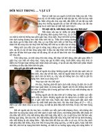
The outer layer of the tear film is the oily layer which serves to decrease evaporative loss.
The middle layer is the aqueous layer which provides the bulk of the tear film.
The inner layer is the mucin layer which allows the aqueous layer to adhere to the hydrophobic
surface of the cornea and conjunctiva.
Abnormalities of any of the three layers can produce either a dry eye or one which overflows with
tears (epiphora).
</div>
<span class='text_page_counter'>(76)</span><div class='page_container' data-page=76></div>
<span class='text_page_counter'>(77)</span><div class='page_container' data-page=77></div>
<span class='text_page_counter'>(78)</span><div class='page_container' data-page=78>
R
R
L
L 1212
</div>
<span class='text_page_counter'>(79)</span><div class='page_container' data-page=79></div>
<span class='text_page_counter'>(80)</span><div class='page_container' data-page=80>
250
250 <sub>25</sub><sub>25</sub>0<sub>0</sub>
7mm
7mm
</div>
<span class='text_page_counter'>(81)</span><div class='page_container' data-page=81>
7mm
7mm 5.5mm5.5mm
</div>
<span class='text_page_counter'>(82)</span><div class='page_container' data-page=82></div>
<span class='text_page_counter'>(83)</span><div class='page_container' data-page=83></div>
<span class='text_page_counter'>(84)</span><div class='page_container' data-page=84></div>
<span class='text_page_counter'>(85)</span><div class='page_container' data-page=85></div>
<span class='text_page_counter'>(86)</span><div class='page_container' data-page=86></div>
<span class='text_page_counter'>(87)</span><div class='page_container' data-page=87>
R
R
L
L 1212
</div>
<span class='text_page_counter'>(88)</span><div class='page_container' data-page=88>
1
1 1111
11
11
1
</div>
<span class='text_page_counter'>(89)</span><div class='page_container' data-page=89></div>
<span class='text_page_counter'>(90)</span><div class='page_container' data-page=90></div>
<span class='text_page_counter'>(91)</span><div class='page_container' data-page=91></div>
<span class='text_page_counter'>(92)</span><div class='page_container' data-page=92></div>
<span class='text_page_counter'>(93)</span><div class='page_container' data-page=93>
REFERENCES
REFERENCES
REFERENCES
REFERENCES
1. TEXTBOOKS
1. TEXTBOOKS
1. TEXTBOOKS
1. TEXTBOOKS
1. Legent F., Perlemuter L., Vandenbrouck Cl.
1. Legent F., Perlemuter L., Vandenbrouck Cl. Cahiers D’anatomie O.R.L., 4Cahiers D’anatomie O.R.L., 4thth<sub> ed, 2 Vol</sub><sub> ed, 2 Vol</sub><sub>. Paris: Masson,</sub><sub>. Paris: Masson,</sub>
1986, pp.71-115.
1986, pp.71-115.
2. Williams PL, Warwick R et al.
2. Williams PL, Warwick R et al. Gray’s Anatomy, 37Gray’s Anatomy, 37thth ed<sub> ed</sub>. NewYork : Churchill Livingstone, 1989, <sub>. NewYork : Churchill Livingstone, 1989, </sub>
pp. 401-22, 499-16.
pp. 401-22, 499-16.
2. Pick TP & Howden R.
2. Pick TP & Howden R. Gray’s Anatomy, The Classic Collector’s edGray’s Anatomy, The Classic Collector’s ed. NewYork : Gramercy, 1977.. NewYork : Gramercy, 1977.
3. Imran T. Khawaja, Barbara A. Phillips.
3. Imran T. Khawaja, Barbara A. Phillips. Obstructive Sleep Apnea : Diagnosis & TreatmentObstructive Sleep Apnea : Diagnosis & Treatment. Hospital . Hospital
Medicine 34(3):33-36, 39-41, 1998.
Medicine 34(3):33-36, 39-41, 1998.
4. Jerald S. Altman, Robert D.H et al.
4. Jerald S. Altman, Robert D.H et al. Effect of UPPP and genial & hyoid advancement on swallowing Effect of UPPP and genial & hyoid advancement on swallowing
In patients with OSAS
In patients with OSAS. The annual meeting of the American Academy of Otolaryngology- Head & . The annual meeting of the American Academy of Otolaryngology- Head &
Neck surgery. Sanfrancisco, CA, Sept, 7-10, 1997
Neck surgery. Sanfrancisco, CA, Sept, 7-10, 1997
5. Sabiston.
5. Sabiston. Textbook of Surgery, 15Textbook of Surgery, 15thth ed<sub> ed</sub>. Philadelphia : W.B. Saunder Company, 1997.<sub>. Philadelphia : W.B. Saunder Company, 1997.</sub>
6.
6. Oxford Textbook of Surgery on CD-ROM, ver 1.0Oxford Textbook of Surgery on CD-ROM, ver 1.0. Oxford University Press, 1995. . Oxford University Press, 1995.
1. Legent F., Perlemuter L., Vandenbrouck Cl.
1. Legent F., Perlemuter L., Vandenbrouck Cl. Cahiers D’anatomie O.R.L., 4Cahiers D’anatomie O.R.L., 4thth ed, 2 Vol<sub> ed, 2 Vol</sub>. Paris: Masson,<sub>. Paris: Masson,</sub>
1986, pp.71-115.
1986, pp.71-115.
2. Williams PL, Warwick R et al.
2. Williams PL, Warwick R et al. Gray’s Anatomy, 37Gray’s Anatomy, 37thth<sub> ed</sub><sub> ed</sub><sub>. NewYork : Churchill Livingstone, 1989, </sub><sub>. NewYork : Churchill Livingstone, 1989, </sub>
pp. 401-22, 499-16.
pp. 401-22, 499-16.
2. Pick TP & Howden R.
2. Pick TP & Howden R. Gray’s Anatomy, The Classic Collector’s edGray’s Anatomy, The Classic Collector’s ed. NewYork : Gramercy, 1977.. NewYork : Gramercy, 1977.
3. Imran T. Khawaja, Barbara A. Phillips.
3. Imran T. Khawaja, Barbara A. Phillips. Obstructive Sleep Apnea : Diagnosis & TreatmentObstructive Sleep Apnea : Diagnosis & Treatment. Hospital . Hospital
Medicine 34(3):33-36, 39-41, 1998.
Medicine 34(3):33-36, 39-41, 1998.
4. Jerald S. Altman, Robert D.H et al.
4. Jerald S. Altman, Robert D.H et al. Effect of UPPP and genial & hyoid advancement on swallowing Effect of UPPP and genial & hyoid advancement on swallowing
In patients with OSAS
In patients with OSAS. The annual meeting of the American Academy of Otolaryngology- Head & . The annual meeting of the American Academy of Otolaryngology- Head &
Neck surgery. Sanfrancisco, CA, Sept, 7-10, 1997
Neck surgery. Sanfrancisco, CA, Sept, 7-10, 1997
5. Sabiston.
5. Sabiston. Textbook of Surgery, 15Textbook of Surgery, 15thth ed<sub> ed</sub>. Philadelphia : W.B. Saunder Company, 1997.<sub>. Philadelphia : W.B. Saunder Company, 1997.</sub>
6.
6. Oxford Textbook of Surgery on CD-ROM, ver 1.0Oxford Textbook of Surgery on CD-ROM, ver 1.0. Oxford University Press, 1995. . Oxford University Press, 1995.
2. WEBSITES
2. WEBSITES
2. WEBSITES
2. WEBSITES
1.
1. />2. />2. />3. />3. />4. />4. />5.
5.
6.
6. Anatomy of the Human Body Henry GrayAnatomy of the Human Body Henry Gray />1.
1. />2. />2. />3. />3. />4. />4. />5.
5.
6.
</div>
<span class='text_page_counter'>(94)</span><div class='page_container' data-page=94>
THANK YOU VERY MUCH FOR
THANK YOU VERY MUCH FOR
YOUR ATTENTION !!!
YOUR ATTENTION !!!
MERCI DE VOTRE ATTENTION !!!
MERCI DE VOTRE ATTENTION !!!
THANK YOU VERY MUCH FOR
THANK YOU VERY MUCH FOR
YOUR ATTENTION !!!
YOUR ATTENTION !!!
MERCI DE VOTRE ATTENTION !!!
</div>
<!--links-->