Báo cáo y học: "Ocular manifestations of Lyme borreliosis in Europe"
Bạn đang xem bản rút gọn của tài liệu. Xem và tải ngay bản đầy đủ của tài liệu tại đây (367.67 KB, 2 trang )
Int. J. Med. Sci. 2009, 6
124
I
I
n
n
t
t
e
e
r
r
n
n
a
a
t
t
i
i
o
o
n
n
a
a
l
l
J
J
o
o
u
u
r
r
n
n
a
a
l
l
o
o
f
f
M
M
e
e
d
d
i
i
c
c
a
a
l
l
S
S
c
c
i
i
e
e
n
n
c
c
e
e
s
s
2009; 6(3):124-125
© Ivyspring International Publisher. All rights reserved
Short Communication
Ocular manifestations of Lyme borreliosis in Europe
Paolo Mora, Arturo Carta
Institute of Ophthalmology, University of Parma - Parma (Italy)
Published: 2009.03.19
Ocular involvement in Lyme borreliosis, even
though possible in every stage of the disease, is most
frequently seen in the late phases (2
nd
and 3
rd
). In a
German series of children affected by Lyme arthritis,
4% also had ocular inflammation consisting of kerati-
tis or uveitis.
[1] In a Finnish cohort of twenty patients
with ocular borreliosis, 10 had uveitis, 5 subjects
showed ocular adnexa inflammation, 4 had
neuro-ophthalmological alterations and one patient
developed branch central retinal vein occlusion.
Contact with a tick was clearly reported in only 13/20
cases.
[2] There are various ocular symptoms of Lyme
disease including: pain, visual impairment, photo-
phobia, myodesospia, diplopia and lack of accom-
modation.
[3]
To observe ocular signs or symptoms it is not
necessary that the site of inoculation of the infection is
close to the eyes (as in Figure 1). It is possible to define
the following ocular findings, from the anterior to the
posterior segment of the eye:
Conjunctivitis: often self-limited; when it ap-
pears in the 1
st
stage of the disease it is associated with
an influenza-like syndrome in 7-11% of the patients. It
is follicular and uni- or bilateral and in the late phases
of disease it may be accompanied by sever eyelid
edema in 3% of subjects.
Keratitis: typical of the 2
nd
and 3
rd
stage of the
disease; it may persist even after appropriate systemic
antibiotic treatment, suggesting an immunological
origin of corneal opacity. It can be disseminated and
potentially bilateral, but the most characteristic pat-
terns are “interstitial” or “ulcerative” with peripheral
neovascolarisation.
Episcleritis/Scleritis: very rare, almost always
related to the late phase of the disease.
Uveitis: the anterior form is infrequently reported
in Lyme disease and is possibly associated with
papillitis. The case observed in our Institute had ex-
actly these features. It consisted in a serologi-
cally-confirmed unilateral papillitis (Figure 2), com-
bined with keratic granulomatous precipitates and
iris-lens sinechyae. Ocular involvement appeared
during the 2
nd
stage of the disease, sixty days after the
removal of a tick from the forearm. Treatment with
amoxicillin for three weeks combined with oral pred-
nisone at decreasing doses was successful in achiev-
ing complete visual recovery.
The intermediate form is the most common form
of uveitis, often associated with iridocyclitis (par-
splanitis). In three cases Borrellia burgdorfori s.l. was
isolated from the vitreous by culture or polymerase
chain reaction. Only a few reports concern panuveitis;
two cases, however, resulted in irreversible visual
loss. Signs of posterior uveitis mostly included
chorio-retinal involvement.
Retinal infection: macular oedema and vasculitis
are the most frequent findings, occasionally compli-
cated by vitreoretinal proliferation and have been
described either during the erythema migrans phase
or in neuroborreliosis. Venular occlusions and
chorio-retinal inflammatory foci are less common
manifestations. A recent report has shown cotton
wool spots as another possible sign of Lyme retinitis.
[4]Neuro-ophthalmological alterations: these repre-
sent early evidence of neuroborreliosis. Diplopia and
visual impairment, with or without meningitis, are
the suggestive signs. In case of optic neuritis the con-
comitant presence of cranial nerve palsies is expected
(mostly VI or VII).
Uncertain reports of orbital myosistis and
Jarisch-Herxheimer reaction have also been proposed
as a consequence of borreliosis.
In conclusion, ocular involvement in Lyme bor-
reliosis is symptomatic and a routine ophthalmologic
evaluation is not recommended in adult patients
(younger patients, on the other hand, should be
Int. J. Med. Sci. 2009, 6
125
screened due to their poor capacity to complain of
ocular disturbances). In order to formulate a rational
suspicion of Lyme disease as the cause of ocular in-
flammation, features must include occurrence in an
endemic zone; and/or the report of contact with a tick
or of previous erythema migrans; positive serology
with presence of IgM in the early stage or high titres
of IgG in the later phases. A clear diagnosis, however,
remains very difficult. The long-term follow-up of
four cases of optic neuritis labelled as Lyme disease
because of the positive serology for Borrelia revealed
that three patients actually developed demyelinating
syndromes.
[5]
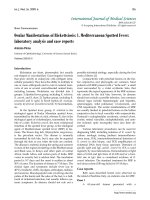
Figure 1: Tick among the eyelashes (courtesy of Dr. N.
Massaro; Agrigento - Italy).
Figure 2: Papillitis with initial macular exudation.
References
1. Huppertz HI, Munchmeir D, Lieb W. Ocular manifestation in
children and adolescents with Lyme arthritis. Br J Ophhalmol
1999; 83:1149-1152
2. Mikkila HO, Seppala IJT, Viljanen MK, Peltomaa MP, Karma A.
The expanding clinical spectrum of ocular Lyme borreliosis.
Ophthalmology 2000; 107:581-587
3. Boyé T. What kind of clinical, epidemiological, and biological
data is essential for the diagnosis of lyme borreliosis? Derma-
tological and ophthalmological courses of Lyme borreliosis.
Médecine et Maladies Infectieuses 2007; 37:S175-188 [French]
4. Klaeger AJ, Herbort CP. Cotton wool spots as possible indica-
toris of retinal vascular pathology in ocular lyme borreliosis. Int
Ophthalmol 2008; 15 [epub]
5. Jacobson DM. Lyme disease and optic neuritis: long-term fol-
low-up of seropositive patients. Neurology 2003; 60:881-882.