IBK – một công cụ mới trong lĩnh vực xử lý ảnh y tế
Bạn đang xem bản rút gọn của tài liệu. Xem và tải ngay bản đầy đủ của tài liệu tại đây (283.71 KB, 8 trang )
Science & Technology Development, Vol 13, No.K5- 2010
Trang 20 Bản quyền thuộc ĐHQG.HCM
IBK – A NEW TOOL FOR MEDICAL IMAGE PROCESSING
Tran Duy Linh, Huynh Quang Linh
University of Technology, VNU- HCM
(Manuscript Received on June 28
th
, 2010, Manuscript Revised October 08
h
, 2010)
ABSTRACT: Along with the rapid development of diagnostic imaging equipment, software for
medical image processing has played an important role in helping doctors and clinicians to reach
accurate diagnoses. In this paper, methods to build a multipurpose tool based on Matlab programming
language and its applications are presented. This new tool features enhancement, segmentation,
registration and 3D reconstruction for medical images obtained from commonly used diagnostic
imaging equipment.
Keywords: IBK, diagnostic imagining, medical image processing.
1.INTRODUCTION
Diagnostic imaging is an invaluable tool
in medicine. In recent years, hospitals in
Vietnam are equipped with more and more
modern diagnostic imaging equipment. The
generation of conventional X-ray machine is
gradually replaced by digital X-ray system and
computed tomography (CT) scanner. Besides,
other imaging equipment such as Magnetic
Resonance Imaging (MRI), Single Photon
Emission Computed Tomography (SPECT),
Positron Emission Tomography (PET), Digital
Subtraction Angiography (DSA) etc. become
more familiar modalities. In current clinical
practice, these imaging modalities allow
medical personnel to look into a living body
with both anatomical and functional
information, in order to diagnose many types of
diseases. Compared with analog imaging
equipment, such digital equipment have many
advantages: no photochemical development of
film is required, image can be displayed on
monitor immediately after the exposure, stored
information is more easily accessible by
magnetic or compact disks, the capacity for
information transmission between local
departments through computer networks
(PACS) or long-distance transmission via
Internet to remotely diagnose (telemedicine),
and especially the feasibility of image
processing: magnify it or change the contrast
level by using image processing tools etc.
Such image processing tools necessitate
the use of computers for processing and
analysis. The computer tasks can be split into
four areas: (1) feature enhancement involved in
noise, artifact removing or contrast increasing;
(2) quantitative analysis by employing
segmentation algorithms; e.g. tumor volume
measurement, localization of pathology, study
of anatomical structure; (3) detection of
medical conditions by applying accurate
registration to structural and functional images
to extract information that was not apparent in
TẠP CHÍ PHÁT TRIỂN KH&CN, TẬP 13, SỐ K5 - 2010
Bản quyền thuộc ĐHQG-HCM Trang 21
an individual dataset and (4) visual
reconstruction: a series of image slices can be
aggregated into 3D representation of patient’s
anatomy. Although hardware-based solutions
for registration are provided by PET/CT and
SPECT/CT scanners, software-based
registration may still be required to correct
misregistration caused by patient motion
between the PET scan and CT scan. There were
a vast number of studies that have reviewed
algorithms of the above techniques [1, 2, 3]. In
this paper, the authors focus on methods which
have been used to built an application named
IBK and its possible applications in clinical
environment.
Numerous foreign software packages are
available for medical image processing and
analysis such as eFilm, 3D-Doctor,
DICOMWorks, BrainSuite etc. The drawbacks
of such packages are their high price and their
user interfaces are in English. Beside of these
packages, equipment manufacturers have their
own built-in software (e.g. Syngo, AVIA,
Volumetrix Suite etc.) which has many
powerful functions. However these software
packages must only be installed on system
manufacturer’s computers. In case we need
register two images obtained from different
firms equipment, these packages can not help.
In Vietnam, Biomedical Electronics Center at
Hanoi University of Technology is a pioneer in
writing medical image processing software.
However their application software, BK-
DICOM has a limited number of
functionalities.
As a result, the authors desire to built a
multi-purpose medical image processing
application featuring enhancement,
segmentation, 3D reconstruction, and
registration of multimodal images obtained
from different equipment. This application has
a user interface in Vietnamese and would be
either used as a flexible illustration tool for
education purpose or distributed free to
medical centers and hospitals in Vietnam in the
future.
2.METHODS
2.1.Approach
Programmed in Matlab 7.7, the application
has been supported by the following
MathWorks toolboxes:
- Graphical User Interface Toolbox
(GUIDE)
- Image Acquisition Toolbox 3.2
- Image Processing Toolbox 6.2
The application is divided into 4 main
modules: image enhancement, image
segmentation, image registration and 3D-
reconstruction. In each module, there are
common modules: image reading, image
information displaying, saving and printing.
After programming process is completed,
the application is tested and then packaged in
an installation file by using Matlab Compiler
tool.
2.2.Enhancement
Medical images are often deteriorated by
noise due to interference and other phenomena
that affect the imaging processes. Image
Science & Technology Development, Vol 13, No.K5- 2010
Trang 22 Bản quyền thuộc ĐHQG.HCM
enhancement is the improvement of image
quality to increase the perception of
information in images for medical specialists.
• Noise Suppression: suitable noise
suppressing algorithm is selected based on
what type of noise presented in the image [4].
Impulse noise (having distribution of extreme
values, only isolated pixels are affected) should
be removed by Mean or Median filter.
Narrowband noise (a few strong frequency
components form the noise) is suppressed by
removing false frequency coefficients from the
discrete two-dimensional spectrum and
reconstructing the image from the new spectral
information.
• Sharpening: enhancing the sharpness by
accentuating edges may contribute to raise
more visible details in an image. Laplacian,
Sobel, Rebert Cross are some algorithms used
to extract edges and thus increase the sharpness
of the image.
• Contrast Enhancement: the appearance
of an image depends significantly on the image
contrast. There are three contrast enhancement
methods: Linear contrast adjustments,
nonlinear contrast adjustments (the brightness
mapping is described by linear or nonlinear
functions) and histogram equalization
(changing pixel intensities so that the
histogram is optimized with respect to even
distribution).
2.3.Segmentation
Image segmentation is the process of
partitioning an image into sets of pixels
corresponding to regions of physiologic
interest. It could be used for evaluating
anatomical areas in diagnosis and treatment.
Segmentation methods can be classified into
two categories [3]:
• Region segmentation: searching for the
regions satisfying a given homogeneity
criterion. Threshold, region growing,
morphological watershed are some common
region segmentation methods.
• Edge-based segmentation: Instead of
locating the interior of the object itself, edge-
based segmentation methods search for edges
between regions with different characteristics.
Sometimes segmentation for color images
is needed, e.g. microscopic images. A color
image is constructed by 3 monochromatic
color components (color spaces). The
segmentation is performed for each color
space.
2.4.Registration
Image registration is the process of
combining images acquired from multiple
sensors (multimodal registration), at different
times (temporal registration), or at different
viewpoints (viewpoint registration).
Information that was not apparent in an
individual dataset can be extracted by
registration. The main task of the registration
algorithm is to find a mapping between two
image sets so that these images can be aligned
into a common coordinate system. The study-
image set is compared with the reference-
image set using a similarity measure. Many
criteria have been used as the basis for
TẠP CHÍ PHÁT TRIỂN KH&CN, TẬP 13, SỐ K5 - 2010
Bản quyền thuộc ĐHQG-HCM Trang 23
similarity measure. Generally, these criteria can
be classified into 3 categories:
• Landmark-based registration uses
corresponding features selected by users. These
features are usually points which can be
anatomical markers attached to the patient in
both image modalities. The transformation that
is required to spatially match the landmarks is
then applied to the image datasets. The number
of identified points determines the type of
transformation (linear conformal, affine,
projective).
• Intensity-based registration operates
directly on the image intensity information. It
is more flexible than landmark-based
registration and can be fully automated. In
practice, it is common to use multi-resolution
approach to speed up the registration process.
Numerous methods for intensity-based
registration have been proposed. These include
correlation-based methods, minimization of
variance of intensity [5, 6], Fourier-based
methods etc.
• Segmentation-based registration
attempts to align anatomical structure (curves,
surfaces etc.) obtained by segmentation. The
transformation is determined by either
corresponding segmented structures of two
images or the segmented structure of one image
to the whole unsegmented second image (in
this case, it is required that the boundary of the
segmented structure matches to edges found in
the second image). Because processed
information is limited on the segmented
structures, this method is faster than the
intensity-based method. However, the
performance of the registration relies on the
accuracy of the segmentation step.
2.5.3D-reconstruction
3D-reconstruction technique creates three-
dimensional (3D) image from a set of two-
dimensional (2D) slices which can be obtained
using various equipment such as CT, MRI,
Ultrasound etc. Generally, the process of 3D-
Reconstruction is composed of the following
steps: (1) 2D slices are read and arranged in the
right spatial order, forming a data volume. (2)
The data volume is then rendered by
multiplanar rendering (MPR), surface
rendering (SR) or volume rendering (VR) to
visualize the images in 3D.
3.RESULTS
IBK version 1.0 has the following built-in
functions:
3.1. Input: Multimodal images: X-ray,
DSA, CT, MRI, Ultrasound, SPECT, CT,
Microscopic image; Multi file formats: JPG,
BMP, PNG, TIF, GIF, DICOM, DICOMDIR.
3.2. Process: 4 features:
Image Enhancement: Resize, Resize
Canvas, Crop, Rotate, Flip, Noise Removal
filters, Brightness/Contrast, Histogram
Equalization, Levels, Desaturation, Invert,
Threshold, Colormap, Grayscale window.
Image Segmentation: Single
thresholding, Double thresholding, Region
growing, Object counting, Distance
measurement, Region area calculation, Region
ratio calculation, Velocity and cardiac output
calculation in Doppler image.
Science & Technology Development, Vol 13, No.K5- 2010
Trang 24 Bản quyền thuộc ĐHQG.HCM
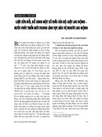
Figure 1. Fibrous tissue (appeared as green region) is segmented to calculate the ratio of its content to non-fibrous
content.
Figure 2:.Red-blue region segmentation & its properties (velocity, flow, distribution) in Doppler ultrasound image
Image Registration: Image Fusion:
manual mode (translate, rotate, resize image by
hand), semi-auto mode (pick points in a pair of
images that identify the same features or
landmarks in the images), automatic mode
(perform automatically by correlation-based
algorithms); Subtraction to analyze temporal
evolution or detect differences: manual and
semi-auto mode; Multi image Registration.
Figure 3. Auto registration mode