Nghiên cứu về lây truyền covid 19 qua dơi ăn quả, chồn, lợn và gà
Bạn đang xem bản rút gọn của tài liệu. Xem và tải ngay bản đầy đủ của tài liệu tại đây (2.18 MB, 39 trang )
The Lancet Microbe
--Manuscript Draft--
iew
ed
Experimental transmission studies of SARS-CoV-2 in fruit bats, ferrets, pigs and
chickens
THELANCETMICROBE-D-20-00091
Article Type:
Article (Original Research)
Keywords:
Sars-Cov-2; animal model; Rousettus fruit bat; ferret; pig; chicken
Corresponding Author:
Martin Beer
Friedrich-Loeffler-Institut
Greifswald-Insel Riems, GERMANY
First Author:
Kore Schlottau
Order of Authors:
Kore Schlottau
ev
Manuscript Number:
Melanie Rissmann
Jacob Schön
Julia Sehl
Claudia Wylezich
Dirk Höper
er
r
Annika Graaf
pe
Thomas C. Mettenleiter
Anne Balkema-Buschmann
Timm Harder
Christian Grund
Donata Hoffmann
ot
Angele Breithaupt
Martin Beer
GERMANY
tn
Manuscript Region of Origin:
Pr
ep
rin
Abstract:
Background
A novel zoonotic SARS-related coronavirus emerged in China at the end of 2019. The
novel SARS-CoV-2 became pandemic within weeks and the number of human
infections and severe cases is increasing. The role of potential animal hosts is still
understudied.
Methods
We intranasally inoculated fruit bats ( Rousettus aegyptiacus ; n=9), ferrets (n=9),
pigs (n=9) and chickens (n=17) with 10 5 TCID 50 of a SARS-CoV-2 isolate per
animal. Animals were monitored clinically and for virus shedding. Direct contact
animals (n=3) were included. Animals were humanely sacrificed for virological and
immune-pathohistological analysis at different time points.
Findings
Under these settings, pigs and chickens were not susceptible to SARS-CoV-2. All
swabs as well as organ samples and contact animals remained negative for viral RNA,
and none of the animals seroconverted. Rousettus aegyptiacus fruit bats experienced
a transient infection, with virus detectable by RT-qPCR, immunohistochemistry (IHC)
and in situ hybridization (ISH) in the nasal cavity, associated with rhinitis. Viral RNA
was also identified in the trachea, lung and lung associated lymphatic tissue. One of
three contact bats became infected. More efficient virus replication but no clinical signs
were observed in ferrets with transmission to all direct contact animals. Prominent viral
RNA loads of up to 10 4 viral genome copies/ml were detected in the upper
respiratory tract. Mild rhinitis was associated with viral antigen detection in the
respiratory and olfactory epithelium. Both fruit bats and ferrets developed SARS-CoV-2
Powered by Editorial Manager® and ProduXion Manager® from Aries Systems Corporation
This preprint research paper has not been peer reviewed. Electronic copy available at: />
Pr
ep
rin
tn
ot
pe
er
r
ev
iew
ed
reactive antibodies reaching neutralizing titers of up to 1:1024.
Interpretation
Pigs and chickens could not be infected intranasally by SARS-CoV-2, whereas fruit
bats showed characteristics of a reservoir host. Virus replication in ferrets resembled a
subclinical human infection with efficient spread. These animals might serve as a
useful model for further studies e.g. testing vaccines or antivirals.
Funding
Intramural funding of the German Federal Ministry of Food and Agriculture provided to
the Friedrich-Loeffler-Institut.
Powered by Editorial Manager® and ProduXion Manager® from Aries Systems Corporation
This preprint research paper has not been peer reviewed. Electronic copy available at: />
Manuscript
Experimental transmission studies of SARS-CoV-2 in fruit bats,
ferrets, pigs and chickens
iew
ed
1
2
3
4
Kore Schlottau*1, Melanie Rissmann*2, Annika Graaf*1, Jacob Schön*1, Julia Sehl3, Claudia
5
Wylezich1, Dirk Höper1, Thomas C. Mettenleiter4, Anne Balkema-Buschmann±,2, Timm
6
Harder±,1, Christian Grund±,1, Donata Hoffmann±,1, Angele Breithaupt#,3 and Martin Beer#,1
pe
er
re
v
7
8
1
Institute of Diagnostic Virology, Friedrich-Loeffler-Institut, Greifswald-Insel Riems, Germany
9
2
Institute of Novel and Emerging Infectious Diseases, Friedrich-Loeffler-Institut, Greifswald-Insel Riems,
10
Germany
11
3
12
Insel Riems, Germany
13
4
Department of Experimental Animal Facilities and Biorisk Managment, Friedrich-Loeffler-Institut, Greifswald-
Friedrich-Loeffler-Institut, Greifswald-Insel Riems, Germany
14
*,± Authors contributed equally to this work
16
# corresponding author
17
18
+49 38351 7 1200
19
20
+49 38351 7 1128
rin
tn
21
ot
15
Lancet Microbe
23
Main Text: max 3500 words: 3579
24
Abstract: max 300 words: 300
References: max 30: 30
Pr
25
ep
22
26
This preprint research paper has not been peer reviewed. Electronic copy available at: />
Abstract
28
Background
29
A novel zoonotic SARS-related coronavirus emerged in China at the end of 2019. The novel
30
SARS-CoV-2 became pandemic within weeks and the number of human infections and severe
31
cases is increasing. The role of potential animal hosts is still understudied.
32
Methods
33
We intranasally inoculated fruit bats (Rousettus aegyptiacus; n=9), ferrets (n=9), pigs (n=9) and
34
chickens (n=17) with 105 TCID50 of a SARS-CoV-2 isolate per animal. Animals were
35
monitored clinically and for virus shedding. Direct contact animals (n=3) were included.
36
Animals were humanely sacrificed for virological and immuno-pathohistological analysis at
37
different time points.
38
Findings
39
Under these settings, pigs and chickens were not susceptible to SARS-CoV-2. All swabs as
40
well as organ samples and contact animals remained negative for viral RNA, and none of the
41
animals seroconverted. Rousettus aegyptiacus fruit bats experienced a transient infection, with
42
virus detectable by RT-qPCR, immunohistochemistry (IHC) and in situ hybridization (ISH) in
43
the nasal cavity, associated with rhinitis. Viral RNA was also identified in the trachea, lung and
44
lung associated lymphatic tissue. One of three contact bats became infected. More efficient
45
virus replication but no clinical signs were observed in ferrets with transmission to all direct
46
contact animals. Mild rhinitis was associated with viral antigen detection in the respiratory and
47
olfactory epithelium. Prominent viral RNA loads of up to 104 viral genome copies/l were
48
detected in the upper respiratory tract of both species, and both species developed SARS-CoV-
49
2 reactive antibodies reaching neutralizing titers of up to 1:1024.
50
Interpretation
51
Pigs and chickens could not be infected intranasally by SARS-CoV-2, whereas fruit bats
53
pe
er
re
v
ot
rin
tn
showed characteristics of a reservoir host. Virus replication in ferrets resembled a subclinical
human infection with efficient spread. These animals might serve as a useful model for further
studies e.g. testing vaccines or antivirals.
Pr
54
ep
52
iew
ed
27
55
Funding
56
Intramural funding of the German Federal Ministry of Food and Agriculture provided to the
57
Friedrich-Loeffler-Institut.
This preprint research paper has not been peer reviewed. Electronic copy available at: />
Research in context
59
Evidence before this study
60
While the first SARS-CoV pandemic could be controlled at an early stage before substantial
61
spread occurred, SARS-CoV-2 has disseminated globally within weeks, and the number of
62
infected humans continues to increase at alarming rates. Although the pandemic is driven by
63
human-to-human transmission, the large number of infected humans also raises the question
64
whether anthropo-zoonotic infections occur by contact of infected humans with animals, which
65
may lead to further spread and endemicity of SARS-CoV-2 in companion and farmed animals.
66
However, contact with zoo and wild animals is also relevant, since bats are considered as
67
reservoir hosts. Infection of ferrets and cats by SARS-CoV has been demonstrated
68
experimentally and naturally. Field infections of pigs were also reported, while poultry did not
69
appear to be affected. In addition to exploring potentially important epidemiological animal
70
reservoirs, suitable animal models for testing vaccines and antiviral drugs are urgently required.
71
For SARS-CoV, non-human primate and ferret models were used. First reports now indicate
72
similar results for SARS-CoV-2. However, data on the susceptibility of bat species, as well as
73
detailed analyzes including viral loads and histopathology of SARS-CoV-2 in ferrets and their
74
contact animals are lacking. Furthermore, the first study on the inoculation of pigs and chickens
75
requires confirmation and extension.
76
Added value of this study
77
In our study, four relevant animal species were intranasally inoculated: fruit bats, ferrets, pigs
78
and chickens. Neither pigs (n = 9) nor chickens (n = 17) showed any signs of infection and none
79
of the contact animals became infected. This is of particular importance for risk analysis in
80
these farmed animals, which are kept in large numbers in contact with humans. Interestingly,
81
this differs to the findings reported after infection of pigs with SARS-CoV. In contrast, the virus
82
replicated in the upper respiratory tract of fruit bats, and was transmitted to contact animals.
83
This indicates that fruit bats, which are kept and bred in captivity can serve as reservoir host
84
model, but also emphasizes the risk to free-living bats e.g. in ecological bat protection
86
pe
er
re
v
ot
rin
tn
programs. Finally, ferret infections resulted in a very high replication rate of SARS-CoV-2 in
the nasal cavity, as confirmed by immunohistochemistry and in situ hybridization. The
transmission to contacts was highly efficient and high virus titers were detected in the nasal
Pr
87
ep
85
iew
ed
58
88
cavity of contacts. We demonstrate by next-generation sequencing that no viral adaptions
89
occurred during infection of ferrets with a human SARS-CoV-2 isolate. Our results suggest that
This preprint research paper has not been peer reviewed. Electronic copy available at: />
the ferret is a highly suitable model for testing vaccines and antiviral treatment for their effect
91
on viral excretion and transmission.
92
Implications of all available evidence
93
Our results are in accordance with all so far available study results, indicating a negligible risk
94
of anthropo-zoonotic transmission to pigs and chickens, but relevant for bats and ferrets. Fruit
95
bats show a different pattern of infection than ferrets, but both can serve as model animals.
96
However, ferrets next to non-human primates, most closely mimic human infection and are
97
therefore suggested as animal model for testing vaccines and antivirals.
pe
er
re
v
iew
ed
90
Pr
ep
rin
tn
ot
98
This preprint research paper has not been peer reviewed. Electronic copy available at: />
Introduction
iew
ed
99
Coronaviruses are enveloped viruses with a large single-stranded RNA genome of positive
101
polarity (ICTV; (1)). While numerous coronaviruses have been identified in animals or humans
102
(2), two recent ß-coronaviruses are remarkable: the Severe Acute Respiratory Syndrome
103
coronavirus (SARS-CoV) (3, 4); and the Middle East Respiratory Syndrome coronavirus
104
(MERS) (5, 6). Both viruses presumably originate from bats (7), but adapted to further animals
105
like palm civets (8) or dromedary camels (6) from which sporadic or sustained spill-over
106
infections occurred resulting in abundant (SARS-CoV) (9), or limited human-to-human
107
infection chains (MERS-CoV) (10), which finally could be controlled.
108
Since the end of 2019, another SARS-CoV-related zoonotic ß-coronavirus - Severe Acute
109
Respiratory Syndrome coronavirus 2 (SARS-CoV-2) – has been spreading pandemically from
110
Wuhan, China. As for SARS-CoV and MERS-CoV, ß-coronaviruses very closely related to
111
SARS-CoV-2 were found in bats (11, 12) and Pangolins (13). Whether the pandemic started by
112
a direct spill-over transmission of the SARS-CoV-2 ancestor from bats to humans or via another
113
intermediate mammalian host providing further adaptation to the human host, is still under
114
debate.
115
Due to the zoonotic origin of SARS-CoV-2 from the likely bat reservoir, several questions
116
concerning the susceptibility of animals arise: (i) susceptibility of putative reservoir hosts like
117
bats, (ii) risk of possible anthropo-zoonotic spill-over infections to farmed animals, and (iii)
118
suitable animal models of human infection to study antivirals and vaccine prototypes. Viral
119
receptor structure may be used as an important predictive factor of susceptibility: Recently it
120
was shown, that SARS-CoV and SARS-CoV-2 employ the same receptor molecule, ACE2 (14),
121
for contact with the receptor-binding-domain (RBD) of the spike (S) protein. Based on
122
molecular studies the ACE2 proteins of human primates, pigs, cats and ferrets closely
123
resembled the human ACE2 receptor. Therefore, these species may be susceptible to SARS-
124
CoV-2 infection as has been shown for SARS-CoV and MERS-CoV (15, 16). During the last
125
influenza A virus H1N1 pandemic in 2009, the virus was transmitted from humans to pigs, and
127
ot
rin
tn
is now endemic in pig holdings worldwide (17), posing a continuous risk of zoonotic spill-back
infections. The potential impact of a SARS-CoV-2 infection of pigs therefore is very high. In
this context, it is also very important to prove that chickens are not susceptible to SARS-CoV-
Pr
128
ep
126
pe
er
re
v
100
129
2. Finally, bats as a major reservoir host of ß-coronaviruses and especially SARS-CoV-related
130
viruses (18) need to be further studied to better understand the viral replication, shedding,
131
transmission or persistence in a putative reservoir host species.
This preprint research paper has not been peer reviewed. Electronic copy available at: />
Here, we intranasally inoculated fruit bats, ferrets, pigs and chickens with SARS-CoV-2 and
133
investigated virus replication and shedding, the clinical course, pathohistological changes as
134
well as transmission.
iew
ed
132
135
136
137
Pr
ep
rin
tn
ot
pe
er
re
v
138
This preprint research paper has not been peer reviewed. Electronic copy available at: />
Materials and methods
140
Ethics
141
The animal experiments were evaluated and approved by the ethics committee of the State
142
Office of Agriculture, Food safety, and Fishery in Mecklenburg – Western Pomerania (LALLF
143
M-V: LVL MV/TSD/7221.3-2-010/18-12). All procedures were carried out in approved
144
biosafety level 3 (BSL3) facilities.
145
Animals & study design
146
Twelve Egyptian fruit bats (Rousettus aegyptiacus, mixed sexes and ages, originating from the
147
FLI breeding colony), twelve ferrets (Mustela putorius, female, nine-twelve month old,
148
originating from the FLI breeding colony), twelve male pigs (Sus scrofa domesticus, nine weeks
149
old; raised by BHZP GmbH (Dahlenburg, Germany)) and twenty chickens (Gallus gallus
150
domesticus (white leghorn, five weeks old, mixed sexes, hatched from SPF-eggs (VALO
151
BioMedia GmbH, Osterholz-Scharmbeck, Germany)) were used. Fruit bats as well as pigs were
152
kept in groups of four and six in different cages and stables, respectively. Ferrets were kept
153
altogether in one cage and chickens were kept in free run conditions with nests and perches. All
154
animals were offered water ad-libitum, and were fed and checked for clinical scores daily and
155
by video supervision during the 21-day study period. All animals tested negative for SARS-
156
CoV-2 genome and antibodies prior to the experiment.
157
Nine fruit bats, ferrets and pigs were infected intranasally while the 17 chickens received oculo-
158
oronasally 105 TCID50 SARS-CoV-2 2019_nCoV Muc-IMB-1 per animal (kindly provided by
159
R. Woelfel, German Armed Forces Institute of Microbiology, Munich, Germany). The
160
inoculum was administered to both nostrils using a pipette (fruit bats, ferrets and chickens) or
161
an intranasal spraying device (pigs) (Teleflex Medical GmbH, Germany). To test viral
162
transmission by direct contact, three naïve sentinel animals were added 24 hours post
163
inoculation. Animals were monitored for body temperature (pigs, fruit bats, ferrets) and body
164
weight (fruit bats, ferrets) throughout the experiment. Viral shedding was tested on nasal
166
pe
er
re
v
ot
rin
tn
washes and rectal swabs (ferrets), oral swabs and pooled feces samples (fruit bats), nasal and
rectal swabs (pigs) or oropharyngeal and cloacal swabs (chicken) on 2, 4, 8, 12, 16, and 21 days
post infection (dpi). On day 4 (animals #1,#2), day 8 (animals #3,#4) and 12 dpi (animals
Pr
167
ep
165
iew
ed
139
168
#5,#6), two or three (chickens) inoculated animals of each species were sacrificed. All
169
remaining animals, including the sentinels, were euthanized on day 21 pi (Fig. 1). All animals
170
were subjected to autopsy. For virus detection and histopathology: nasal conchae, trachea, lung,
This preprint research paper has not been peer reviewed. Electronic copy available at: />
tracheobronchial lymph node (not for chicken), heart, liver, spleen, duodenum, colon/cecum,
172
pancreas, kidney, adrenal gland, skeletal muscle, skin, brain were collected.
173
Further materials and methods
174
For details on virus, cells, virus titration, RNA extraction, RT-qPCR, next-generation
175
sequencing,
176
hybridization, please refer to the materials&methods section in the supplement.
177
Role of the funding source
178
The funder of the study had no role in study design, data collection, data analysis, data
179
interpretation, or writing of the report. MB had full access to all the data in the study and had
180
final responsibility for the decision to submit for publication.
detection,
histopathology,
immunohistochemistry
and
in-situ
pe
er
re
v
antibody
iew
ed
171
Pr
ep
rin
tn
ot
181
This preprint research paper has not been peer reviewed. Electronic copy available at: />
Results
183
Egyptian fruit bats
184
No clinical signs (such as anorexia or respiratory signs), elevated temperatures, body weight
185
loss or mortality were observed in any of the bats.
186
Oral virus shedding was observed in infected bats from 2 to 12 dpi, with one out of the three
187
remaining infected bats still virus positive at 12 dpi (fruit bat #8), the other ones were sacrificed
188
as scheduled on 4 and 8 dpi. Oral shedding was also detected in two out of three contact animals
189
until 8 dpi (fruit bats #10 & #11, Fig 2A). Virus was isolated from one oral swab on day 2 pi
190
(101.75 TCID50/ml, fruit bat #8) (Fig 3A). Fecal shedding was observed in all three cages at 2
191
and 4 dpi with Cq values ranging from 29.54 to 36.43 (data not shown). SARS-CoV-2 genome
192
(Cq values between 23.16 and 38.97; 1.96x10⁴ to 1.32x101 genome copies/µl RNA) was
193
detected in the nasal epithelium in seven of nine infected bats sacrificed at 4, 8 and 21 dpi, with
194
one animal each giving negative results at 8 and 12 dpi respectively. Interestingly, the nasal
195
epithelium of one contact animal contained viral RNA on day 21 pi (Cq value 32.89; 3.12
196
genome copies/µl RNA). At 4 dpi, genome was also detected in respiratory tissues (trachea
197
(2/2), lung (1/2) and lung associated lymphatic tissue (2/2)) and at lower levels in heart, skin,
198
duodenum and adrenal gland (one animal at 4 dpi) and in duodenum, skin and adrenal gland
199
(one animal) on 8 dpi (Fig 2C). Virus could be cultivated from the trachea (102.25 TCID50/ml)
200
and the nasal epithelium (101.75 TCID50/ml) of fruit bat #2 at 4 dpi. For all other RT-qPCR
201
positive samples, cultivation of replicating virus was impossible.
202
SARS-CoV-2 reactive antibodies were observed in all inoculated bats by iIFA starting from 8
203
dpi as well as in one contact bat (#10) on day 21 with titers ≥16. Only a slight increase in
204
antibody levels could be observed between day 8 and day 21 (with varying titers between 16
205
and 64). Neutralizing antibodies could be detected in the same fruit bats with titers up to 64
206
(Table 1A).
207
Necropsy revealed no pathological alterations in any of the inoculated or contact bats. At 4 dpi,
209
pe
er
re
v
ot
rin
tn
minimal to mild rhinitis was found, with epithelial necrosis, edema, infiltrating lymphocytes
and neutrophils, and intraluminal cellular debris (Fig 4A). Immunohistochemistry (IHC)
revealed viral antigen, restricted to foci in the nasal respiratory epithelium and single cells of
Pr
210
ep
208
iew
ed
182
211
the non-respiratory, stratified epithelium (fruit bats #1,2; Fig. 4B,C), confirmed by in situ
212
hybridization (ISH). Although viral antigen was absent at later time points, moderate rhinitis
213
was detected at 8 dpi (fruit bats #3, #4), 12 dpi (fruit bat #6), and to a milder extent at 21 dpi
214
(fruit bats #7, #11), indicating previous replication sites. Despite the detection of viral RNA by
This preprint research paper has not been peer reviewed. Electronic copy available at: />
RT-qPCR, no viral antigen was detectable in the lung. However, single infected animals at 4, 8
216
and 12 dpi as well as one contact animal presented with interstitial, mixed cellular infiltrates
217
and in one case also with perivascular lymphocytic cuffs (Table S.1, Fig. S.1A-C). Minimally
218
increased numbers of alveolar macrophages were found at all time points. None of the other
219
organs were found positive for viral antigen and no further relevant morphological changes
220
were detected.
221
Ferrets
222
None of the ferrets showed clinical signs or loss of body weight during the study period. Body
223
temperatures remained normal.
224
Viral shedding was detected in nasal washes in eight out of nine infected ferrets between day 2
225
and day 8 pi with Cq values ranging from 21.77 to 36.35 (8.44x10³ to 0.34 genome copies/µl
226
RNA). Virus isolation was successful from nasal washes collected on days 2 pi (ferret #2,3,4:
227
102.5 – 102.875 TCID50/ml) and 4 pi (ferret #4; 102.75 TCID50/ml) (Fig. 3B). All three naïve ferrets
228
were infected by direct contact to the other inoculated ferrets. The first RT-qPCR positive nasal
229
wash sample in a contact ferret was observed on 8 dpi. Ferret #12 showed viral shedding on 8
230
and 12 dpi (Cq values 37.03 and 28.59, respectively). Ferret #11 had positive results in nasal
231
washes between day 12 and 21 pi (Cq values 37.39, 26.15 and 36.93) and ferret #10 on day 16
232
and 21 pi (Cq values 28.04 and 30.00) (Fig. 2B). Analysis of the rectal swabs showed minor
233
amounts of viral RNA in individual ferrets at singular time points with Cq values between 33.97
234
and 38.45 (data not shown).
235
The two ferrets (ferret #1,#2) sacrificed at 4 dpi were RT-qPCR positive in different tissues
236
(lung, muscle, skin, trachea, lung lymph node and colon) with the highest viral genome load in
237
the nasal conchae (Cq values 24.31 and 26.21; 1.93x10³ – 5.26x10² genome copies/µl RNA).
238
The two ferrets euthanatized at 8 dpi (ferret #3,#4) were positive in the nasal conchae (Cq values
239
34.77 and 21.57; 1.61 – 1.21x10⁴ genome copies/µl RNA). On 12 dpi, one of two ferrets was
240
also positive in the nasal conchae (ferret #6, Cq value 29.26). The last three inoculated ferrets
241
were sacrificed at 21 dpi. These animals showed only very weak RT-qPCR positivity in the
243
pe
er
re
v
ot
rin
tn
cerebrum (ferret #7, Cq value 37.78) and in the caecum (ferret #9, Cq value 37.47). The three
contact ferrets euthanized on the same day (21 dpi) were all positive in the nasal conchae (Cq
values between 26.29 and 36.51). In addition, RT-qPCR positive samples were collected from
Pr
244
ep
242
iew
ed
215
245
muscle, lung, cerebrum, cerebellum and trachea tissue, which were all positive in ferret #10 and
246
#11 whereas lung lymph node, skin and adrenal gland were only positive in one animal (Fig.
247
2D).
This preprint research paper has not been peer reviewed. Electronic copy available at: />
Antibodies against SARS-CoV-2 were detected by iIFA from day 8 pi in all inoculated ferrets
249
with varying titers (64 to 8192). One of three contact animals also showed high antibody titers
250
(ferret #12, highest reactive serum dilution 8192), whereas the others remained negative.
251
Neutralizing antibodies were observed in three inoculated ferrets (ferret #7 128; ferret #8 1024
252
and ferret #9 1024 as the highest effective serum dilution) sacrificed on day 21 pi and one
253
contact animal (ferret #12, 256) by VNT (Table 1B).
254
Post mortem examination did not identify relevant pathological alterations. At 4 dpi, viral
255
antigen was associated with rhinitis, showing epithelial degeneration and necrosis, intraluminal
256
cellular debris and mild inflammation (Fig. 4D-F). A more pronounced rhinitis developed at
257
day 8 and 12 pi. At 21 dpi, rhinitis was only slightly detectable (ferret #7) or absent (ferret
258
#8,#9). We also observed an antigen associated rhinitis in the contact ferrets (#10,#11). Viral
259
antigen was detected in the nasal cavity at days 4 pi (ferret #1,#2), 8 pi (ferret #3), and 21 pi
260
(contact ferret #10#11) in the nasal respiratory and olfactory epithelium. Remarkably, the
261
olfactory epithelium of the vomero-nasal organ was affected (ferret #11; Fig. S2A-C). IHC
262
results were confirmed by ISH (Fig. S3A-B). No viral antigen was identified in the lung. Single
263
infected animals at days 4 and 8 pi and all contact animals showed interstitial, mixed cellular
264
infiltrates and in some cases also perivascular lymphocytic cuffs (Table S1, Fig. S1D-F).
265
Minimally increased numbers of alveolar macrophages were found at all time-points. None of
266
the other organs was found positive for viral antigen, and no further relevant morphological
267
alterations were detected.
268
Pigs and chickens
269
No clinical signs, including elevated body temperatures, were observed in any of the 12 pigs or
270
20 chickens. All collected samples were negative for SARS-CoV-2 genome. SARS-CoV-2
271
reactive antibodies were not detected. Histopathology was inconspicuous (animals sacrificed at
272
4, 8, and 12 dpi) or not performed on tissues obtained from animals sacrificed at 21 dpi. Three
273
porcine cell lines (PK-15, SK-6 and ST) as well as embryonated chicken eggs inoculated with
274
SARS-CoV-2 proved to be non-permissive (data not shown).
Pr
ep
rin
tn
ot
pe
er
re
v
iew
ed
248
This preprint research paper has not been peer reviewed. Electronic copy available at: />
Discussion
276
Our study focused on four animal species, which are potentially relevant as models (fruit bats,
277
ferrets) or could pose a risk as a viral reservoir following anthropo-zoonotic spill-over
278
infections into food-producing animals (pigs, chickens).
279
Neither pigs (n = 9) nor chickens (n = 17) were susceptible to SARS-CoV-2 by intranasal or
280
oculo-oronasal infection. All swabs as well as organ samples and contact animals (three animals
281
in direct contact) remained negative for SARS-CoV-2 RNA and did not seroconvert. Non-
282
permissiveness of chickens to SARS-CoV-2 infection parallel previous reports on the lack of
283
susceptibility of chicken to SARS-CoV (20) and confirm recently reported results (21). We
284
showed that this extends to embryonated chicken eggs, which are a classical substrate for
285
isolation and propagation of a plethora of zoonotic viruses. The chicken data are also in
286
agreement with studies on the chicken ACE2 receptor (22) that contains alterations in three of
287
five critical residues (K31E; E35R, M82R). In contrast, similar predictions suggested that pigs
288
as well as ferrets would likely be susceptible to SARS-CoV-2 due to their matching ACE2
289
receptor-binding site (22). In contrast to such in silico predictions, our study as well as the report
290
by Shi et al (21) ruled out any susceptibility of pigs by the intranasal inoculation route. We
291
extend these findings further by showing non-permissiveness of three universal porcine cell
292
lines (PK-15, SK-6 and ST cells).
293
On the other hand, we present here to our knowledge first data on the intranasal inoculation of
294
nine Rousettus aegyptiacus fruit bats, which resulted in a transient infection in the respiratory
295
tract and virus shedding. SARS-CoV-2 genome could be detected by RT-qPCR in nasal
296
conchae, trachea, lung and lung lymph node in fruit bat #1 and fruit bat #2 as well as in skin
297
and duodenum of fruit bat #2, dissected on day 4 pi (Fig. 2C). Infectious virus was isolated
298
from nasal conchae and trachea tissues from the same animal. Virus shedding was detectable
299
by RT-qPCR in oral swabs up to day 12 pi, but infectious virus could only be isolated from fruit
300
bat #2 at 2 dpi (Fig 2A and 3A). In total, seven out of nine inoculated fruit bats had viral genome
301
in their nasal cavity, as confirmed by IHC and ISH at 4 dpi. Rhinitis was the detectable lesion
303
pe
er
re
v
ot
rin
tn
associated with presence of viral antigen, mainly in the respiratory epithelium. Despite the
absence of viral antigen at later time points, rhinitis was still identifiable, indicating earlier
replication sites. Some infected animals as well as contact fruit bat #10 presented with mild
Pr
304
ep
302
iew
ed
275
305
inflammation in the lung. Its occurrence and significance should be addressed in future studies,
306
because no lesion-associated antigen was detectable. Starting from 8 dpi, a weak immune-
307
response developed as demonstrated in iIFA and VNT. The virus was transmitted to one out of
This preprint research paper has not been peer reviewed. Electronic copy available at: />
the three naïve contact fruit bats (fruit bat #10). The other two naïve contact animals remained
309
seronegative. Interestingly, in fruit bat #10 an early pregnancy was determined during necropsy.
310
Several studies show a higher virus detection rate in bats during the reproductive phase,
311
probably due to the associated immunosuppression (23). ß-coronaviruses were shown to infect
312
a variety of bat species with limited clinical signs even during active virus shedding (24).
313
Moreover, low antibody titers are typical for bats (25). Although Egyptian fruit bats express
314
ACE2 in the intestine and respiratory tract, an earlier study revealed very limited evidence of
315
virus replication and seroconversion after infection with SARS-like coronaviruses, however,
316
serum samples of some of these bats, collected prior to the infection, turned out to be already
317
reactive with SARS S or N proteins (26). In the present study, SARS-CoV-2 transiently
318
replicated in particular in the respiratory epithelium as shown by RT-qPCR, IHC and ISH. Our
319
data suggest that intranasal infection of Rousettus aegyptiacus could reflect reservoir host
320
status. Furthermore, we demonstrate that bat-to-bat transmission is possible. Consequently, bats
321
are at risk of being infected anthropo-zoonotically by SARS-CoV-2. It is therefore highly
322
recommended, that during the pandemic, all contacts to bats, e.g. during research programs or
323
ecological analyses should be avoided.
324
SARS-CoV-2 replicated most efficiently in ferrets. Eight of nine intranasally infected ferrets
325
shed virus between day 2 and 8 pi. Viral genome was detected by RT-qPCR in nasal washes
326
and infectious virus isolated from two animals at 2 and 4 dpi (Fig. 2B and 3B). Only ferret #5
327
remained RT-qPCR negative during the observation period and developed only a weak iIFA
328
titer. All other inoculated ferrets showed increasing SARS-CoV-2 reactive antibodies starting
329
from day 8 pi. In general, the measured antibody levels were much higher in ferrets than in bats
330
(Table 1), indicating a more prominent virus dissemination in the infected animals. For iIFA,
331
this might also be explained by the use of different secondary antibodies. Neutralizing
332
antibodies were only detected at later time points (21 dpi), but also with high titers of up to
333
1024 in ferrets, while we detected neutralizing antibodies in bats from day 8 dpi at comparably
334
low titers of 16 – 64 (Table 1B). This might represent a reservoir host infection, which deserves
335
more detailed analysis in future studies.
pe
er
re
v
ot
rin
tn
SARS-CoV-2 was efficiently transmitted to three naïve ferrets by direct contact. In those
animals, viral RNA was present in nasal washes starting from day 12 pi and detected by RT-
Pr
337
ep
336
iew
ed
308
338
qPCR mostly in the nasal conchae, but also lung, trachea, lung lymph node or cerebrum and
339
cerebellum (Fig. 2D). Viral antigen within the upper respiratory tract was confirmed by strong
340
positive IHC and ISH in the nasal cavity. In the case of SARS-CoV, the virus was found to
341
replicate in the upper and lower respiratory tract, and the animals developed no or mild clinical
This preprint research paper has not been peer reviewed. Electronic copy available at: />
disease characterized by nasal discharge, sneezing and fever (27). We also used high-
343
throughput sequencing to analyze the complete genome of the used virus inoculum as well of
344
samples from the inoculated ferrets. Complete sequence identity demonstrates that the virus did
345
not adapt during ferret inoculation and that no additional mutations were required for an
346
efficient infection of these animals with a human SARS-CoV-2 isolate.
347
Our results are in line with two recent reports that were also able to show productive SARS-
348
CoV-2 infection of ferrets with no, or only mild clinical signs (21, 28). However, histopathology
349
and tissue tropism data were very limited in both studies. Our report adds important detailed
350
histopathology substantiating the restriction of the main SARS-CoV-2 replication site to the
351
nasal cavity. Presence of viral antigen in the nasal respiratory and olfactory epithelium,
352
including the vomero-nasal organ, was associated with rhinitis. Interestingly, the lesions were
353
still present at later time points despite absence of viral antigen. Nevertheless, no viral antigen
354
was identified in the lung, although several animals showed pulmonary inflammation.
355
In general, RT-qPCR detected viral genome in a significantly broader spectrum of tissues as
356
IHC. The differences could be explained by (i) a higher sensitivity of RT-qPCR, (ii) the
357
restriction of labelling to cell associated antigen whereas RT-qPCR detects viral RNA in blood,
358
secretions and excretions (i.e. tracheal and bronchial mucus, saliva on the fur), and not least
359
(iii) viral antigen was found in restricted foci of the nasal cavity only, that might be missed in
360
tissue sections although several areas have been analyzed. Although less sensitive, IHC is an
361
excellent tool to localize and identify infected target cells. To avoid cross contamination at
362
necropsy, instruments were washed in sodium hypochlorite-based reagents after each tissue
363
sample. Numerous extraction controls were executed and questionable results were confirmed
364
by a second RT-qPCR assay. Therefore, we assume that our RT-qPCR results are highly
365
reliable. Testing a broader tissue spectrum, including salivary glands, the lower urinary tract,
366
full gastrointestinal tract and the cerebrospinal fluid will help to increased understanding of the
367
source of viral RNA in secretions, excretions as well as in the brain.
368
In summary, farmed animals like chickens and pigs were resistant against intranasal SARS-
370
pe
er
re
v
ot
rin
tn
CoV-2 inoculation under our experimental conditions. This is relevant for risk assessment and
epidemiology of the infection. Furthermore, our study demonstrated that ferrets and Rousettus
fruits bats could be productively infected. Especially SARS-CoV-2 infection in ferrets, which
Pr
371
ep
369
iew
ed
342
372
resembles a mild infection of humans, might serve as a useful animal model for testing
373
prototypic COVID-19 vaccines and antivirals.
This preprint research paper has not been peer reviewed. Electronic copy available at: />
Acknowledgment
375
The authors are very grateful to Roman Wölfel (German Armed Forces Institute of
376
Microbiology) for providing the SARS-CoV-2 isolate used in this study. We thank Bernd
377
Köllner for generating the anti-bat monoclonal antibody. We also acknowledge Mareen Lange,
378
Christian Korthase, Silvia Schuparis, Gabriele Czerwinski and Patrick Zitzow for their
379
excellent technical assistance and Frank Klipp, Doreen Fiedler, Harald Manthei, René Siewert,
380
Christian Lipinski, Ralf Henkel and Domenique Lux for their excellent support in the animal
381
experiments.
382
Author’s contribution
383
KS, MR, AG, JS, DHo, ABB, TH and ChG performed the animal experiments. KS; MR; AG
384
and JS did molecular, serological and classical virological analyses. AB and JuS did animal
385
necropsies, AB did histopathology, immunohistochemistry and in situ hybridization analysis.
386
DHö, CW and BH added sequencing and quantification data. KS; AG, DHo, TH, TM, ABB
387
and MB designed the study. KS; MR; AG; AB and MB wrote the manuscript. All authors
388
critically evaluated and approved the manuscript.
pe
er
re
v
iew
ed
374
389
Declaration of Interests
391
All authors declare no competing interest.
ot
390
Pr
ep
rin
tn
392
This preprint research paper has not been peer reviewed. Electronic copy available at: />
References
394
395
396
397
398
399
400
401
402
403
404
405
406
407
408
409
410
411
412
413
414
415
416
417
418
419
420
421
422
423
424
425
426
427
428
429
430
431
432
433
434
435
436
437
438
439
440
441
442
1.
Masters PS, Perlmen S. Fields Virology: Chaper 28: Coronaviridae. In: Knipe DM, Howley PM,
eds. 2013;Philadelphia: Lippincott Williams & Wilkins:825-58.
2.
Fehr AR, Perlman S. Coronaviruses Methods and Protocols: Coronaviruses: An Overview of
Their Replication and Pathogenesis. In: Maier HJ, Bickerton E, Britton P, eds. 2015;Springer
Protocols:1-27.
3.
Tsang KW, Ho PL, Ooi GC, Yee WK, Wang T, Chan-Yeung M, et al. A cluster of cases of severe
acute respiratory syndrome in Hong Kong. The New England journal of medicine. 2003;348(20):197785.
4.
Peiris JSM, Lai ST, Poon LLM, Guan Y, Yam LYC, Lim W, et al. Coronavirus as a possible cause
of severe acute respiratory syndrome. Lancet. 2003;361(9366):1319-25.
5.
Zaki AM, van Boheemen S, Bestebroer TM, Osterhaus ADME, Fouchier RAM. Isolation of a
Novel Coronavirus from a Man with Pneumonia in Saudi Arabia. New Engl J Med. 2012;367(19):181420.
6.
Haagmans BL, Al Dhahiry SHS, Reusken CBEM, Raj VS, Galiano M, Myers R, et al. Middle East
respiratory syndrome coronavirus in dromedary camels: an outbreak investigation. Lancet Infectious
Diseases. 2014;14(2):140-5.
7.
Li WD, Shi ZL, Yu M, Ren WZ, Smith C, Epstein JH, et al. Bats are natural reservoirs of SARSlike coronaviruses. Science. 2005;310(5748):676-9.
8.
Song HD, Tu CC, Zhang GW, Wang SY, Zheng K, Lei LC, et al. Cross-host evolution of severe
acute respiratory syndrome coronavirus in palm civet and human. Proceedings of the National
Academy of Sciences of the United States of America. 2005;102(7):2430-5.
9.
Cherry JD, Krogstad P. SARS: the first pandemic of the 21st century. Pediatr Res.
2004;56(1):1-5.
10.
Drosten C, Meyer B, Muller MA, Corman VM, Al-Masri M, Hossain R, et al. Transmission of
MERS-Coronavirus in Household Contacts. New Engl J Med. 2014;371(9):828-35.
11.
Zhou P, Yang XL, Wang XG, Hu B, Zhang L, Zhang W, et al. A pneumonia outbreak associated
with a new coronavirus of probable bat origin. Nature. 2020.
12.
Wu F, Zhao S, Yu B, Chen YM, Wang W, Song ZG, et al. A new coronavirus associated with
human respiratory disease in China. Nature. 2020;579(7798):265-+.
13.
Zhang T, Wu Q, Zhang Z. Probable Pangolin Origin of SARS-CoV-2 Associated with the COVID19 Outbreak. Curr Biol. 2020;30(7):1346-51 e2.
14.
Hoffmann M, Kleine-Weber H, Schroeder S, Kruger N, Herrler T, Erichsen S, et al. SARS-CoV-2
Cell Entry Depends on ACE2 and TMPRSS2 and Is Blocked by a Clinically Proven Protease Inhibitor.
Cell. 2020.
15.
Gretebeck LM, Subbarao K. Animal models for SARS and MERS coronaviruses. Current
opinion in virology. 2015;13:123-9.
16.
Chen W, Yan M, Yang L, Ding B, He B, Wang Y, et al. SARS-associated coronavirus transmitted
from human to pig. Emerging infectious diseases. 2005;11(3):446-8.
17.
Novel Swine-Origin Influenza AVIT, Dawood FS, Jain S, Finelli L, Shaw MW, Lindstrom S, et al.
Emergence of a novel swine-origin influenza A (H1N1) virus in humans. The New England journal of
medicine. 2009;360(25):2605-15.
18.
Wang LF, Shi ZL, Zhang SY, Field H, Daszak P, Eaton BT. Review of bats and SARS. Emerging
infectious diseases. 2006;12(12):1834-40.
19.
Belser JA, Katz JM, Tumpey TM. The ferret as a model organism to study influenza A virus
infection. Dis Model Mech. 2011;4(5):575-9.
20.
Swayne DE, Suarez DL, Spackman E, Tumpey TM, Beck JR, Erdman D, et al. Domestic poultry
and SARS coronavirus, southern China. Emerging infectious diseases. 2004;10(5):914-6.
21.
Shi J, Wen Z, Zhong G, Yang H, Wang C, Liu R, et al. Susceptibility of ferrets, cats, dogs, and
different domestic animals to SARS-coronavirus-2. Science. 2020.
Pr
ep
rin
tn
ot
pe
er
re
v
iew
ed
393
This preprint research paper has not been peer reviewed. Electronic copy available at: />
iew
ed
22.
Wan YS, Shang J, Graham R, Baric RS, Li F. Receptor Recognition by the Novel Coronavirus
from Wuhan: an Analysis Based on Decade-Long Structural Studies of SARS Coronavirus. Journal of
virology. 2020;94(7).
23.
Drexler JF, Corman VM, Wegner T, Tateno AF, Zerbinati RM, Gloza-Rausch F, et al.
Amplification of Emerging Viruses in a Bat Colony. Emerging infectious diseases. 2011;17(3):449-56.
24.
Shi ZL, Hu ZH. A review of studies on animal reservoirs of the SARS coronavirus. Virus
research. 2008;133(1):74-87.
25.
Schountz T, Baker ML, Butler J, Munster V. Immunological Control of Viral Infections in Bats
and the Emergence of Viruses Highly Pathogenic to Humans. Front Immunol. 2017;8.
26.
van Doremalen N, Schafer A, Menachery VD, Letko M, Bushmaker T, Fischer RJ, et al. SARSLike Coronavirus WIV1-CoV Does Not Replicate in Egyptian Fruit Bats (Rousettus aegyptiacus).
Viruses. 2018;10(12).
27.
Enkirch T, von Messling V. Ferret models of viral pathogenesis. Virology. 2015;479:259-70.
28.
Kim YI, Kim SG, Kim SM, Kim EH, Park SJ, Yu KM, et al. Infection and Rapid Transmission of
SARS-CoV-2 in Ferrets. Cell Host Microbe. 2020.
29.
Wylezich C, Papa A, Beer M, Hoper D. A Versatile Sample Processing Workflow for
Metagenomic Pathogen Detection. Scientific reports. 2018;8(1):13108.
30.
Corman VM, Landt O, Kaiser M, Molenkamp R, Meijer A, Chu DKW, et al. Detection of 2019
novel coronavirus (2019-nCoV) by real-time RT-PCR. Eurosurveillance. 2020;25(3):23-30.
pe
er
re
v
443
444
445
446
447
448
449
450
451
452
453
454
455
456
457
458
459
460
461
462
463
464
Pr
ep
rin
tn
ot
465
This preprint research paper has not been peer reviewed. Electronic copy available at: />
Tables
467
Table 1: Serological evidence of SARS-CoV-2 infection in A) fruit bats and B) ferrets.
B)
477
< 1:16
472
1:128
473
1:1024
1: 1024
474
< 1:16
475
< 1:16
1:256
476
Pr
ep
478
iIFA
< 1:16
< 1:16
1:128
1:512
1:64
1:4096
1:4096
1:8192
1:4096
< 1:16
< 1:16
1:8192
rin
tn
ferret #1, day 4
ferret #2, day 4
ferret #3, day 8
ferret #4, day 8
ferret #5, day 12
ferret #6, day 12
ferret #7, day 21
ferret #8, day 21
ferret #9, day 21
ferret #10, day 21
ferret #11, day 21
ferret #12, day 21
VNT
< 1:16
< 1:16
1:32
1:32
1:32
1:16
1:64
1:32
1:32
1:16
< 1:16
< 1:16
468
VNT
< 1:16
469
< 1:16
< 1:16
470
< 1:16
471
< 1:16
pe
er
re
v
fruit bat #1, day 4
fruit bat #2, day 4
fruit bat #3, day 8
fruit bat #4, day 8
fruit bat #5, day 12
fruit bat #6, day 12
fruit bat #7, day 21
fruit bat #8, day 21
fruit bat #9, day 21
fruit bat #10, day 21
fruit bat #11, day 21
fruit bat #12, day 21
iIFA
< 1:16
< 1:16
1:16
1:16
1:16
1:32
1:64
1:32
1:64
1:16
< 1:16
< 1:16
ot
A)
iew
ed
466
This preprint research paper has not been peer reviewed. Electronic copy available at: />
Figures
iew
ed
479
pe
er
re
v
480
481
482
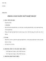
I - infection; C - contact animals; S - swabbing; A - Autopsy
483
Figure 1: Outline of the in vivo experiments with an observation period of 21 days.
485
Procedure of the trials with fruit bats, ferrets, domestic pigs and chickens are shown. Black-
486
colored animals (n=9 for each species, except chickens n=17) were inoculated intranasally (or
487
oculo-oronasally for chicken) with 105 TCID50; grey animals (n=3 for each species) depict
488
direct contact animals associated after day 1 post inoculation; black- and grey-colored animals
489
on the right were found not susceptible; red animals became infected and showed strong viral
490
shedding; rose/pink animals were infected but displayed only minute shedding of virus.
Pr
ep
rin
tn
ot
484
This preprint research paper has not been peer reviewed. Electronic copy available at: />
r
e
e
p
t
491
e
r
P
r
p
t
in
d
e
w
e
i
v
e
r
o
n
492
Figure 2: SARS-CoV-2 viral genome loads in A) oral swabs of fruits bats, B) nasal washes of ferrets, tissues collected from C) fruit bats and D) ferrets
493
experimentally infected with SARS-CoV-2 and the contact animals, respectively. Genome copies per µl RNA template were calculated based on a quantified
494
standard RNA. Red-colored animals became infected and showed strong viral shedding; rose/pink animals were infected but displayed only minute shedding of
495
virus. Organs with positive IHC results were marked with an orange ring.
This preprint research paper has not been peer reviewed. Electronic copy available at: />
iew
ed
496
Figure 3: Shedding of infectious SARS-CoV-2 in A) fruit bat oral swabs and B) ferret
498
nasal washes. Given are TCID50/ml values for every day with a RT-qPCR positive result. All
499
other samples were <101TCID50/ml for fruit bats or <102.5TCID50/ml for ferrets.
pe
er
re
v
497
500
Pr
ep
rin
tn
ot
501
This preprint research paper has not been peer reviewed. Electronic copy available at: />
iew
ed
pe
er
re
v
502
Figure 4. SARS-CoV-2 associated rhinitis and antigen detection at day 4 pi in a bat (A-C)
504
and a ferret (D-F). (A) Bat, rhinitis, with intraluminal debris (black arrow), slight mucosal
505
edema and minimal inflammation (green arrow), (B) Bat, nasal respiratory epithelium,
506
intralesional viral antigen mainly within intraluminal debris, (C) Bat, non-respiratory
507
epithelium, with single antigen positive cells, no inflammation. (D) Ferret, rhinitis, with
508
degeneration and necrosis of the respiratory epithelium (black arrow), slight mucosal edema
509
and numerous infiltrates (green arrow), (E) Ferret, nasal respiratory epithelium, intralesional,
510
abundant viral antigen, (F) Ferret, olfactory epithelium, multifocal, intralesional viral antigen
511
(A, D) Histopathology, H&E stain, bar 20 µm (B, C, E, F) Immunohistochemistry, ABC
512
method, AEC chromogen (red-brown), Mayer’s hematoxylin counter stain (blue), bar 20 µm.
Pr
515
rin
tn
514
ep
513
ot
503
This preprint research paper has not been peer reviewed. Electronic copy available at: />
Supplementary
517
Supplementary Material & Methods
518
Virus and cells
519
SARS-CoV-2 isolate 2019_nCoV Muc-IMB-1 was kindly provided by German Armed Forces
520
Institute of Microbiology (Munich, Germany). The complete sequence of this isolate is
521
available through GISAID under the accession ID_EPI_ISL_406862 and name “hCoV-
522
19/Germany/BavPat1/2020”. The virus was propagated once in Vero E6 in a mixture of equal
523
volumes of Eagle MEM (Hanks’ balanced salts solution) and Eagle MEM (Earle’s balanced
524
salts solution) supplemented with 2mM L-Glutamine, nonessential amino acids, adjusted to 850
525
mg/L, NaHCO3, 120 mg/L sodium pyruvate, 10% fetal bovine serum (FBS), pH 7.2. No
526
contaminants were detected within the virus stock preparation and the sequence identity of the
527
passaged virus (study accession number: PRJEB37671) was confirmed by metagenomics
528
analysis employing previously published high throughput sequencing procedures using
529
Illumina MiSeq sequencing (29). The virus was harvested after 72h, titrated on Vero E6 cells
530
and stored at -80°C until further use.
531
RNA extraction and detection of SARS-CoV-2
532
Total RNA was extracted from oral, nasal and rectal samples, nasal washes, fecal samples and
533
tissue samples collected at different time points using the NucleoMagVet kit
534
(Macherey&Nagel, Düren, Germany) according to the manufacturer’s instructions. Tissue
535
samples were homogenized in 1 ml cell culture medium and a 5 mm steel bead in a TissueLyser
536
(Qiagen, Hilden, Germany). Fecal samples were vortexed in sterile NaCl and the supernatant
537
was sterile filtered (22 µm) after centrifugation. Swab samples were transferred into 0.5-1 ml
538
of serum-free tissue culture media and further processed after 30 min shaking.
539
SARS-CoV-2 RNA was detected by the “E-gene Sarbeco FAM” published by Corman et al.
540
(30). The RT-qPCR reaction was prepared using the AgPath-ID-One-Step RT-PCR kit (Thermo
541
Fisher Scientific, Waltham, Massachusetts, USA) in a volume of 12.5 µl including 1 µl of E-
543
pe
er
re
v
ot
rin
tn
gene Sarbeco FAM mix, 1 µl of ß-Actin-mix2-HEX as internal control) and 2.5 µl of extracted
RNA. The reaction was performed for 10 min at 45°C for reverse transcription, 5 min at 95°C
for activation, and 42 cycles of 15 sec at 95°C for denaturation, 20 sec at 57°C for annealing
Pr
544
ep
542
iew
ed
516
545
and 30 sec at 72°C for elongation. Fluorescence was measured during the annealing phase. All
546
RT-qPCRs were performed on a BioRad real-time CFX96 detection system (Bio-Rad,
547
Hercules, USA). Absolute quantification was done using a standard quantified by the QX200
This preprint research paper has not been peer reviewed. Electronic copy available at: />