Tài liệu Photoreceptor Sensory Cilium and Associated Disorders pptx
Bạn đang xem bản rút gọn của tài liệu. Xem và tải ngay bản đầy đủ của tài liệu tại đây (920.46 KB, 18 trang )
Chapter 2
© 2012 Li et al., licensee InTech. This is an open access chapter distributed under the terms of the Creative
Commons Attribution License ( which permits unrestricted use,
distribution, and reproduction in any medium, provided the original work is properly cited.
Photoreceptor Sensory Cilium
and Associated Disorders
Linjing Li, Ozge Yildiz, Manisha Anand and Hemant Khanna
Additional information is available at the end of the chapter
1. Introduction
The primary cilium is a microtubule-based extension of the plasma membrane, which is
present in almost all cell types. Ciliary microtubules extend from a basal body (or mother
centriole), which docks at the apical membrane. Elegant studies have been carried out to
determine the mechanism that regulates the docking of the mother centriole at the
membrane for cilia formation. Cilia function as antennae of the cell to detect chemical and
physical changes of the microenvironment [1-5]. Owing to their near-ubiquitous nature, cilia
are involved in diverse cellular functions, such as patterning of left-right asymmetry (nodal
cilia), limb development, bone morphogenesis, and neurosensory functions
(mechanosensation, olfaction, and photoreception). Cilia are also implicated in several
developmental cascades, such as Wnt signaling, sonic hedgehog signaling, and platelet
derived growth factor receptor signaling pathways. Such functions of cilia are brought
about by the ability of the ciliary membrane to concentrate a specific subset of membrane
proteins in the ciliary compartment as compared to the rest of the cell membrane [6-8].
Cilia are generated by an elaborate process of formation of multiple protein complexes and
molecular motor dependent transport of membrane cargo from the proximal to the distal tip,
thereby extending the microtubule-based axoneme and the ciliary membrane. Such transport,
called Intraflagellar Transport, was initially identified in green alga Chlamydomonas reinhardtii
and is composed of more than 20 IFT subunits arranged in two distinct complexes, IFT-A and
IFT-B [9-10]. They interact with motors and transport cargo along axoneme [11]. Microtubules
are polarized with a plus end (growing tip), and a minus end (at the proximal end of cilia). The
anterograde motor Kinesin (heterotrimeric Kinesin-2 or homodimeric Kif17) mobilizes
proteins to the distal (plus) end while cytoplasmic dynein 2 carries cargos to the proximal end
of cilia [12-15]. Similarly, IFT-A and IFT-B play complementary roles in ciliary transport. The
complex B, contributing to anterograde transport, is indispensable for the ciliogenesis and
Ocular Diseases
44
maintenance. In contrast, complex A, involved in the retrograde transport, does not play
essential role in ciliary assembly [11]. Defects in IFT disturb the ciliogenesis or ciliary
maintenance. Even slight defects in the composition of the ciliary membrane or in the
generation and/or maturation of cilia result in developmental and degenerative disorders in
humans, such as Bardet-Biedl Syndrome (BBS), Joubert Syndrome (JBTS), Meckel-Gruber
Syndrome (MKS), Senior-Løken Syndrome (SLSN), Usher Syndrome (USH), renal cystic
diseases, and photoreceptor degeneration and blindness [6-7, 16-18].
2. Photoreceptor sensory cilium and its components
In photoreceptors (rods and cones), cilia are highly specialized and modified into a very
distinct part of the cell, which consists multiple membranous discs and initiates
phototransduction cascade in response to light. The details of the phototransduction cascade
in photoreceptors have been elegantly described elsewhere and will not be covered in this
chapter.
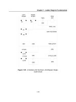
There are three major compartments that compose the sensory cilia of photoreceptor: the outer
segment (OS), transition zone (TZ) and basal body (Figure 1). Like other primary cilia,
photoreceptor cilia are 9+0 microtubule-based structures that are nucleated from the basal
body. The mother centriole consists of triplet microtubules and recruits proteins and initiates
axoneme assembly. The region adjacent to the basal body is TZ (also called connecting cilium;
CC), and consists of doublet microtubules [19-20]. These microtubules are linked to the plasma
membrane via transition fibers and Y-linkers, the two distinct structures of TZ [21]. TZ, is a
narrow conduit between OS and IS [22]. It is estimated to be 200~500 nm long and 170 nm in
diameter. TZ carries out critical transport function by acting as a gate between the IS and the
OS. The sensory OS of photoreceptors is enriched in membrane proteins, such as rhodopsin,
the cyclic nucleotide gated (CNG) channel, membrane guanylyl cyclases, and peripherin-2 [22-
25]. Moreover, the TZ is the only link between the two segments and all proteins need to be
transported via this narrow bridge-like structure to the OS. Hence, the TZ serves as a
bottleneck as well as a track to generate and maintain the sensory cilium. Several proteins,
most of which are associated with human retinal degenerative diseases, are enriched at the TZ
of photoreceptors. These include RPGR (retinitis pigmentosa GTPase regulator), CEP290, and
Nephrocystin-1 (NPHP1). The microtubules then extend in the form of axoneme. Depending
upon the species and cell-type examined, the axoneme can extend to half or full length of the
OS. The axoneme is recognized by the fact that it consists of singlet microtubules. Not much is
known about the specific function of the axoneme. However, functional analysis of RP1
(retinitis pigmentosa 1) protein that localizes specifically to the axoneme of photoreceptors
indicated that it might be involved in stabilizing the OS discs. The membranous discs arranged
in a perpendicular orientation to the axoneme and axoneme is believed to prove a structural
support to the OS discs.
In addition to maintaining a specific composition of the OS, the photoreceptors also undergo
massive protein trafficking. In fact, photoreceptors are most active neurons in the human
body and have high-energy demands. This is due to the fact that photoreceptors shed their
Photoreceptor Sensory Cilium and Associated Disorders
45
distal discs at a high rate. It is estimated that 10% of the distal tips of the OS is shed every
day by undergoing phagocytosis by the overlying retinal pigmented epithelium (RPE) cells
[26]. As no protein synthesis occurs in the OS, all components necessary for the renewal of
OS discs are synthesized in the IS and transported to the OS at a very high rate.
Approximately 2000 opsins transported to the OS per second in a normal human
photoreceptor. Even slight disturbances in the synthesis and transport of proteins to the OS
results in photoreceptor degeneration and blindness.
Figure 1. Schematic representation of a rod photoreceptor cell. The membranous discs in the outer
segment are enclosed in the plasma membrane. The photoreceptors are rich in mitochondria, which are
concentrated around the apical inner segment
3. Docking of cargo and selection at the TZ of photoreceptors
Even though the OS proteins can be targeted to the cilia, they are first docked at the basal
body or adjacent membrane. Multiple models have been proposed for the site of docking of
the cargo vesicles [27]. These propose docking directly at the basal body, docking at the
lateral plasma membrane and then movement of vesicles in the plasma membrane towards
to the ciliary compartment, or docking at a privileged domain of the apical plasma
membrane. In vertebrate photoreceptors, such a privileged domain was identified as
periciliary ridge. Opsin-laden vesicles were identified at this privileged region as well as
Ocular Diseases
46
transiently in the TZ or CC of photoreceptors [25]. More recently, several ciliary disease
proteins mutated in Usher Syndrome, were identified at the periciliary ridge and are thought
to make a connecting link between the apical plasma membrane and the ciliary membrane [28-
29]. If such a domain plays a direct role in cargo docking awaits further investigations,
specifically geared towards ascertaining the composition of this microdomain.
After gaining access to the periciliary ridge, the cargo is transported into the TZ, which acts
as a ‘check post’. Due to its elegant meshwork-like structure with Y-shaped linkers that
connect the axonemal microtubules to the plasma membrane, its composition of this
structure has been the subject of many recent studies. Remarkable studies identified a
network of multiprotein complexes of ciliary disease proteins that are found at the TZ and
act as diffusion barrier to limit the trafficking of membrane cargo into the ciliary
compartment [22, 30-33]. These proteins include RPGR, RPGR-interacting protein 1
(RPGRIP1) [34-35], CEP290/NPHP6 [36-37], MKS-associated proteins and other JBTS and
NPHP-associated proteins [6, 38]. Interestingly, these proteins exist in discrete multiprotein
complexes at the TZ. A direct role of TZ proteins in acting as a barrier was established when
Witman and colleagues showed that mutation in Chlamydomonas CEP290 causes
accumulation of non-ciliary membrane proteins to enter cilia and vice versa [39]. However,
such a function of CEP290 in photoreceptors still needs to be investigated.
4. Ciliary disorders of retina (retinal ciliopathies)
As the OS of photoreceptors is a sensory cilium, the degenerative diseases that affect the
formation or function of the OS can be categorized as a ciliary disorder. However, for
simplicity, we will discuss only those cilia-dependent retinopathies that occur due to defects
in ciliary TZ proteins and result in defective trafficking of proteins to the OS. Inactivation of
the IFT in conditional Kif3a
-/-
mice and Tg737
orpk
, a hypomorphic allele of IFT88, results in
opsin accumulation in the IS [40-41]. Mutations in rhodopsin that affect its trafficking to OS
are associated with degenerative blindness disorders of the retina [42-47]. Moreover,
ablation of IFT subunit IFT20, which localizes to Golgi and cilia, also results in entrapment
of opsins in the IS [48]. Ciliary proteins RP1 and RPGRIP1, mutations in which result in
RP/LCA are required for cilia-dependent OS generation [35, 49-50]. Pleiotropic disorders,
such as Senior-Loken Syndrome, Joubert Syndrome, and Bardet-Biedl Syndrome, are also
caused by mutations in ciliary proteins and share retinal degeneration as a common
phenotype [51-53] (Table 1). In this chapter, we will specifically discuss RPGR and RP2,
which are mutated in X-linked forms of retinopathies and CEP290, which is a frequent cause
of Leber congenital amaurosis (LCA), a childhood blindness disorder (Figure 2).
4.1. Non-syndromic retinal ciliopathies
Retinitis Pigmentosa (RP). RP, detected in 1:3000 people worldwide, is a group of severe
blindness disorders that is caused by progressive loss of rod and cone photoreceptors. It is
inherited in autosomal recessive, autosomal dominant as well as X-linked manner. Patients
exhibit symptoms of night blindness and loss of peripheral vision (due to rod death) in the
Photoreceptor Sensory Cilium and Associated Disorders
47
first two decades of life, which is followed by complete blindness due to loss of cone
photoreceptors [54-55]. Loss of cones can either be due to the fact that the causative gene is
also expressed in cone photoreceptors or due to starvation or loss of availability of trophic
factors secreted from the rods (majority cell type in photoreceptor layer; 95-97%) if the
mutation is in a rod-specific gene [56-57].
Figure 2. Schematic representation of the localization of the ciliary proteins being discussed in this
chapter. As shown, RPGR localizes to the transition zone and basal body and RP1 is concentrated at the
distal axoneme, which extends into the outer segment. RPGRIP1 tethers RPGR at the transition zone.
RP2 is detected at the Golgi as well as transition zone in photoreceptors. CEP290/NPHP6 is detected at
the transition zone, basal body, as well as in the cytosol.
Some forms of RP are caused by defects in genes that encode for ciliary proteins. These
include RPGR, RP1, RP2, and TOPORS [50, 56-60]. RP1 and TOPORS are two ciliary proteins
mutated in adRP. However, they localize to distinct ciliary compartments: RP1 localizes to
the axoneme whereas TOPORS is concentrated in the basal body and transition zone of
photoreceptors (Figure 2). The RPGR and RP2 genes are mutated in X-linked forms of RP
and together account for more than 90% of XLRP cases [61-64]. Among these, RPGR
mutations are found in 70-80% of XLRP and more than 25% of simplex RP males with no
family history. On the other hand, RP2 mutations account for 6-10% of XLRP cases. There is
considerable clinical heterogeneity among cases of XLRP, which has affected the ability to
differentiate between RPGR and RP2 patients in the clinic. This has prompted investigations
into genotype-phenotype correlation studies. Such studies are relatively well documented
for RPGR patients owing to their majority occurrence as compared to RP2 mutations [65-67].
Ocular Diseases
48
Nonetheless, recently, a comprehensive analysis of a large group of RP2 patients revealed
interesting observations: a majority of RP2 patients seem to exhibit an early involvement of
the macula (the central region of the retina) [68].
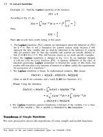
RPGR: The RPGR gene consists of 19 exons and encodes for multiple alternatively spliced
transcripts. There are two major transcripts: RPGR
1-19
and RPGR
ORF15
. As the name suggests,
the RPGR
1-19
isoform consists of exons 1-19 whereas RPGR
ORF15
isoform consists of exons 1-15
and terminates in intron 15. Both these isoforms therefore, contain a common amino-terminal
part comprising of exons 1-15. A part of this region, encoded by exons 2-11 contains a domain
of the protein that is homologous to RCC1 (regulator of chromosome condensation 1), a
guanine nucleotide exchange factor (GEF) for small GTPases involved in nucleocytoplasmic
trafficking of proteins. This domain of RPGR is termed RCC1-like domain (RLD). The
carboxyl-terminal region is distinct between these two isoforms. While the RPGR
1-19
isoform
possesses an isoprenylation motif at the extreme carboxyl-terminus, the RPGR
ORF15
isoform
encodes for an unusual stretch of Glutamic acid and Glycine rich (Glu/Gly rich) domain
(Figure 3). At DNA level, the terminal exon of this isoform contains purine-rich repeats [60, 62,
64, 69]. Ablation of the Rpgr gene in mice affects opsin trafficking and results in photoreceptor
degeneration, starting at around 6 months of age [70]. Similar phenotype was detected in two
naturally occurring canine models of RPGR mutation, although the severity of disease was
different in the two mutants [71]. First direct correlation of a function of RPGR in cilia was
obtained when it was shown that RPGR localizes predominantly to the TZ of photoreceptors
and interacts with other ciliary and transport proteins [72-73]. More recently, it was found that
silencing of rpgr in zebrafish embryos results in shorter cilia and developmental anomalies,
reminiscent of ciliary dysfunction [74-75]. These findings indicate that RPGR is involved in
regulating the trafficking of proteins at the TZ. Mechanistic insights into RPGR function were
obtained when it was shown that RPGR possesses enzymatic activity. RPGR acts as a GEF for
the small GTPase RAB8A, which is involved in cilia formation and maturation. As a GEF,
RPGR catalyzes the conversion of the inactive, GDP-bound RAB8A to active GTP-RAB8A to
facilitate the trafficking of cargo vesicles [76]. The precise function of RPGR as a GEF in
photoreceptors still needs to be delineated.
RP2: The RP2 gene is composed of 5 exons and encodes a protein of 350 amino acids. The
structure of RP2 reveals two major domains: an amino-terminal domain homologous to
tubulin binding cofactor C (TBCC) homology domain and a carboxyl-terminal nucleoside
diphosphate kinase domain [77-79] (Figure 3). The purified RP2 protein possesses GTPase
activating protein (GAP) activity towards the small GTPase ARL3 (ADP Ribosylation Factor-
Like protein 3). As a GAP, RP2 assists in the conversion of GTP-bound ARL3 to ARL3-GDP
[80]. Although some human mutations affect this association or activity, the precise role of
RP2 as a GAP in photoreceptors is still not clear. The amino terminus of RP2 is palmitoylated
and myristoylated and hence, may associate with cell membrane. In fact, RP2 has been found
to associate with the plasma membrane of cells and of photoreceptors [81]. In addition, RP2 is
also present at the basal body of primary cilia and undergoes trafficking into the cilia like IFT
[59]. RP2 interacts with ciliary protein polycystin-2 and assists in the trafficking of polycystin-
2 to the cilia. Recent studies have shown that ciliary localization of RP2 is regulated by
Photoreceptor Sensory Cilium and Associated Disorders
49
importins, proteins involved in nucleocytoplasmic trafficking [82]. These data suggest a
potential role of such machinery in regulating protein import into the cilia. Silencing of RP2
in cells results in the swelling of the distal tip of the cilium but spares the rate of trafficking of
the IFT machinery. Further investigation revealed that RP2 is involved in the secretion of
polycystin-2 from ciliary tip to the external microenvironment. One possible scenario is that
RP2 may not be directly involved in the secretion rather assists in the trafficking and delivery
of an accessory cargo that is required for the secretion of polysystin-2 and other such proteins
from the ciliary tip. In photoreceptors, RP2 also localizes to the basal body, TZ as well as
Golgi. Silencing of RP2 was also shown to fragment Golgi and may affect Golgi to cilia
trafficking in cells [89]. The in vivo effect of ablation of RP2 in photoreceptors will provide
critical clues to its involvement in cilia formation, function and protein trafficking.
Leber congenital amaurosis (LCA). LCA is considered the most severe form of retinal
degenerative disease that occurs in the childhood or early adulthood, with an incidence of 1
in 30,000 births worldwide. Defective retina exhibits perturbations in the initial
development of photoreceptors [83]. Like RP, LCA also exhibits considerable genetic and
clinical heterogeneity. To date, mutations in 18 genes have been identified to cause LCA
(RetNet, Of these, four genes, CEP290, RPGR-
interacting protein 1 (RPGRIP1), LCA5 or lebercilin and Tubby-like protein 1, encode for
ciliary proteins. We will discuss CEP290 and RPGRIP1 below.
CEP290. Mutations in the cilia-centrosomal protein CEP290 are frequently observed in LCA,
with an incidence of 22-25% cases [37]. The CEP290 gene consists of 55 exons and encodes a
protein of 2,479 amino acids (Figure 3). The CEP290 is a multidomain protein and consists of
several coiled-coil domains. Involvement of CEP290 in early onset retinal degeneration was
determined when a naturally occurring mouse model called rd16 (retinal degeneration 16) was
identified to carry an in frame deletion in the Cep290 gene. The rd16 mouse exhibits early onset
severe retinal degeneration, characteristic of LCA in humans, and is accompanied by partial
mislocalization of RPGR to the IS. The domain of CEP290 that is deleted in the rd16 mouse is
termed DRD (deleted in rd16 domain) [84]. The deletion renders the CEP290 protein prone to
degradation; however, expression of truncated CEP290 protein can be detected in the retina
and other tissues in the rd16 mouse [36]. CEP290 localizes predominantly to the CC/TZ of
photoreceptors and interacts with selected ciliary and transport assemblies, including retinal
disease proteins Retinitis Pigmentosa GTPase Regulator (RPGR) and RPGR-interacting protein
(RPGRIP1), which are mutated in RP and LCA, respectively [36].
In cell culture studies, CEP290 has been shown to regulate cilia assembly program by
modulating the localization of RAB8A and Pericentriolar Material 1 (PCM1) [85-86].
Additionally, studies using Chlamydomonas CEP290 indicated that it is involved in the
stabilization of the diffusion barrier formed by the Y-linkers [39]. It was recently
demonstrated that CEP290 interacts with a novel ciliary protein RKIP (Raf-1 Kinase
Inhibitory Protein) and modulates its intracellular protein levels. Silencing of cep290 in
zebrafish or mutation in the rd16 retina results in aberrant accumulation of RKIP; high levels
of RKIP subsequently result in mislocalization of RAB8A [84]. Moreover, CEP290 interacts
with BBS6; relative dosage of the two proteins seems to be critical in modulating the
Ocular Diseases
50
formation of OS, cochlear cilia, and olfactory cilia [87]. These studies further demonstrated
the diverse roles of CEP290 in modulating the formation, maturation, and function of cilia.
Figure 3. Schematic representation of the primary structure of RPGR, RP2, and CEP290. The two major
isoforms of RPGR: RPGR
1-19
and RPGR
ORF15
are depicted. The RCC1-like Domain (RLD) is encoded by
exons 2-11 of RPGR. The RPGR
1-19
isoform possesses a carboxyl terminal isoprenylation (IsoPr) site. The
RP2 protein consists of amino terminal myristoylation /palmitoylation (My/Pa) site, tubulin binding
cofactor C (TBCC) domain and a nucleoside diphosphate kinase (NDK) domain. The CEP290 protein is
a multidomain molecule. Both human and mouse CEP290 protein are shown. In rd16 mouse, there is a
deletion in the myosin-tail homology domain of the CEP290 protein. SMC: Structural Maintenance of
Chromosomes; CC: coiled coil.
Photoreceptor Sensory Cilium and Associated Disorders
51
RPGRIP1. RPGRIP1 is a ciliary protein that associates directly with the TZ microtubules.
Mutations in RPGRIP1 have been identified in a small percentage of LCA cases. In mice,
ablation of the Rpgrip1 gene results in defective OS development and early onset retinal
degeneration. RPGRIP1 was identified as an interacting partner of RPGR in photoreceptors.
Like rd16 retina, the Rpgrip1
-/-
mouse retina exhibits mislocalization of RPGR to the IS of
photoreceptors and its absence from the TZ. These studies indicate that RPGRIP1 tethers
RPGR to the TZ. In addition to RPGR, RPGRIP1 also directly interacts with NPHP4; disease-
causing mutations in both these proteins perturb this interaction [35, 88].
Syndromic Ciliopathies. In addition to non-syndromic retinal cilipathies described above,
photoreceptor degeneration is a common feature in multiple syndromic ciliopathies, such as
Senior-Løken Syndrome (cystic kidneys and retinopathy), Joubert Syndrome (cerebellar
vermis hypoplasia, cystic kidneys, and retinal coloboma) and Bardet-Biedl Syndrome (BBS;
obesity, mental retardation, polydactyly and retinal degeneration) [6]. Interestingly, some of
the proteins described above are also mutated in syndromic ciliopathies and/or associate
with other ciliopathy proteins in the cilia. For example, some RPGR patients exhibit extra-
retinal phenotypes, such as hearing defects, respiratory infections, sperm dysfunction, and
primary cilia dyskinesia. CEP290, on the other hand, is also mutated in syndromic
ciliopathies JBTS, MKS, and BBS.
Joubert Syndrome (JBTS). JBTS is an autosomal recessive disorder characterized by cerebellar
vermis hypoplasia and retinal coloboma. A characteristic clinical feature of JBTS is the
appearance of ‘molar tooth sign’, which represents a malformation of midbrain-hindbrain
junction. Mutations in several ciliary proteins, such as CEP290/NPHP6, NPHP3,
RPGRIP1L/NPHP8, AHI1, MKS3, and NPHP1 are associated with JBTS.
Meckel-Gruber Syndrome (MKS). MKS is characterized by embryonic lethality as a result of
malformation or malfunction of multiple organs during development. Some characteristic
clinical features include microphthalmia (small eye), renal dysplasia, polydactyly, and situs
inversus. Interestingly, some of the genes that are mutated in JBTS are also associated with
MKS. These include CEP290/NPHP6, RPGRIP1L/NPHP8, MKS1, MKS3, CC2D2A, and
TMEM216. It has now been demonstrated that the type of mutation, location of the mutation
and the relative combination of the different alleles can determine the outcome of the
disorder.
Senior-Løken and Bardet-Biedl Syndromes. Senior-Løken Syndrome (SLSN) is characterized by
renal cystic disease Nephronophthisis (NPHP) and retinal degeneration. Mutations in
NPHP5 (or nephroretinin) are associated with SLSN; 100% of NPHP5 patients exhibit retinal
degeneration. It was demonstrated that NPHP5 localizes to the cilia and interacts with
RPGR in the retina. The retinal phenotype is partly attributed to the perturbed interaction of
NPHP5 with RPGR in photoreceptors. Bardet-Biedl Syndrome (BBS), on the other hand,
involves retinal degeneration, cystic renal disease, cognitive impairment, obesity, infertility
and polydactyly as some of the main features. To date, mutations in 16 genes, all of which
encode for ciliary proteins have been identified in BBS. These include BBS1-BBS12, MKS1,
CEP290, SDCCAG8, and SEPT7.
Ocular Diseases
52
In addition to the above-mentioned syndromic ciliopathies, there are several other disorders
that have been elegantly described elsewhere and are not discussed in this chapter. All these
disorders result from defective ciliary development or function. As cilia are involved in
regulating numerous signaling cascades, including Wnt signaling, planar cell polarity,
hedgehog signaling and cell cycle control, defects in these pathways have also been
implicated as a cause of associated disorders. The involvement of signaling cascades in
photoreceptor ciliary development and function is not completely understood.
Table 1. This table depicts selected diseases classified as cilia dependent retinopathies, including non-
syndromic as well as syndromic forms. Notably, retinal degeneration is a commonly occurring
phenotype in all these disorders.
5. Conclusion
As a number of retinal ciliopathy proteins have now been identified the TZ of
photoreceptors, the next step is now to delineate the mechanism by which these proteins
modulate the function of the TZ and regulate photoreceptor OS development and function.
The existence of discrete multiprotein complexes at the TZ indicates that these complexes
are involved in the selection and trafficking of specific cargo moieties to the OS. Mutations
in the constituent proteins may impair the function of some of the complexes and trafficking
Photoreceptor Sensory Cilium and Associated Disorders
53
of cognate cargo while other complexes may function normally to extend the life of the
photoreceptor. However, if the ciliary protein mutated in disease were involved in the
trafficking of proteins regulating the development of OS discs, such as rhodopsin, one
would expect a severe and early onset retinal degeneration. It has been shown that RPGR,
NPHP proteins and BBS proteins (BBSome) exist in multiprotein complexes and regulate
ciliary trafficking.
Some of the TZ proteins possess enzymatic activity. As discussed above, RPGR is a GEF for
RAB8A while RP2 is a GAP for ARL3. Such activity of these proteins may impart specificity
to the cargo vesicle docking and fusion to the ciliary membrane for crossing the TZ barrier.
Moreover, modulating the activity of these proteins may provide insights into developing
therapeutic paradigms for associated disorders. It should however, be noted that some TZ
proteins are also present in other subcellular compartments of the cell. For example, some
RPGR isoforms are detected in the basal body and Golgi; RP2 localizes to Golgi and CEP290
localizes to the cytosol and basal body, in addition to the TZ. These observations beg the
question: Are these proteins also involved in extraciliary functions or are these proteins
participate in alternative pathways to ultimately regulate cilia dependent cascades. It was
recently shown that CEP290 interacts with RKIP, which is involved in modulating MAP
Kinase signaling cascades. In addition, CEP290 modulates intracellular levels of RKIP and
likely controls its degradation. Hence, CEP290’s involvement in intracellular signaling and
in protein degradation pathways may be linked to cilia formation or function. However,
further investigations are necessary to establish such links and to further delineate the roles
of TZ proteins in regulating protein trafficking and photoreceptor OS development and
function.
Author details
Linjing Li, Ozge Yildiz, Manisha Anand and Hemant Khanna
*
Department of Ophthalmology, University of Massachusetts Medical School, Worcester, MA, USA
Acknowledgement
This work is supported by grants from the National Institutes of Health, Foundation
Fighting Blindness, and Worcester foundation (to HK).
6. References
[1] Pazour, G. J. and Bloodgood, R. A. Targeting proteins to the ciliary membrane Curr Top
Dev Biol 2008; 85 115-149
[2] Gherman, A., Davis, E. E. and Katsanis, N. The ciliary proteome database: an integrated
community resource for the genetic and functional dissection of cilia Nat Genet 2006;
38(9) 961-962
*
Corresponding author
Ocular Diseases
54
[3] Li, J. B., Gerdes, J. M., Haycraft, C. J., Fan, Y., Teslovich, T. M., May-Simera, H., Li, H.,
Blacque, O. E., Li, L., Leitch, C. C., Lewis, R. A., Green, J. S., Parfrey, P. S., Leroux, M. R.,
Davidson, W. S., Beales, P. L., Guay-Woodford, L. M., Yoder, B. K., Stormo, G. D.,
Katsanis, N. and Dutcher, S. K. Comparative genomics identifies a flagellar and basal
body proteome that includes the BBS5 human disease gene Cell 2004; 117(4) 541-552
[4] Pazour, G. J. Comparative genomics: prediction of the ciliary and basal body proteome
Curr Biol 2004; 14(14) R575-577
[5] Sorokin, S. Centrioles and the formation of rudimentary cilia by fibroblasts and smooth
muscle cells J Cell Biol 1962; 15(363-377
[6] Hildebrandt, F., Benzing, T. and Katsanis, N. Ciliopathies N Engl J Med 2011; 364(16)
1533-1543
[7] Singla, V. and Reiter, J. F. The primary cilium as the cell's antenna: signaling at a sensory
organelle Science 2006; 313(5787) 629-633
[8] Yoder, B. K. More than just the postal service: novel roles for IFT proteins in signal
transduction Dev Cell 2006; 10(5) 541-542
[9] Kozminski, K. G., Johnson, K. A., Forscher, P. and Rosenbaum, J. L. A motility in the
eukaryotic flagellum unrelated to flagellar beating Proc Natl Acad Sci U S A 1993;
90(12) 5519-5523
[10] Rosenbaum, J. Intraflagellar transport Curr Biol 2002; 12(4) R125
[11] Ishikawa, H. and Marshall, W. F. Ciliogenesis: building the cell's antenna Nat Rev Mol
Cell Biol 2011; 12(4) 222-234
[12] Blacque, O. E., Cevik, S. and Kaplan, O. I. Intraflagellar transport: from molecular
characterisation to mechanism Front Biosci 2008; 13(2633-2652
[13] Scholey, J. M. Intraflagellar transport motors in cilia: moving along the cell's antenna J
Cell Biol 2008; 180(1) 23-29
[14] Cole, D. G. Intraflagellar transport in the unicellular green alga, Chlamydomonas
reinhardtii Protist 2003; 154(2) 181-191
[15] Pedersen, L. B. and Rosenbaum, J. L. Intraflagellar transport (IFT) role in ciliary
assembly, resorption and signalling Curr Top Dev Biol 2008; 85(23-61
[16] Badano, J. L., Mitsuma, N., Beales, P. L. and Katsanis, N. The ciliopathies: an emerging
class of human genetic disorders Annu Rev Genomics Hum Genet 2006; 7(125-148
[17] Sloboda, R. D. A healthy understanding of intraflagellar transport Cell Motil
Cytoskeleton 2002; 52(1) 1-8
[18] Murga-Zamalloa, C. A., Swaroop, A. and Khanna, H. RPGR-containing protein
complexes in syndromic and non-syndromic retinal degeneration due to ciliary
dysfunction J Genet 2009; 88(4) 399-407
[19] Besharse, J. C., Hollyfield, J. G. and Rayborn, M. E. Photoreceptor outer segments:
accelerated membrane renewal in rods after exposure to light Science 1977; 196(4289)
536-538
[20] De Robertis, E. Electron microscope observations on the submicroscopic organization of
the retinal rods J Biophys Biochem Cytol 1956; 2(3) 319-330
[21] Silverman, M. A. and Leroux, M. R. Intraflagellar transport and the generation of
dynamic, structurally and functionally diverse cilia Trends Cell Biol 2009; 19(7) 306-316
Photoreceptor Sensory Cilium and Associated Disorders
55
[22] Insinna, C. and Besharse, J. C. Intraflagellar transport and the sensory outer segment of
vertebrate photoreceptors Dev Dyn 2008; 237(8) 1982-1992
[23] Huttl, S., Michalakis, S., Seeliger, M., Luo, D. G., Acar, N., Geiger, H., Hudl, K., Mader,
R., Haverkamp, S., Moser, M., Pfeifer, A., Gerstner, A., Yau, K. W. and Biel, M.
Impaired channel targeting and retinal degeneration in mice lacking the cyclic
nucleotide-gated channel subunit CNGB1 J Neurosci 2005; 25(1) 130-138
[24] Baehr, W., Karan, S., Maeda, T., Luo, D. G., Li, S., Bronson, J. D., Watt, C. B., Yau, K. W.,
Frederick, J. M. and Palczewski, K. The function of guanylate cyclase 1 and guanylate
cyclase 2 in rod and cone photoreceptors J Biol Chem 2007; 282(12) 8837-8847
[25] Papermaster, D. S., Schneider, B. G. and Besharse, J. C. Vesicular transport of newly
synthesized opsin from the Golgi apparatus toward the rod outer segment.
Ultrastructural immunocytochemical and autoradiographic evidence in Xenopus
retinas Invest Ophthalmol Vis Sci 1985; 26(10) 1386-1404
[26] Young, R. W. The renewal of photoreceptor cell outer segments J Cell Biol 1967; 33(1)
61-72
[27] Nachury, M. V., Seeley, E. S. and Jin, H. Trafficking to the ciliary membrane: how to get
across the periciliary diffusion barrier? Annu Rev Cell Dev Biol 2010; 26 59-87
[28] Maerker, T., van Wijk, E., Overlack, N., Kersten, F. F., McGee, J., Goldmann, T., Sehn, E.,
Roepman, R., Walsh, E. J., Kremer, H. and Wolfrum, U. A novel Usher protein network
at the periciliary reloading point between molecular transport machineries in vertebrate
photoreceptor cells Hum Mol Genet 2008; 17(1) 71-86
[29] Yang, J., Liu, X., Zhao, Y., Adamian, M., Pawlyk, B., Sun, X., McMillan, D. R., Liberman,
M. C. and Li, T. Ablation of whirlin long isoform disrupts the USH2 protein complex
and causes vision and hearing loss PLoS Genet 2010; 6(5) e1000955
[30] Besharse, J. C., Forestner, D. M. and Defoe, D. M. Membrane assembly in retinal
photoreceptors. III. Distinct membrane domains of the connecting cilium of developing
rods J Neurosci 1985; 5(4) 1035-1048
[31] Horst, C. J., Forestner, D. M. and Besharse, J. C. Cytoskeletal-membrane interactions: a
stable interaction between cell surface glycoconjugates and doublet microtubules of the
photoreceptor connecting cilium J Cell Biol 1987; 105(6 Pt 2) 2973-2987
[32] Rohlich, P. The sensory cilium of retinal rods is analogous to the transitional zone of
motile cilia Cell Tissue Res 1975; 161(3) 421-430
[33] Horst, C. J., Johnson, L. V. and Besharse, J. C. Transmembrane assemblage of the
photoreceptor connecting cilium and motile cilium transition zone contain a common
immunologic epitope Cell Motil Cytoskeleton 1990; 17(4) 329-344
[34] Dryja, T. P., Adams, S. M., Grimsby, J. L., McGee, T. L., Hong, D. H., Li, T., Andreasson,
S. and Berson, E. L. Null RPGRIP1 alleles in patients with Leber congenital amaurosis
Am J Hum Genet 2001; 68(5) 1295-1298
[35] Zhao, Y., Hong, D. H., Pawlyk, B., Yue, G., Adamian, M., Grynberg, M., Godzik, A. and
Li, T. The retinitis pigmentosa GTPase regulator (RPGR)- interacting protein:
subserving RPGR function and participating in disk morphogenesis Proc Natl Acad Sci
U S A 2003; 100(7) 3965-3970
Ocular Diseases
56
[36] Chang, B., Khanna, H., Hawes, N., Jimeno, D., He, S., Lillo, C., Parapuram, S. K., Cheng,
H., Scott, A., Hurd, R. E., Sayer, J. A., Otto, E. A., Attanasio, M., O'Toole, J. F., Jin, G.,
Shou, C., Hildebrandt, F., Williams, D. S., Heckenlively, J. R. and Swaroop, A. In-frame
deletion in a novel centrosomal/ciliary protein CEP290/NPHP6 perturbs its interaction
with RPGR and results in early-onset retinal degeneration in the rd16 mouse Hum Mol
Genet 2006; 15(11) 1847-1857
[37] den Hollander, A. I., Koenekoop, R. K., Yzer, S., Lopez, I., Arends, M. L., Voesenek, K.
E., Zonneveld, M. N., Strom, T. M., Meitinger, T., Brunner, H. G., Hoyng, C. B., van den
Born, L. I., Rohrschneider, K. and Cremers, F. P. Mutations in the CEP290 (NPHP6)
gene are a frequent cause of Leber congenital amaurosis Am J Hum Genet 2006; 79(3)
556-561
[38] Sang, L., Miller, J. J., Corbit, K. C., Giles, R. H., Brauer, M. J., Otto, E. A., Baye, L. M.,
Wen, X., Scales, S. J., Kwong, M., Huntzicker, E. G., Sfakianos, M. K., Sandoval, W.,
Bazan, J. F., Kulkarni, P., Garcia-Gonzalo, F. R., Seol, A. D., O'Toole, J. F., Held, S.,
Reutter, H. M., Lane, W. S., Rafiq, M. A., Noor, A., Ansar, M., Devi, A. R., Sheffield, V.
C., Slusarski, D. C., Vincent, J. B., Doherty, D. A., Hildebrandt, F., Reiter, J. F. and
Jackson, P. K. Mapping the NPHP-JBTS-MKS Protein Network Reveals Ciliopathy
Disease Genes and Pathways Cell 2011; 145(4) 513-528
[39] Craige, B., Tsao, C. C., Diener, D. R., Hou, Y., Lechtreck, K. F., Rosenbaum, J. L. and
Witman, G. B. CEP290 tethers flagellar transition zone microtubules to the membrane
and regulates flagellar protein content J Cell Biol 2010; 190(5) 927-940
[40] Marszalek, J. R., Liu, X., Roberts, E. A., Chui, D., Marth, J. D., Williams, D. S. and
Goldstein, L. S. Genetic evidence for selective transport of opsin and arrestin by kinesin-
II in mammalian photoreceptors Cell 2000; 102(2) 175-187
[41] Pazour, G. J., Baker, S. A., Deane, J. A., Cole, D. G., Dickert, B. L., Rosenbaum, J. L.,
Witman, G. B. and Besharse, J. C. The intraflagellar transport protein, IFT88, is essential
for vertebrate photoreceptor assembly and maintenance J Cell Biol 2002; 157(1) 103-113
[42] Deretic, D., Schmerl, S., Hargrave, P. A., Arendt, A. and McDowell, J. H. Regulation of
sorting and post-Golgi trafficking of rhodopsin by its C-terminal sequence QVS(A)PA
Proc Natl Acad Sci U S A 1998; 95(18) 10620-10625
[43] Deretic, D., Williams, A. H., Ransom, N., Morel, V., Hargrave, P. A. and Arendt, A.
Rhodopsin C terminus, the site of mutations causing retinal disease, regulates
trafficking by binding to ADP-ribosylation factor 4 (ARF4) Proc Natl Acad Sci U S A
2005; 102(9) 3301-3306
[44] Mazelova, J., Astuto-Gribble, L., Inoue, H., Tam, B. M., Schonteich, E., Prekeris, R.,
Moritz, O. L., Randazzo, P. A. and Deretic, D. Ciliary targeting motif VxPx directs
assembly of a trafficking module through Arf4 EMBO J 2009; 28(3) 183-192
[45] Sung, C. H., Makino, C., Baylor, D. and Nathans, J. A rhodopsin gene mutation
responsible for autosomal dominant retinitis pigmentosa results in a protein that is
defective in localization to the photoreceptor outer segment J Neurosci 1994; 14(10)
5818-5833
Photoreceptor Sensory Cilium and Associated Disorders
57
[46] Tai, A. W., Chuang, J. Z., Bode, C., Wolfrum, U. and Sung, C. H. Rhodopsin's carboxy-
terminal cytoplasmic tail acts as a membrane receptor for cytoplasmic dynein by
binding to the dynein light chain Tctex-1 Cell 1999; 97(7) 877-887
[47] Colley, N. J., Cassill, J. A., Baker, E. K. and Zuker, C. S. Defective intracellular transport
is the molecular basis of rhodopsin-dependent dominant retinal degeneration Proc Natl
Acad Sci U S A 1995; 92(7) 3070-3074
[48] Keady, B. T., Le, Y. Z. and Pazour, G. J. IFT20 is required for opsin trafficking and
photoreceptor outer segment development Mol Biol Cell 2011; 22(7) 921-930
[49] Liu, Q., Lyubarsky, A., Skalet, J. H., Pugh, E. N., Jr. and Pierce, E. A. RP1 is required for
the correct stacking of outer segment discs Invest Ophthalmol Vis Sci 2003; 44(10) 4171-
4183
[50] Liu, Q., Zuo, J. and Pierce, E. A. The retinitis pigmentosa 1 protein is a photoreceptor
microtubule-associated protein J Neurosci 2004; 24(29) 6427-6436
[51] Badano, J. L., Mitsuma, N., Beales, P. L. and Katsanis, N. The Ciliopathies: An Emerging
Class of Human Genetic Disorders Annu Rev Genomics Hum Genet 2006;
[52] Badano, J. L., Teslovich, T. M. and Katsanis, N. The centrosome in human genetic
disease Nat Rev Genet 2005; 6(3) 194-205
[53] Sayer, J. A., Otto, E. A., O'Toole, J. F., Nurnberg, G., Kennedy, M. A., Becker, C.,
Hennies, H. C., Helou, J., Attanasio, M., Fausett, B. V., Utsch, B., Khanna, H., Liu, Y.,
Drummond, I., Kawakami, I., Kusakabe, T., Tsuda, M., Ma, L., Lee, H., Larson, R. G.,
Allen, S. J., Wilkinson, C. J., Nigg, E. A., Shou, C., Lillo, C., Williams, D. S., Hoppe, B.,
Kemper, M. J., Neuhaus, T., Parisi, M. A., Glass, I. A., Petry, M., Kispert, A., Gloy, J.,
Ganner, A., Walz, G., Zhu, X., Goldman, D., Nurnberg, P., Swaroop, A., Leroux, M. R.
and Hildebrandt, F. The centrosomal protein nephrocystin-6 is mutated in Joubert
syndrome and activates transcription factor ATF4 Nat Genet 2006; 38(6) 674-681
[54] Fishman, G. A., Farber, M. D. and Derlacki, D. J. X-linked retinitis pigmentosa. Profile
of clinical findings Arch Ophthalmol 1988; 106(3) 369-375
[55] Heckenlively, J. R., Yoser, S. L., Friedman, L. H. and Oversier, J. J. Clinical findings and
common symptoms in retinitis pigmentosa Am J Ophthalmol 1988; 105(5) 504-511
[56] Punzo, C., Kornacker, K. and Cepko, C. L. Stimulation of the insulin/mTOR pathway
delays cone death in a mouse model of retinitis pigmentosa Nat Neurosci 2009; 12(1) 44-
52
[57] Leveillard, T., Mohand-Said, S., Lorentz, O., Hicks, D., Fintz, A. C., Clerin, E., Simonutti,
M., Forster, V., Cavusoglu, N., Chalmel, F., Dolle, P., Poch, O., Lambrou, G. and Sahel, J.
A. Identification and characterization of rod-derived cone viability factor Nat Genet
2004; 36(7) 755-759
[58] Chakarova, C. F., Khanna, H., Shah, A. Z., Patil, S. B., Sedmak, T., Murga-Zamalloa, C.
A., Papaioannou, M. G., Nagel-Wolfrum, K., Lopez, I., Munro, P., Cheetham, M.,
Koenekoop, R. K., Rios, R. M., Matter, K., Wolfrum, U., Swaroop, A. and Bhattacharya,
S. S. TOPORS, implicated in retinal degeneration, is a cilia-centrosomal protein Hum
Mol Genet 2011; 20(5) 975-987
[59] Hurd, T., Zhou, W., Jenkins, P., Liu, C. J., Swaroop, A., Khanna, H., Martens, J.,
Hildebrandt, F. and Margolis, B. The retinitis pigmentosa protein RP2 interacts with
Ocular Diseases
58
polycystin 2 and regulates cilia-mediated vertebrate development Hum Mol Genet
2010; 19(22) 4330-4344
[60] Murga-Zamalloa, C., Swaroop, A. and Khanna, H. Multiprotein Complexes of Retinitis
Pigmentosa GTPase Regulator (RPGR), a Ciliary Protein Mutated in X-Linked Retinitis
Pigmentosa (XLRP) Adv Exp Med Biol 2010; 664(105-114
[61] Meindl, A., Dry, K., Herrmann, K., Manson, F., Ciccodicola, A., Edgar, A., Carvalho, M.
R., Achatz, H., Hellebrand, H., Lennon, A., Migliaccio, C., Porter, K., Zrenner, E., Bird,
A., Jay, M., Lorenz, B., Wittwer, B., D'Urso, M., Meitinger, T. and Wright, A. A gene
(RPGR) with homology to the RCC1 guanine nucleotide exchange factor is mutated in
X-linked retinitis pigmentosa (RP3) Nat Genet 1996; 13(1) 35-42
[62] Roepman, R., van Duijnhoven, G., Rosenberg, T., Pinckers, A. J., Bleeker-Wagemakers,
L. M., Bergen, A. A., Post, J., Beck, A., Reinhardt, R., Ropers, H. H., Cremers, F. P. and
Berger, W. Positional cloning of the gene for X-linked retinitis pigmentosa 3: homology
with the guanine-nucleotide-exchange factor RCC1 Hum Mol Genet 1996; 5(7) 1035-
1041
[63] Schwahn, U., Lenzner, S., Dong, J., Feil, S., Hinzmann, B., van Duijnhoven, G.,
Kirschner, R., Hemberger, M., Bergen, A. A., Rosenberg, T., Pinckers, A. J., Fundele, R.,
Rosenthal, A., Cremers, F. P., Ropers, H. H. and Berger, W. Positional cloning of the
gene for X-linked retinitis pigmentosa 2 Nat Genet 1998; 19(4) 327-332
[64] Vervoort, R., Lennon, A., Bird, A. C., Tulloch, B., Axton, R., Miano, M. G., Meindl, A.,
Meitinger, T., Ciccodicola, A. and Wright, A. F. Mutational hot spot within a new RPGR
exon in X-linked retinitis pigmentosa Nat Genet 2000; 25(4) 462-466
[65] Breuer, D. K., Yashar, B. M., Filippova, E., Hiriyanna, S., Lyons, R. H., Mears, A. J.,
Asaye, B., Acar, C., Vervoort, R., Wright, A. F., Musarella, M. A., Wheeler, P.,
MacDonald, I., Iannaccone, A., Birch, D., Hoffman, D. R., Fishman, G. A., Heckenlively,
J. R., Jacobson, S. G., Sieving, P. A. and Swaroop, A. A comprehensive mutation
analysis of RP2 and RPGR in a North American cohort of families with X-linked
retinitis pigmentosa Am J Hum Genet 2002; 70(6) 1545-1554
[66] Sharon, D., Bruns, G. A., McGee, T. L., Sandberg, M. A., Berson, E. L. and Dryja, T. P. X-
linked retinitis pigmentosa: mutation spectrum of the RPGR and RP2 genes and
correlation with visual function Invest Ophthalmol Vis Sci 2000; 41(9) 2712-2721
[67] Sharon, D., Sandberg, M. A., Rabe, V. W., Stillberger, M., Dryja, T. P. and Berson, E. L.
RP2 and RPGR mutations and clinical correlations in patients with X-linked retinitis
pigmentosa Am J Hum Genet 2003; 73(5) 1131-1146
[68] Jayasundera, T., Branham, K. E., Othman, M., Rhoades, W. R., Karoukis, A. J., Khanna,
H., Swaroop, A. and Heckenlively, J. R. RP2 phenotype and pathogenetic correlations in
X-linked retinitis pigmentosa Arch Ophthalmol 2010; 128(7) 915-923
[69] Renault, L., Kuhlmann, J., Henkel, A. and Wittinghofer, A. Structural basis for guanine
nucleotide exchange on Ran by the regulator of chromosome condensation (RCC1) Cell
2001; 105(2) 245-255
[70] Hong, D. H., Pawlyk, B. S., Shang, J., Sandberg, M. A., Berson, E. L. and Li, T. A retinitis
pigmentosa GTPase regulator (RPGR)-deficient mouse model for X-linked retinitis
pigmentosa (RP3) Proc Natl Acad Sci U S A 2000; 97(7) 3649-3654
Photoreceptor Sensory Cilium and Associated Disorders
59
[71] Zhang, Q., Acland, G. M., Wu, W. X., Johnson, J. L., Pearce-Kelling, S., Tulloch, B.,
Vervoort, R., Wright, A. F. and Aguirre, G. D. Different RPGR exon ORF15 mutations in
Canids provide insights into photoreceptor cell degeneration Hum Mol Genet 2002;
11(9) 993-1003
[72] Hong, D. H., Pawlyk, B., Sokolov, M., Strissel, K. J., Yang, J., Tulloch, B., Wright, A. F.,
Arshavsky, V. Y. and Li, T. RPGR isoforms in photoreceptor connecting cilia and the
transitional zone of motile cilia Invest Ophthalmol Vis Sci 2003; 44(6) 2413-2421
[73] Khanna, H., Hurd, T. W., Lillo, C., Shu, X., Parapuram, S. K., He, S., Akimoto, M.,
Wright, A. F., Margolis, B., Williams, D. S. and Swaroop, A. RPGR-ORF15, which is
mutated in retinitis pigmentosa, associates with SMC1, SMC3, and microtubule
transport proteins J Biol Chem 2005; 280(39) 33580-33587
[74] Ghosh, A. K., Murga-Zamalloa, C. A., Chan, L., Hitchcock, P. F., Swaroop, A. and
Khanna, H. Human retinopathy-associated ciliary protein retinitis pigmentosa GTPase
regulator mediates cilia-dependent vertebrate development Hum Mol Genet 2010; 19(1)
90-98
[75] Shu, X., Zeng, Z., Gautier, P., Lennon, A., Gakovic, M., Patton, E. E. and Wright, A. F.
Zebrafish Rpgr is required for normal retinal development and plays a role in dynein-
based retrograde transport processes Hum Mol Genet 2010; 19(4) 657-670
[76] Murga-Zamalloa, C. A., Atkins, S. J., Peranen, J., Swaroop, A. and Khanna, H.
Interaction of retinitis pigmentosa GTPase regulator (RPGR) with RAB8A GTPase:
implications for cilia dysfunction and photoreceptor degeneration Hum Mol Genet
2010; 19(18) 3591-3598
[77] Bartolini, F., Bhamidipati, A., Thomas, S., Schwahn, U., Lewis, S. A. and Cowan, N. J.
Functional overlap between retinitis pigmentosa 2 protein and the tubulin-specific
chaperone cofactor C J Biol Chem 2002; 277(17) 14629-14634
[78] Evans, R. J., Hardcastle, A. J. and Cheetham, M. E. Focus on molecules: X-linked
Retinitis Pigmentosa 2 protein, RP2 Exp Eye Res 2006; 82(4) 543-544
[79] Kuhnel, K., Veltel, S., Schlichting, I. and Wittinghofer, A. Crystal structure of the human
retinitis pigmentosa 2 protein and its interaction with Arl3 Structure 2006; 14(2) 367-378
[80] Veltel, S., Gasper, R., Eisenacher, E. and Wittinghofer, A. The retinitis pigmentosa 2
gene product is a GTPase-activating protein for Arf-like 3 Nat Struct Mol Biol 2008;
15(4) 373-380
[81] Chapple, J. P., Grayson, C., Hardcastle, A. J., Bailey, T. A., Matter, K., Adamson, P.,
Graham, C. H., Willison, K. R. and Cheetham, M. E. Organization on the plasma
membrane of the retinitis pigmentosa protein RP2: investigation of association with
detergent-resistant membranes and polarized sorting Biochem J 2003; 372(Pt 2) 427-433
[82] Hurd, T. W., Fan, S. and Margolis, B. L. Localization of retinitis pigmentosa 2 to cilia is
regulated by Importin beta2 J Cell Sci 2011; 124(Pt 5) 718-726
[83] den Hollander, A. I., Roepman, R., Koenekoop, R. K. and Cremers, F. P. Leber
congenital amaurosis: genes, proteins and disease mechanisms Prog Retin Eye Res 2008;
27(4) 391-419
[84] Murga-Zamalloa, C. A., Ghosh, A. K., Patil, S. B., Reed, N. A., Chan, L. S., Davuluri, S.,
Peranen, J., Hurd, T. W., Rachel, R. A. and Khanna, H. Accumulation of the Raf-1 kinase
Ocular Diseases
60
inhibitory protein (Rkip) is associated with Cep290-mediated photoreceptor
degeneration in ciliopathies J Biol Chem 2011; 286(32) 28276-28286
[85] Kim, J., Krishnaswami, S. R. and Gleeson, J. G. CEP290 interacts with the centriolar
satellite component PCM-1 and is required for Rab8 localization to the primary cilium
Hum Mol Genet 2008; 17(23) 3796-3805
[86] Tsang, W. Y., Bossard, C., Khanna, H., Peranen, J., Swaroop, A., Malhotra, V. and
Dynlacht, B. D. CP110 suppresses primary cilia formation through its interaction with
CEP290, a protein deficient in human ciliary disease Dev Cell 2008; 15(2) 187-197
[87] Rachel, R. A., May-Simera, H. L., Veleri, S., Gotoh, N., Choi, B. Y., Murga-Zamalloa, C.,
McIntyre, J. C., Marek, J., Lopez, I., Hackett, A. N., Brooks, M., den Hollander, A. I.,
Beales, P. L., Li, T., Jacobson, S. G., Sood, R., Martens, J. R., Liu, P., Friedman, T. B.,
Khanna, H., Koenekoop, R. K., Kelley, M. W. and Swaroop, A. Combining Cep290 and
Mkks ciliopathy alleles in mice rescues sensory defects and restores ciliogenesis J Clin
Invest 2012; 122(4) 1233-1245
[88] Roepman, R., Letteboer, S. J., Arts, H. H., van Beersum, S. E., Lu, X., Krieger, E.,
Ferreira, P. A. and Cremers, F. P. Interaction of nephrocystin-4 and RPGRIP1 is
disrupted by nephronophthisis or Leber congenital amaurosis-associated mutations
Proc Natl Acad Sci U S A 2005; 102(51) 18520-18525
[89] Evans RJ, Schwarz N, Nagel-Wolfrum K, Wolfrum U, Hardcastle AJ, Cheetham ME.
The retinitis pigmentosa protein RP2 links pericentriolar vesicle transport between the
Golgi and the primary cilium. Hum Mol Genet. 2010 Apr 1;19(7):1358-67