Draft Report on Alternative (Non-Animal) Methods for Cosmetics Testing: current status and future prospects – 2010 doc
Bạn đang xem bản rút gọn của tài liệu. Xem và tải ngay bản đầy đủ của tài liệu tại đây (1.31 MB, 45 trang )
Working Group 5: Reproductive Toxicity DRAFT FOR CONSULTATION 14.07.10
1
1
2
3
4
5
Draft Report on Alternative (Non-Animal) Methods for Cosmetics Testing: 6
current status and future prospects – 2010: 7
8
9
Chapter 5 10
Reproductive Toxicity 11
12
13
Compiled by Workgroup 5 14
14 July 2010 15
16
17
Sarah Adler
1
, Thomas Broschard
2
, Susanne Bremer
3
, Mark Cronin
4
, George Daston
5
, 18
Elise Grignard
3
, Aldert Piersma
6
, Guillermo Repetto
7
and Michael Schwarz
8
19
20
21
1
Centre for Documentation and Evaluation of Alternatives to Animal Experiments (ZEBET), Federal 22
Institute for Risk Assessment (BfR), Berlin, Germany; 23
2
Merck KGaA, Darmstadt, Germany; 24
3
Institute for Health & Consumer Protection, Joint Research Centre, European Commission, Ispra, 25
Italy; 26
4
School of Pharmacy and Chemistry Liverpool, John Moores University, Liverpool, England; 27
5
Miami Valley Innovation Center, The Procter and Gamble Company, Cincinnati, USA; 28
6
Laboratory for Health Protection Research, National Institute for Public Health and the Environment 29
RIVM, Bilthoven, The Netherlands; 30
7
National Institute of Toxicology and Forensic Sciences, University Pablo de Olavide, Sevilla, Spain; 31
8
Institute of Pharmacology und Toxicology, University of Tuebingen, Germany 32
33
Working Group 5: Reproductive Toxicity DRAFT FOR CONSULTATION 14.07.10
2
1. Executive Summary 34
In the last decades, significant efforts have been undertaken to develop alternative methods to 35
assess reproductive toxicity. However, despite the impressive number of alternative tests that 36
have been published and are listed in this report, the majority of these tests have not yet 37
gained regulatory acceptance. There are a number of reasons for the relatively slow progress 38
in the implementation of alternative methods for reproductive toxicity safety evaluations, 39
these include: the lengthy research and development phase; a lack of understanding of the 40
mode of actions of reproductive toxicants; and the huge number of physiological mechanisms 41
involved in mammalian reproduction which can be affected by xenobiotics. Among the 42
various stages in the reproductive cycle, embryo-foetal development is considered as one of 43
the most critical steps. Substantial effort has been spent in the development of promising in 44
vitro assays, such as the Zebrafish embryo test and pluripotent embryonic stem cell models, to 45
allow for the detection of the teratogenic potential of substances. However, besides their 46
current role as mechanistic support and screening tools, the role of alternative methods as part 47
of integrated testing strategies for regulatory toxicity evaluations has to be defined further. 48
The complexity of mammalian reproduction requires integrated testing strategies to fulfil all 49
needs for hazard identification and risk assessment. A promising way forward is the use of 50
recently established comprehensive databases in which toxicological information derived 51
from standardised animal experimentations is collected. These databases will allow for the 52
identification of the most sensitive targets of reproductive toxicants. This priority setting of 53
sensitive endpoints is the first step to obtain a detailed understanding of the toxicological 54
relevance of the in vitro tests described in this report and how they can be used in integrated 55
testing strategies. Furthermore, this mapping exercise will also support the identification of 56
information gaps where further efforts in test development are necessary to design specific 57
alternative methods covering identified sensitive endpoints. 58
According to the Cosmetics Directive 76/768/EEC only alternatives leading to full 59
replacement of animal experiments are of relevance for safety evaluations of cosmetic 60
ingredients. Regardless, the retrospective analysis of available in vivo data allowing the 61
detection of the most sensitive endpoints, the definition of a tool-box of alternative methods 62
as well as the eventual need to develop additional alternatives to cover the missing building 63
blocks in the testing strategy will need more than 10 years to complete. 64
65
66
67
68
Working Group 5: Reproductive Toxicity DRAFT FOR CONSULTATION 14.07.10
3
2. Introduction 69
2.1. Complexity of the Reproductive Cycle 70
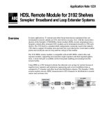
Reproductive toxicity refers to a wide variety of toxicological effects that may occur in 71
different phases within the reproductive cycle (figure 1). This includes effects on fertility, 72
sexual behaviour, embryo implantation, embryonic/foetal development, parturition, postnatal 73
adaptation, and subsequent growth and development into sexual maturity. An enormous 74
variety of mechanisms at the molecular, cellular and tissue levels cooperate in a concerted and 75
genetically programmed way to regulate these processes. The sensitivity to chemical insults 76
may differ extensively between processes. In addition, different temporal windows of 77
sensitivity have been observed for different processes. As an example, neural tube closure 78
occurs early in pregnancy, and most effects on this process can only be determined after 79
exposure during this critical period of time. 80
81
82
83
84
85
86
87
Figure 1: The main stages in the mammalian reproductive cycle. 88
89
Postnatal
development
Birth
Fetogenesis
Growth and
development
Fertilisation
Gamete
production
Transport of
the zygote
Implantation
Embryogenesis
Sexual
maturation
Working Group 5: Reproductive Toxicity DRAFT FOR CONSULTATION 14.07.10
4
90
91
2.2. Alternatives for Reproductive Toxicity Testing 92
Over the last two decades, a wealth of ex vivo and in vitro assays have been proposed as 93
alternative test systems for testing toxic effects on the various processes in reproduction and 94
development. Individual in vitro models are reductionistic in nature and are therefore unable to 95
cover all aspects of the reproductive cycle since reproduction requires a complex interplay of 96
integrated functions. However, parts of the reproductive cycle can be mimicked by in vitro 97
systems and it is conceivable that a panel of well-designed and validated in vitro tests could 98
replace a substantial proportion of in vivo testing procedures. This chapter gives an inventory 99
of the current state of development of alternative test systems for reproductive toxicity hazard 100
assessment. 101
Although not applicable for cosmetic ingredients, reduction of animal studies is a more 102
feasible goal than replacement, one example being the current OECD activity towards an 103
extended-1-generation study protocol, which, if it would replace the current 2-generation 104
study, would reduce animal use by roughly 40% in each study [1]. The addition of relevant 105
parameters to this novel study protocol represents a good example of refined testing. 106
107
3. Information Requirements for the Safety Assessment of Cosmetic 108
Within the EU the safety of cosmetic products is regulated by the Cosmetics Product 109
Directive 76/768/EEC [2] which will be replaced stepwise by the new EU Cosmetics 110
Regulation 1223/2009. According to Article 2 of Directive 76/768/EEC, a “cosmetic product 111
put on the market must not cause damage to human health when applied under normal or 112
reasonably foreseeable conditions of use”. In addition, Article 7a of the same Directive states 113
that the safety evaluation of a finished product should be based on the general toxicological 114
profile, the chemical structure and the level of exposure of each ingredient. This implies that 115
a quantitative risk assessment is required for each single ingredient of a cosmetic product. 116
Being responsible for the safety of its cosmetic product, the producer performs a risk 117
assessment based on the data of all ingredients used. Therefore, a pre-market approval is not 118
necessary for most ingredients used for cosmetics. However, certain ingredients listed in 119
positive lists of the Cosmetics Directive such as colorants (Annex IV), preservatives (Annex 120
VI), UV filters (Annex VII) and, most recently, hair dyes require approval of their safety prior 121
to marketing by the EU commission which require the submission of a full dossier[3]. 122
Specific requirements for the evaluation of the safety of a cosmetic ingredient are not further 123
Working Group 5: Reproductive Toxicity DRAFT FOR CONSULTATION 14.07.10
5
specified in the Directive either with regard to reproductive toxicity or to any other 124
toxicological endpoint. However, information on data requirements with regard to the safety 125
evaluation of cosmetic ingredients are provided in the Notes of Guidance of the former SCCP 126
(now SCCS) [4]: For substances which are submitted for inclusion in the positive lists of the 127
Cosmetics Directive, a comprehensive dossier must be provided for evaluation by the SCCS. 128
The dossier includes data on acute toxicity (if available), dermal and mucous membrane 129
irritation, dermal penetration, skin sensitization, repeated dose toxicity, genotoxicity, and 130
phototoxicity (if the cosmetic product is intended to be used on sunlight-exposed skin). 131
Further, it is stated that when considerable oral intake is expected, or when dermal penetration 132
data suggest a significant systemic absorption, information on toxicokinetics, carcinogenicity 133
and reproductive toxicity “may become necessary”. Additional recommendations on specific 134
in vivo
or in vitro reproductive toxicity studies to be submitted with a dossier are not described 135
in the Notes on Guidance. From the SCCS/SCCP opinions published within recent years 136
(2000 – 2009) ( it can 137
be concluded that in most cases an in vivo developmental toxicity study in the rat (OECD TG 138
414) - submitted by the manufacturer as the only study on reproductive toxicity - was 139
considered sufficient by the SCCS. In only a few cases additional data from a 1- or 2-140
generation study (OECD TG 415 and 416) were included in a dossier [5]. 141
For substances, which are not listed in one of the Annexes of the Cosmetic Directive, data on 142
reproductive toxicity are not explicitly asked for in the Notes of Guidance. However, some 143
indications of adverse effects on the fertility could be obtained e.g. from repeated dose 144
toxicity studies, if available (e.g. histopathologic effects on reproductive organs, effects on the 145
endocrine system). 146
147
148
149
4. Inventory of Animal Test Methods Currently Used for the Evaluation of 150
Developmental and Reproductive Toxicity 151
In the following, OECD test guidelines for the regulatory investigation of the developmental 152
and reproductive toxicity of chemicals are described. The list comprises an inventory of the 153
main study protocols. However, not all of them are used for testing cosmetic ingredients. 154
155
4.1. OECD Test Guideline 414: Prenatal Development Toxicity Study for the Testing of 156
Chemicals 157
This Guideline provides general information concerning the effects of prenatal exposure on 158
the pregnant test animal and on the developing organism; this may include assessment of 159
Working Group 5: Reproductive Toxicity DRAFT FOR CONSULTATION 14.07.10
6
maternal effects as well as death, structural abnormalities, or altered growth in the foetus. The 160
guideline is not intended to examine solely the period of organogenesis, (e.g. days 5-15 in the 161
rodent, and days 6-18 in the rabbit) but also effects from preimplantation, when appropriate, 162
through the entire period of gestation to the day before caesarean section. Functional deficits, 163
although an important part of development, are not a part of this Guideline. They may be 164
tested for in a separate study or as an adjunct to this study using the Guideline for 165
developmental neurotoxicity. 166
The test substance is normally administered to pregnant animals at least from implantation to 167
one day prior to the day of scheduled kill, which should be as close as possible to the normal 168
day of delivery. The Guideline is intended for use with rodent (preferably rat) and non-rodent 169
(preferably rabbit). Each test and control group should contain a sufficient number of females 170
to result in approximately 20 female animals with implantation sites at necropsy. Three 171
concentrations, at least, should be used. The test substance or vehicle is usually administered 172
orally by intubation. A limit test may be performed if no effects are expected at a dose of 173
1000 mg/kg bw/d. The study includes measurements (weighing) and clinical daily 174
observations. One day prior to the expected day of delivery the females are killed, the uterine 175
contents are examined, and the foetuses are evaluated for soft tissue and skeletal changes. In 176
any study which demonstrates an absence of toxic effects, further investigation to establish 177
absorption and bioavailability of the test substance should be considered [6]. 178
179
4.2. OECD Test Guideline 415: One-Generation Reproduction Toxicity Study 180
This Test Guideline provides general information concerning the effects of a test substance on 181
male and female reproductive performance, such as gonadal function, oestrous cycle, mating 182
behaviour, conception, parturition, lactation and weaning. The study may also provide 183
preliminary information about developmental toxic effects of the test substance, such as 184
neonatal morbidity, mortality, behaviour and teratogenesis and to serve as a guide for 185
subsequent tests. The test substance is administered orally in graduated doses to several 186
groups of males and females. 187
Males should be dosed during growth and for at least one complete spermatogenic cycle; 188
females of the Parent generation should be dosed for at least two complete oestrous cycles. 189
The animals are then mated. The test substance is administered to both sexes during the 190
mating period and thereafter only to females during pregnancy and for the duration of the 191
nursing period. This Test Guideline is intended primarily for use with the rat or mouse. Each 192
test and control group should contain a sufficient number of animals to yield about 20 193
Working Group 5: Reproductive Toxicity DRAFT FOR CONSULTATION 14.07.10
7
pregnant females at, or near, term. Three test groups, at least, should be used. It is 194
recommended that the test substance be administered in the diet or drinking water. A limit test 195
may be performed if no effects would be expected at a dose of 1000 mg/kg bw/d. The results 196
of this study include measurements (weighing, food consumption) and daily and detailed 197
observations, each day preferably at the same time, as well as gross necropsy and 198
histopathology. The findings of a reproduction toxicity study should be evaluated in terms of 199
the observed effects, necropsy and microscopic findings [7]. 200
201
4.3. OECD Test Guideline 416: Two-Generation Reproduction Toxicity 202
This Guideline provides general information concerning the effects of a substance on the 203
integrity and performance of the male and female reproductive systems, and on the growth 204
and development of the offspring, including gonadal function, the oestrus cycle, mating 205
behaviour, conception, gestation, parturition, lactation, and weaning, and the growth and 206
development of the offspring. The study may also provide information about the effects on 207
neonatal morbidity, mortality, and preliminary data on prenatal and postnatal developmental 208
toxicity as well as serving as a guide for subsequent tests. In addition to studying growth and 209
development of the F1 generation, this Guideline is also intended to assess the integrity and 210
performance of the male and female reproductive systems as well as growth and development 211
of the F2 generation. For further information on developmental toxicity and functional 212
deficiencies, either additional study segments can be incorporated into this protocol, utilising 213
the Guidelines for developmental toxicity and/or developmental neurotoxicity, or these 214
endpoints could be studied in separate studies. 215
The test substance is administered daily in graduated doses to several groups of males and 216
females. Males and females of the Parent generation (5-9 weeks old) should be dosed during 217
growth, their mating, the resulting pregnancies and through the weaning of their first 218
generation offspring. The administration of the substance is continued to first generation 219
offspring during their growth into adulthood, mating and production of a second generation 220
(until the weaning). The rat is the preferred species for testing. Each test and control group 221
should contain a sufficient number of animals to yield preferably not less than 20 pregnant 222
females at or near parturition. At least three dose levels and a concurrent control shall be used. 223
A limit test may be performed if no effects would be expected at a dose of 1000 mg/kg bw/d. 224
The results of this study include: measurements (weighing, sperm parameters, oestrus cycle 225
parameters and offspring parameters), clinical daily observations, as well as gross necropsy 226
and histopathology. The findings of this two-generation reproduction toxicity study should be 227
Working Group 5: Reproductive Toxicity DRAFT FOR CONSULTATION 14.07.10
8
evaluated in terms of the observed effects including necropsy and microscopic findings. A 228
properly conducted reproductive toxicity test should provide a satisfactory estimation of a no-229
effect level and an understanding of adverse effects on reproduction, parturition, lactation, 230
postnatal development including growth and sexual development [8]. 231
232
4.4. OECD Test Guideline 421: Reproduction/Developmental Toxicity Screening Test 233
This Guideline generates limited information concerning the effects of a substance on male 234
and female reproductive performance such as gonadal function, mating behaviour, 235
conception, development of the conceptus and parturition. It is not an alternative to, nor does 236
it replace the existing Test Guidelines 414, 415 and 416. This Screening Test Guideline can 237
be used to provide initial information on possible effects on reproduction and/or development. 238
This test does not provide complete information on all aspects of reproduction and 239
development. In particular, it offers only limited means of detecting post-natal manifestations 240
of prenatal exposure, or effects that may be induced during post-natal exposure. Due (amongst 241
other reasons) to the relatively small numbers of animals in the dose groups, the selectivity of 242
the end points, and the short duration of the study, this method will not provide evidence for 243
definite claims of no effects. However, positive results are useful for initial hazard assessment 244
and contribute to decisions with respect to the necessity and timing of additional testing. 245
246
The test substance is administered in graduated doses to several groups of male and female 247
rats. Males should be dosed for a minimum of four weeks. Females should be dosed 248
throughout the study, so approximately 54 days. It is recommended that each group be started 249
with at least 10 animals of each sex. Generally, at least three test groups and a control group 250
should be used. Dose levels may be based on information from acute toxicity tests or on 251
results from repeated dose studies. The test substance is administered orally and daily. The 252
limit test corresponds to one dose level of at least 1000 mg/kg body weight. The results of this 253
study include measurements (weighing, food/water consumption) and daily and detailed 254
observations, preferably each day at the same time, as well as gross necropsy and 255
histopathology. The findings of this toxicity study should be evaluated in terms of the 256
observed effects, necropsy and microscopic findings. Because of the short period of treatment 257
of the male, the histopathology of the testis and epididymus must be considered, along with 258
the fertility data, when assessing male reproductive effects [9]. 259
260
4.5. OECD Test Guideline 422: Combined Repeated Dose Toxicity Study with the 261
Reproduction/Developmental Toxicity Screening Test 262
Working Group 5: Reproductive Toxicity DRAFT FOR CONSULTATION 14.07.10
9
The test may be particularly useful as part of the initial screening for the assessment of 263
chemicals for which little or no toxicological information is available and can serve as an 264
alternative to conducting two separate tests for repeated dose toxicity (Guideline 407) and 265
reproduction/developmental toxicity (Guideline 421), respectively. It can also be used as a 266
dose range finding study for more extensive reproduction/developmental studies, or when 267
otherwise considered relevant. 268
The method comprises the basic repeated dose toxicity study that may be used for chemicals 269
on which a 90-day study is not warranted (e.g. when the production volume does not exceed 270
certain limits) or as a preliminary study to a long-term study. It further comprises a 271
reproduction/developmental toxicity screening test and, therefore, can also be used to provide 272
initial information on possible effects on male and female reproductive performance such as 273
gonadal function, mating behaviour, conception, development of the conceptus and 274
parturition, either at an early stage of assessing the toxicological properties of chemicals. This 275
test does not provide complete information on all aspects of reproduction and development. In 276
particular, it offers only limited means of detecting postnatal manifestations of prenatal 277
exposure, or effects that may be induced during postnatal exposure. Due (amongst other 278
reasons) to the selectivity of the endpoints and the short duration of the study, this method 279
will not provide evidence for definite claims of no reproduction/developmental effects. 280
Although, as a consequence, negative data do not indicate absolute safety with respect to 281
reproduction and development, this information may provide some reassurance if actual 282
exposures were clearly less than the dose related to the No Observed Adverse Effect Level 283
(NOAEL). The Guideline also places emphasis on neurological and immunological effects. 284
The test substance is administered in graduated doses to several groups of male and female 285
rats. Males should be dosed for a minimum of four weeks; females should be dosed 286
throughout the study (approximately 54 days). Normally, matings of "one male to one female" 287
should be used in this study. It is recommended that the test substance be administered orally 288
by gavage. Each group should be started with at least 10 animals of each sex. Generally at 289
least three test groups and a control group should be used. Dose levels should be selected 290
taking into account any existing toxicity and (toxico-) kinetic data available. The limit test 291
corresponds to one dose level of at least 1000 mg/kg body weight. The results of this study 292
include measurements (weighing, food/water consumption) and daily detailed observations 293
(including sensory reactivity to stimuli), preferably each day at the same time, as well as gross 294
necropsy and histopathology. The findings of this toxicity study should be evaluated in terms 295
of the observed effects, necropsy and microscopic findings. The evaluation will include the 296
Working Group 5: Reproductive Toxicity DRAFT FOR CONSULTATION 14.07.10
10
relationship between the dose of the test substance and the presence or absence of 297
observations. Because of the short period of treatment of the male, the histopathology of the 298
testis and epididymus must be considered along with the fertility data, when assessing male 299
reproduction effects [10]. 300
301
4.6. OECD Test Guideline 426: Developmental Neurotoxicity Study
302
The developmental neurotoxicity study provides information on the potential functional and 303
morphological effects on the developing nervous system of the offspring of repeated exposure 304
to a substance during in utero and early postnatal development. 305
A developmental neurotoxicity study can be conducted as a separate study, incorporated into 306
a reproductive toxicity and/or adult neurotoxicity study (e.g., Test Guidelines 415, 416, 424), 307
or added onto a prenatal developmental toxicity study (e.g., Test Guideline 414). When the 308
developmental neurotoxicity study is incorporated within or attached to another study, it is 309
imperative to preserve the integrity of both study types. 310
The test substance is administered daily, generally orally, to mated females (rats are preferred) 311
from the time of implantation (GD 6) throughout lactation (PND 21). At least three dose 312
levels and a concurrent control should be used and a total of 20 litters are recommended at 313
each dose level. Dams are tested to assess effects in pregnant and lactating females and may 314
also provide comparative information. Offspring are randomly selected from within litters for 315
neurotoxicity evaluation. All dams and all offspring should be carefully observed at least once 316
daily with respect to their health, including morbidity and mortality. The evaluation consists 317
of observations to detect gross neurological and behavioural abnormalities, and the evaluation 318
of brain weights and neuropathology during postnatal development and adulthood. The report 319
should include the body weight, the food/water consumption; the detailed clinical 320
observations, the necropsy findings, a detailed description of all behavioural, the number of 321
animals at the start and at the end of the study and the toxic response data by sex and dose 322
level [11]. 323
324
4.7. OECD Test Guideline 440: Uterotrophic Bioassay in Rodents: A short-term 325
screening test for oestrogenic properties 326
The Uterotrophic Bioassay is an in vivo short-term screening test. It evaluates the ability of a 327
chemical to elicit biological endocrine disruption activities consistent with agonists or 328
antagonists of natural oestrogens (e.g. 17ß-estradiol). It is based on the increase in uterine 329
weight or uterotrophic response. The uterus responds to oestrogens in two ways. An initial 330
response is an increase in weight due to water imbibition. This response is followed by a 331
Working Group 5: Reproductive Toxicity DRAFT FOR CONSULTATION 14.07.10
11
weight gain due to tissue growth. The uterus responses in rats and mice are comparable 332
qualitatively. 333
This bioassay serves as an in vivo screening assay and its application should be seen in the 334
context of the “OECD Conceptual Framework for the Testing and Assessment of Endocrine 335
Disrupting Chemicals”. In this Conceptual Framework the Uterotrophic Bioassay is contained 336
in Level 3 as an in vivo assay providing data about a single endocrine mechanism, i.e. 337
oestrogenicity. 338
The Uterotrophic Bioassay relies for its sensitivity on an animal test system in which the 339
hypothalamic-pituitary-ovarian axis is not functional. Two oestrogen sensitive states in the 340
female rodent meet this requirement: i) immature females after weaning and prior to puberty 341
and ii) young adult females after ovariectomy with adequate time for uterine tissues to 342
regress. 343
The test substance is administered daily by oral gavage or subcutaneous injection. Each 344
treated and control group should include at least 6 animals. Graduated test substance doses are 345
administered to a minimum of two treatment groups of experimental animals using one dose 346
level per group and an administration period of three consecutive days for immature method 347
and a minimum administration period of three consecutive days for ovx-adult method. The 348
animals are necropsied approximately 24 hours after the last dose. For oestrogen agonists, the 349
mean uterine weight of the treated animal groups relative to the vehicle group is assessed for a 350
statistically significant increase. A statistically significant increase in the mean uterine weight 351
of a test group indicates a positive response in this bioassay. The report should include: the 352
daily body weights, the daily record of status of animal, the wet and blotted uterine weight, 353
the daily food consumption [12]. 354
355
4.8. OECD Test Guideline 441: Hershberger Bioassay in Rats: A Short-term Screening 356
Assay for (Anti-) Androgenic Properties
357
The Hershberger Bioassay is an in vivo short-term screening test. It evaluates the ability of a 358
chemical to elicit biological endocrine disruption activities consistent with androgen agonists, 359
antagonists or 5 α-reductase inhibitors. The current bioassay is based on the changes in weight 360
of five androgen-dependent tissues in the castrate-peripubertal male rat: the ventral prostate, 361
seminal vesicle (plus fluids and coagulating glands), levator ani-bulbocavernosus muscle, 362
paired Cowper's glands and the glans penis. 363
In order to establish whether a test substance can have androgenic or antiandrogenic action, 364
two - respectively three - dose groups of the test substance, plus positive and vehicle 365
(negative) controls are normally sufficient. The test substance is administered by gavage or 366
Working Group 5: Reproductive Toxicity DRAFT FOR CONSULTATION 14.07.10
12
subcutaneous injection daily for 10 consecutive days. To test for antiandrogens, the test 367
substance is administered together with a reference androgen agonist. Each treated and control 368
group should include a minimum of 6 animals. The animals are necropsied approximately 24 369
hours after the last administration of the test substance. The tissues are excised and their fresh 370
weights determined. A statistically significant increase (androgenic) or decrease 371
(antiandrogenic) in the weights of two of the five tissues indicates a positive response in this 372
assay [13]. 373
374
4.9. OECD Test Guideline 455: The Stably Transfected Human Estrogen Receptor-α 375
Transcriptional Activation Assay for Detection of Estrogenic Agonist-Activity of Chemicals
376
This Test Guideline describes an in vitro assay, which provides mechanistic information, and 377
can be used for screening and prioritization purposes. This assay evaluates Transcriptional 378
Activation mediated by the hERα of estrogen responsive genes, a process considered to be 379
one of the key mechanisms of possible endocrine disruption related health hazards. 380
The test system utilises the hERα-HeLa-9903 cell line derived from a human cervical tumor 381
and stably transfected. This cell line can measure the ability of a test chemical to induce 382
hERα-mediated transactivation of luciferase gene expression. The cells are exposed to 7 non-383
cytotoxic concentrations of the test chemical for 20-24 hours to induce the reporter gene 384
products. Four reference chemicals should be included in each experiment: a strong estrogen 385
(17ß-estradiol), a weak estrogen (17α-estradiol), a very weak estrogen (17α-386
methyltestosterone) and a negative control (corticosterone). The activity of the luciferase 387
enzyme is measured in a luminometer. A test chemical is considered to be positive if the 388
maximum response induced is equal to or exceeds 10% of the response of the positive control 389
(1 nM 17α-estradiol) in at least two of two or two of three runs [14]. 390
391
4.10. Draft OECD Test Guideline Extended One Generation Reproductive Toxicity Study 392
In the extended one-generation study [1] males and females are exposed 4 and 2 weeks 393
premating respectively, and females are exposed throughout the mating period, pregnancy, 394
and weaning of their pups. Male exposure is continued for 10 weeks to cover the entire 395
spermatogenic cycle, followed by necropsy. F1 animals are kept until adulthood and 396
developmental landmarks are assessed. F1 are divided into up to three cohorts, one to assess 397
their reproductive capacity, one for developmental immune toxicity (DIT) assessment, and 398
one for developmental neurotoxicity (DNT) assessment. Discussion is ongoing as to whether 399
each of these cohorts should be mandatory or not. The DIT and DNT cohorts have been 400
considered mandatory by the OECD expert group on the study design. The reproductive 401
Working Group 5: Reproductive Toxicity DRAFT FOR CONSULTATION 14.07.10
13
toxicity cohort, which would generate an F2 generation, was proposed by Cooper et al. to 402
need to be triggered dependent on findings in the earlier part of the study. A comprehensive 403
retrospective analysis of existing 2-generation studies is currently ongoing to assess whether 404
and in which cases in the past the F2 generation has given unique and crucial information on 405
reproductive toxicity parameters that has impacted on the risk assessment or classification & 406
labelling of the compound tested. Depending on the outcome of this analysis, an OECD 407
expert group will advise in the fall of 2010 about the necessity of the reproductive cohort in 408
the extended one-generation study. The extended one generation study may eventually replace 409
the 2-generation study in current testing strategies. This replacement will possibly reduce the 410
number of animals with 40% in each study. It will also result in considerable refinement of 411
the study design through the addition of a series of novel parameters and the assessment of 412
many parameters in more animals per litter than currently prescribed in the 2-generation 413
study. 414
415
416
417
418
419
5. Inventory of Alternative Methods 420
5.1. Developmental Toxicity 421
5.1.1. Whole Embryo Tests 422
5.1.1.1. The Rodent Whole Embryo Culture Test 423
Rodent postimplantation whole embryo culture (WEC) is the only available ex vivo test that 424
covers the critical phase of organogenesis in a complete mammalian embryo. It is widely used 425
both in mechanistic studies and as a screening test for developmental toxicants. Gestation day 426
10-12 rat embryos are cultured during organogenesis in vitro and treated with test chemicals. 427
End points used in the WEC are a series of well defined morphological end points: all tissues 428
receive a score dependent on their developmental stage, and all scores added up give the so-429
called Total Morphological Score (TMS). Besides this score, malformations and size 430
measurements are noted, the latter comprising of yolk sac diameter, head length and crown-431
rump length [15]. 432
The protocol of the WEC was standardized [16] and scientifically validated according to the 433
ECVAM validation criteria [17]. However, the predictability and applicability domains of the 434
WEC are not sufficiently defined yet to allow regulatory implementation. The WEC is 435
currently used by many laboratories in academia and industry. 436
Working Group 5: Reproductive Toxicity DRAFT FOR CONSULTATION 14.07.10
14
437
5.1.1.2. The Zebrafish Embryo Teratogenicity Assay
438
The zebrafish (Danio rerio) embryo is an in vitro model to investigate the developmental 439
toxicity potential of substances on the developing vertebrate organism [18]. Primary 440
endpoints are lethality, malformations and growth retardation. The development of the 441
zebrafish embryo is very similar to the embryogenesis in higher vertebrates, including 442
humans, and many molecular pathways are evolutionary conserved between zebrafish and 443
humans [19]. This method is used not only as a screening tool for teratogenicity [20;21], but 444
also as a means of investigating specific mechanisms related to the teratogenic potential of 445
certain substances [22;23]. 446
In principle, the fertilized fish eggs are exposed to different concentrations of a test substance. 447
At different time points, the exposed developing fish embryos are observed and scored for 448
lethal, embryotoxic and/or teratogenic effects. Several protocols have been published 449
differing in e.g., (i) the start and duration of exposure to the test substance (ii) the use of 450
complete or dechorionated fish embryos (iii) the presence or absence of a metabolic activation 451
system [24] or (iv) the scoring system and observation intervals. 452
The zebrafish embryo teratogenicity assay is currently used by many laboratories in academia 453
and industry. An important step forward would be the agreement on a common standard 454
protocol, which is the prerequisite of a successful prevalidation. 455
456
5.1.1.3. Frog Embryo Teratogenesis Assay Xenopus (FETAX)
457
The FETAX is a whole embryo screening assay, based on the South African clawed frog 458
Xenopus laevis,
to identify substances that may pose a developmental hazard in humans [25]. 459
According to the American Society for Testing and Methods (ASTM) guidelines [26], 460
fertilized eggs in the mid- to late-blastula stage are incubated in media containing the test 461
substance for 96 h. The embryos are scored for lethality, growth retardation and 462
malformations at different timepoints. Similar to the zebrafish embryo teratogenicity assay, 463
FETAX encompasses organogenesis and does not include later events of development. 464
In an interlaboratory validation study using 12 compounds, FETAX yielded repeatable and 465
reliable data. However, transferability is still an issue of concern. The inclusion of a 466
mammalian metabolic activation system was essential for the correct prediction of the 467
teratogenic potential of substances. However, FETAX still requires further development [25]. 468
Efforts have to be made to improve the predictability of this assay [27]. 469
470
5.1.1.4. The Chicken Embryotoxicity Screening Test (CHEST)
471
Working Group 5: Reproductive Toxicity DRAFT FOR CONSULTATION 14.07.10
15
The chicken embryotoxicity screening test (CHEST) was first described in 1976 by Jelinek et 472
al
. as a fast and cheap teratogenicity test [28]. In the first protocol described CHEST 473
comprised two phases of testing, i.e. CHEST I, which determines the toxic dose range in very 474
early administration time (24 hours) and CHEST II that determines the teratogenic dose range 475
and covers late effects on the embryo development (days 2, 3 and 4). Recently adaptations of 476
this protocol were developed [29]. 477
The main endpoints assessed using the modified CHEST are mortality, malformations, 478
embryo development, blood vessel development and blood vessel coloration. Compounds or 479
mixtures can easily be administered to the windowed eggs and effects on the developing 480
embryo can be investigated. Moreover, the chick embryo possesses its own basic metabolic 481
capacity providing the possibility to screen for metabolites [30]. Studies of Bernshausen et al. 482
revealed metabolic activities of CYP and GST in 72 h old chicken embryo sub cellular 483
fractions [31]. 484
However, the chick embryo in ovo system has been criticised for not being able to distinguish 485
general toxicity from specific developmental effects and the absence of mammalian maternal-486
foetal relations [32]. In addition, CHEST produces a high rate of false positives especially 487
among irritant and corrosive substances that show an evident effect on the blood vessels of the 488
chick embryo [29]. 489
Several studies have evaluated the CHEST and similar protocols [33-38] and CHEST was 490
demonstrated to be a reproducible test system that delivered quantifiable data for evaluation. 491
At the present time several laboratories in academia and industry are using CHEST. 492
Furthermore, the chemical industry employs a highly standardized and in-house validated 493
adapted protocol for CHEST for routine embryotoxicity screening purposes. 494
495
5.1.2. The Micromass Test 496
The micromass test (MM) is making use of cell cultures of the limb bud and/or neuronal cells 497
[39;40]. The cells are isolated from the limb or the cephalic tissues of mid-organogenesis 498
embryos. After preparing a single cell solution the cells are seeded in a high density and 499
undergo differentiation into chondrocytes and neurons without additional stimulation. The 500
differentiation after exposure to test chemicals is analysed by using defined toxicological 501
endpoints [41]. 502
The protocol using micromass cultures of the limb buds has been validated in an ECVAM 503
validation study [17]. Data on intra- and interlaboratory variability, transferability and in 504
vivo
/in vitro comparisons are available. The number of laboratories currently using the MM is 505
limited.
506
Working Group 5: Reproductive Toxicity DRAFT FOR CONSULTATION 14.07.10
16
507
5.1.3. Pluripotent Stem Cell Based in vitro Tests 508
The potential of embryonic stem cells to differentiate into all cell types of the mammalian 509
organism (pluripotency) provides the scientific rationale to assess adverse effects on the 510
differentiating embryonic stem cells that might be relevant for embryotoxicity in vivo. In 2002 511
the embryonic stem cell test (EST) that is based on the cytotoxicity assessment as well as the 512
evaluation of differentiation inhibition into cardiomyocytes was scientifically validated [42]. 513
However, in post validation evaluations, it has been demonstrated that this test is limited in its 514
applicability domain and its predictive capacity [43]. Nevertheless, various industrial sectors 515
are still using method involving ES cells differentiation for predicting embryotoxicity. These 516
embryonic stem cell tests vary in their readouts but also in the target cell differentiation [44]. 517
Depending on the area of application, adverse effects on differentiating neural cells, 518
cardiomyocytes and cells of the skeletal cells have been investigated. Effects on the quantity 519
of differentiated target cells have been assessed by using immunological methods such as 520
flow cytometry [45] or molecular biological methods such as RT-PCRs [46]. Several of the 521
methodologies could also be automated in order to increase the throughput of substances and 522
make the test available for screening purposes [47]. 523
In addition, the establishment of human embryonic stem cells based tests should contribute to 524
a detailed understanding on mechanisms leading to human developmental toxicity which 525
should substantially contribute to a better hazard identification/characterisation for humans. 526
However, these approaches are still in their infant status. 527
The generation of genetically engineered embryonic stem cell lines allows an easy monitoring 528
of toxic effects in medium throughput applications. For example the generation of transgenic 529
cell lines that are using a heart cell specific promoter/enhancer controlling the expression of 530
reporter genes allows measuring quantitatively side effects on differentiating heart cells 531
through a reduction in fluorescence [48]. Another class of reporter gene assays such as the 532
ReProGlo assay detects chemical induced alterations in the canonical Wnt/_-catenin 533
signalling pathway, which is involved in the regulation of early embryonic development [49]. 534
The development of additional genetically engineered embryonic stem cell lines evaluating 535
biologically significant perturbations in key toxicity pathways of embryotoxicity might follow 536
and will provide a mechanistic understanding on developmental toxicity. Nevertheless, also 537
these tests are still in its research and developmental phase. 538
The establishment of stable differentiation protocols into toxicological relevant cell types is 539
challenging and requires additional scientific work. Huge scientific/technical efforts are 540
Working Group 5: Reproductive Toxicity DRAFT FOR CONSULTATION 14.07.10
17
currently ongoing to stabilise stem cell differentiation. Due to the growing knowledge in stem 541
cell technologies progress can be expected in the next couple of years. First indications that 542
successful tests can be developed have been published [50-53]. 543
544
5.2. Placental Toxicity and Transport 545
5.2.1. The Placental Perfusion Assay 546
Understanding the placental transport of compounds provided to the pregnant mother is 547
essential to reduce the risks of fetal exposure to harmful substances during pregnancy. The 548
placenta serves as the interface between the maternal and fetal circulations during pregnancy. 549
Ex vivo
placental perfusion provides an opportunity to carry out research without ethical 550
difficulties. It takes around 30 min following the birth to set up a perfusion and the perfusion 551
conditions allow for continued placental tissue viability for several hours. Viability of the 552
placenta during the experiments is verified by monitoring leakage from the fetal compartment, 553
oxygen transfer, and glucose consumption. Appropriate antipyrine transfer between the 554
maternal and fetal circulations confirms proper experimental set up and can be used to 555
normalize differences between placentas. Other advantages of placental perfusion 556
experiments include the retention of in vivo placental organization and assessment of binding 557
to placental tissue [54;55]. However, the application of this assay is limited due to placenta to 558
placenta variations and the limited relevance of the term placenta for the period of embryonic 559
development. In addition, only preliminary data based on a limited number of substances are 560
available. 561
562
5.2.2. Trophoblast Cell Assay 563
In this assay the BeWo cell line is used which represents an immortalized trophoblastic line of 564
human origin. The cells form polarized, confluent monolayers and have proven useful in 565
transport studies. The assay based on BeWo cells serves as an in vitro model of the rate-566
limiting barrier to maternal–fetal exchange. The BeWo b30 model consists predominantly of 567
cytotrophoblast cells which form a confluent monolayer with tight junctions, but they do not 568
spontaneously differentiate to syncytiotrophoblasts, and the model lacks the connective tissue 569
which is present in vivo [56]. 570
571
5.3. Preimplantation Toxicity 572
5.3.1. Male Fertility 573
5.3.1.1. Computer Assisted Sperm Analysis 574
Working Group 5: Reproductive Toxicity DRAFT FOR CONSULTATION 14.07.10
18
The computer assisted sperm analysis (CASA) allows to monitor adverse effects of chemicals 575
on spermatozoa with possible implications on fertility potential viability, motility, velocity, 576
motion, and morphology of mammalian semen will be analysed in real-time. This allows the 577
detection of reversible and irreversible damages (recovery effect) to the mature sperm as well 578
as repeated dose effects. For reproductive medicine fully automated semen analysers are 579
available. Several chemicals have already been tested in different laboratories and an 580
INVITTOX protocol is available. The test has been evaluated by two independent laboratories 581
by testing more than 35 test chemicals [57]. 582
The lower sensitivity of mature sperm in comparison to earlier stages of spermatogenesis 583
must be considered and may limit the relevance of this test. 584
585
5.3.1.2. Leydig Cell Assay 586
A disturbance of the endocrine system due to effects of chemicals on steroidogenesis or due to 587
specific cytotoxic effects on Leydig cells leads to a decreased development of spermatozoa 588
and impaired fertility since Leydig cells nurture the developing sperm cell. A new Leydig cell 589
line, BLT1-L17, that responds very well and quite robustly to luteinizing hormone (LH) or its 590
analogue, chorionic gonadotropin (hCG), has been characterized. 591
In the assay the MTT test serves as a general toxicity endpoint and testosterone production as 592
the Leydig cell-specific endpoint. BLT1-L17 cells were exposed to 15 chemicals and the data 593
obtained with this set of test chemicals indicate that the cell line is a good candidate for 594
further development into a rigorous test applicable for in vitro reproductive toxicity 595
assessment acting via interference with testosterone production [57]. 596
597
5.3.1.3. Sertoli Cell Assay 598
Sertoli cells form the basis of the blood-testis barrier and divide the tubular area into 599
adluminal and basal compartments protecting the maturing germ cells from chemical insults. 600
In the assay, rat primary cultures and the SerW3 line are used. The Sertoli cell assay was 601
developed by pharmaceutical industry and transferred to a second laboratory. General 602
cytotoxicity and the secretion of inhibin B are measured. These two endpoints allow a 603
classification of test chemicals as positive or negative for testicular toxicity. In addition, the 604
integrity of tight junctions forming the blood-testis barrier can be studied in the SerW3 cell 605
line, providing a new endpoint to study the mechanism of action of testicular toxicants. 606
Further studies are needed to fully understand the utility of this test [57]. 607
608
5.3.1.4. ReProComet Assay 609
Working Group 5: Reproductive Toxicity DRAFT FOR CONSULTATION 14.07.10
19
The ReProComet assay (Repair Proficient Comet assay) was developed to detect chemically induced 610
DNA damage in sperm cells. In order to circumvent the intrinsic repair deficiency of the sperm cells a 611
strategy is deployed involving a supplementation with protein extract from somatic cells after the 612
chemical treatment. Liquid nitrogen frozen bull sperm is used for the analysis. Bull sperm is incubated 613
with the test chemicals for 2 hours. A SYBR-14/Propidium iodide flow cytometric analysis is used to 614
evaluate sperm viability in addition to the four Comet assay endpoints tail length, tail moment, 615
fraction of tail DNA, fraction of head DNA [57;58]. The rationale of the test design needs further 616
clarification. 617
618
5.3.2. Female Fertility 619
5.3.2.1. Follicle Culture Bioassay (FBA) 620
The FBA allows multiparametric in vitro analysis of effects of chemicals on the ovarian 621
function such as folliculogenesis, steroidogenesis and oogenesis. Mouse ovarian pre-antral 622
follicles are grown in vitro until the preovulatory stage followed by in vitro ovulation induction 623
and mature oocyte retrieval. During the in vitro growth period (12 days) the follicles develop 624
with theca cell proliferation, granulose cell proliferation and differentiation, meanwhile 625
supporting oocyte growth and maturation. In the FBA the in vitro growing follicles are 626
exposed to chemicals in a chronically or acute manner and effects on the different biological 627
processes of folliculogenesis, steroidogenesis and oogenesis are analyzed with morphological, 628
biochemical and functional parameters. The FBA is still in the phase of development. It 629
requires further standardisation and transferability to other laboratories has to be addressed 630
[57;59]. 631
632
5.3.2.2. In vitro Bovine Oocyte Maturation Assay (bIVM) 633
The bIVM assay focuses on the use of bovine oocytes for toxicity testing during the process 634
of oocyte maturation in vitro. The test screens for potential adverse effects on the process of 635
oocyte maturation after exposure of cumulus-oocyte complexes to test substances, with 636
special reference to nuclear configuration changes within the oocyte as compared to control 637
non-exposed oocytes. Endpoint is the successful achievement of the maturation stage 638
metaphase II (completion of meiosis up to the metaphase II). The inter-laboratory variability 639
and the transferability of the bIVM test was analyzed for a set of eight chemicals, and the 640
statistical analysis of the data obtained from the two laboratories demonstrated that there was 641
a good concordance of results across the laboratories [57;60;61]. Testing of additional 642
compounds is necessary in order to assess the predictability of this test. 643
644
Working Group 5: Reproductive Toxicity DRAFT FOR CONSULTATION 14.07.10
20
645
5.3.2.3. In vitro Bovine Fertilisation Test (bIVF)
646
The bIVF assay focuses on the use of bovine oocytes and sperms for toxicity testing during 647
the process of in vitro fertilization. The purpose of the test is to (1) screen for adverse effects 648
of chemicals on the process of oocyte fertilisation and (2) investigate the mechanism of action 649
of reproductive toxicants. Both oocytes and sperms are exposed to test chemicals; therefore, 650
the adverse effects on the function of both gametes can be monitored. Specific endpoints are 651
(1) Penetration of capacitated bull spermatozoa into matured oocytes and (2) formation of the 652
female and male pronuclei [60]. This test is still in a very early phase of development and 653
further investigations are necessary to assess its toxicological relevance [57]. 654
655
5.3.2.4. Mouse Peri-Implantation Assay (MEPA) 656
The mouse peri-implantation assay is an in vitro bioassay that allows studying the effect of 657
compounds on the development of the pre-implantation embryo and its capacity to survive 658
upon hatching around the implantation period. The assay is based on the in vitro culture of 659
mouse zygotes. The zygotes are cultured in groups of 10 for 7 days with daily observation and 660
scoring of embryo development. These daily morphological observations allow pinpointing 661
potential deviations of the timely regulated pre-implantation embryo. The bioassay is highly 662
reproducible in one laboratory. It allows the characterization of the sensitive stage of embryo 663
development [62]. The MEPA is still in the phase of development. It requires further 664
standardisation and transferability to other laboratories has to be addressed [57]. 665
666
5.4. In vitro Tests for Assessing Effects on the Endocrine System 667
5.4.1. Ishikawa Cell Test 668
The human endometrium is a fertility-determining factor. Its receptivity during the 669
implantation window may be altered by chemicals. The Ishikawa cell test aims to identify 670
chemicals which alter the expression of embryo-implantation-associated target genes in 671
human endometrial adenocarcinoma Ishikawa cells. Ishikawa cells are cultured to 672
subconfluency and incubated for 0.5 to 24 hours with test substances. This test system is a 673
tissue specific model to detect estrogenic activity of chemicals which up-regulate 674
progesterone receptor (PR) mRNA in the human endometrium. The Ishikawa model is 675
informative regarding the mode of action of positive tested chemicals and provides guidance 676
for prioritization for further testing [63]. The Ishikawa cell test is still in the phase of 677
development. It requires further standardisation and transferability to other laboratories has to 678
be addressed. 679
Working Group 5: Reproductive Toxicity DRAFT FOR CONSULTATION 14.07.10
21
680
5.4.2. Cell Proliferation Based Assays for Testing Estrogen Activity 681
Estrogenic activity of substances can be assessed by measuring in vitro proliferation of cell 682
lines containing the ER-α and ER-β estrogens receptor such as the human breast cancer cell 683
line MCF-7. The binding of the natural hormone or other estrogen like xenobiotics leads to 684
conformational changes that allow the estrogen-ligand complex to proceed from inactive 685
proteins to active transcriptional regulators that induce transcription of estrogen responsive 686
genes which lead to an estrogen dependent proliferation of cells [64]. 687
688
5.4.3. Receptor Binding Assays 689
Relevant hormonal receptors can be isolated either from primary tissues such as rat prostate 690
[65] or generated with recombinant technologies [66]. Nevertheless, all tests rely on the same 691
principles assessing the competitive binding of a substance to a receptor of interest. 692
Most advanced are receptor binding tests based on the estrogen receptor. Chemical 693
interactions with the estrogen receptor might affect the development of female secondary 694
sexual characteristics and/or the regulation of the menstrual cycle. Several tests such as the 695
uterine cytosol (ER-Rat Uterine Cytosol) assay [67] or the human recombinant full length 696
estrogen receptor-alpha binding assay [68] have been intensively evaluated in (pre)validation 697
trials under the lead of the US-EPA. The regulatory acceptance of estrogen receptor binding 698
tests is in preparation. 699
Another important receptor for the endocrine system is the androgen receptor. Androgens are 700
mainly concerned in the development and maintenance of male secondary sexual 701
characteristics. Several receptor binding tests based on isolated proteins from the cytosol of 702
the rat prostate [69] or recombinant proteins [66;70] have been developed and optimized. The 703
validation of other androgen receptor binding tests has been taken up in the work programme 704
of the OECD. 705
Another highly relevant receptor in the context of receptor mediated reproductive toxicity is 706
the progesterone receptor. As for the previous receptors also for the progesterone, receptor 707
binding assays have been developed in order to assess effects that might have influence on the 708
menstrual cycle, the pregnancy and/or embryogenesis. Currently several developed assays are 709
available that are using for example rabbit uterine cytosol [71], recombinant receptor [72] or 710
even whole cells [73]. 711
The thyroid hormone receptor is highly relevant for the development of the central nervous 712
system. Tests monitoring the binding of the thyroid hormone triiodothyronine (T3) to its 713
receptor are in the development phase, using recombinant proteins [74]. 714
Working Group 5: Reproductive Toxicity DRAFT FOR CONSULTATION 14.07.10
22
Other hormonal receptors playing a key role are binding hormones produced by the 715
hypothalamus (Gonadotropin Releasing Hormone) or pituitary gland (Follicle-Stimulating 716
Hormone, Luteinizing Hormone). These tests are still in the phase of research and 717
development but need to be considered since these hormones are involved in the feedback 718
loop controlling the reproductive system. Even if biologically highly relevant, further 719
assessments are needed if they act as major target for xenobiotics (toxicological relevance). 720
721
5.4.4. Transcriptional Tests 722
In contrary to the receptor binding tests which only provide information on the binding 723
capacity of a substance to a particular hormone receptor, the so-called transcriptional 724
activation assays are able to distinguish between agonist and antagonistic effects of 725
xenobiotics. The basic principal of transcriptional assays relies on genetically engineered cells 726
which express hormone receptors as well as reporter genes driven by hormone responsive 727
genes. The intensity of the receptor binding can be measured for example by using 728
spectrophotometric techniques. 729
This basic principle has been used for the development of several transcriptional tests 730
involving various hormones which are in different stages of standardisation and validation. 731
The estrogen receptor (ER) transcriptional assays for example quantify the induction of a 732
reporter gene product by the test substance or reference estrogen. The antagonism is measured 733
by the inhibition of the reference estrogen induction of the reporter gene, or cell proliferation. 734
Most advanced in the class of transcriptional assays is for example the “LUMI-CELL” test 735
that is currently undergoing a formal validation study by ICCVAM. The process of regulatory 736
acceptance of this test is already included in the OECD work plan 2009 of the Test Guidelines 737
Programme. Other tests that will certainly also contribute to a performance based test 738
guideline are tests named “MELN” [75] and “ERα CALUX” [76]. Anti-estrogenic activities 739
can also be mediated through the activation of the aryl hydrocarbon receptor. Transcriptional 740
activation assays of this receptor are in optimization phase, using different cell lines [77;78]. 741
Similar to ER transcriptional assays, androgen receptor transcriptional assays have been 742
designed [79; 80]. A Japanese Stably Transfected Transcriptional Activation (STTA) Assay 743
for the detection of androgenic and anti-androgenic activity of chemicals is under 744
consideration by OECD (included in work plan 2009 of the Test Guidelines Programme). 745
Other transcriptional assays following the same scientific principle are, tests e.g. assessing the 746
progesterone transcriptional activity [81;82] or the interaction with the thyroid receptor 747
Working Group 5: Reproductive Toxicity DRAFT FOR CONSULTATION 14.07.10
23
[83;84]. These tests are in their early phase of their evolution and additional work is necessary 748
to optimize the tests. 749
750
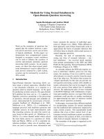
5.4.5. Tests Assessing Steroidogenesis 751
In the past years significant progress has been made by developing in vitro cell-based assays 752
aiming to detect substances that affect the synthesis of the sex steroid hormones. These tests 753
are at different stages in their development. Nevertheless, all tests are designed to identify 754
xenobiotics that have as their target sites components that perturbed biochemical pathways. 755
The complexity of possible target enzymes is demonstrated in figure 2. 756
757
Working Group 5: Reproductive Toxicity DRAFT FOR CONSULTATION 14.07.10
24
758
Figure 2: gonadal steroidogenesis pathway ( 759
760
Furthermore, several receptors regulating steroidogenesis are involved and need to be 761
considered as possible target for endocrine effects (GnRH, LH, and FSH Receptors). Different 762
assays measuring the gonadotrophin-stimulated steroidogenesis are under development, e.g. 763
FSH [85] or LH [86]. 764
A cell based assay on steroidogenesis, using the H295R cells, designed to measure effects on 765
estradiol and testosterone production has been validated and a draft test guideline is currently 766
Working Group 5: Reproductive Toxicity DRAFT FOR CONSULTATION 14.07.10
25
under discussion [87]. Other tests focusing on the aromatase enzyme (CYP19) are under 767
development, e.g. using human placental microsomes [88]. 768
769
5.5. Application of In Silico Techniques to Reproductive Toxicology 770
5.5.1. Existing Data 771
There are a number of international efforts to bring together existing toxicological 772
information on reproductive toxicology in an electronic format. For data that are publicly 773
available (i.e. not Confidential Business Information), they may be released via the internet. 774
Therefore, a number of searchable resources have been developed. These resources can be 775
used in at least two ways: to provide existing information on a substance such that testing may 776
not be required; or as a source of data for further in silico modelling. 777
There are a number of important issues when developing and using toxicological databases. 778
The first is ensuring the quality of the information within the database. There are different 779
issues to determination of data quality. The chemical structure and its identifiers (e.g. name, 780
CAS, 2-D or 3-D structure) must be consistent and correct. The structure and ontology of the 781
database must be sophisticated enough to capture the required information regarding a 782
toxicological test i.e. species, test, duration, dose, purity, effects etc. In addition, the transfer 783
of information e.g. from the open literature requires checking and quality assurance. Finally, 784
there is the issue of the quality of the individual data. This last consideration of assigning data 785
quality, is a process that may be undertaken by the database user. 786
There are a number of (meta-) databases that can be searched for toxicological information 787
(including reproductive effects) on single chemicals. There has been excellent development in 788
these databases in the past few years and these meta-databases have the capability to search 789
numerous data resources and compile the information together. Of particular note are: 790
• United States Environmental Protection Agency (US EPA) Aggregated Computational 791
Toxicological Resource (ACToR) available from 792
/> 793
• Organisation for Economic Cooperation Development (OECD) eChemPortal available 794
from 795
• United States Environmental Protection Agency (US EPA) TOXREF: EPA Toxicology 796
Reference Database in Support of ToxCast
TM
Program available from 797
/> 798
799
There are a number of other databases and toxicological resources. For the development of 800
(Q)SARs for reproductive toxicity these have, historically, been relatively limited in terms of 801