Báo cáo khoa học: Autocatalytic processing of procathepsin B is triggered by proenzyme activity doc
Bạn đang xem bản rút gọn của tài liệu. Xem và tải ngay bản đầy đủ của tài liệu tại đây (284.14 KB, 9 trang )
Autocatalytic processing of procathepsin B is triggered by
proenzyme activity
Jerica Rozman Pungerc
ˇ
ar
1,
*, Dejan Caglic
ˇ
1,
*, Mohammed Sajid
3
, Marko Dolinar
2
, Olga Vasiljeva
1
,
Urs
ˇ
ka Poz
ˇ
gan
1
, Dus
ˇ
an Turk
1
, Matthew Bogyo
4
, Vito Turk
1
and Boris Turk
1
1 Department of Biochemistry and Molecular and Structural Biology, Joz
ˇ
ef Stefan Institute, Ljubljana, Slovenia
2 Faculty of Chemistry and Chemical Technology, University of Ljubljana, Slovenia
3 Biochemistry and Molecular Biology Core, Sandler Center for Basic Research in Parasitic Diseases, University of California, San Francisco,
CA, USA
4 Department of Pathology, Stanford University School of Medicine, CA, USA
Cysteine cathepsins comprise a group of papain-like
cysteine proteases found predominantly in lysosomes.
Cathepsin B (EC 3.4.22.1) is one of the most abundant
and thoroughly studied. It plays an important role in
nonselective protein degradation inside lysosomes, and
is involved in the processing of other proteins and hor-
mones such as trypsinogen and thyroglobulin [1–3].
Secreted cathepsin B is often associated with patho-
logical conditions such as cancer progression [3–5],
rheumatoid arthritis and osteoarthritis [3,6].
Cysteine cathepsins, including cathepsin B, are syn-
thesized as inactive proenzymes, which are activated
by other proteases or by autocatalytic processing in
the acidic environment of late endosomes and lyso-
somes [1,2]. From the crystal structures of procathep-
sins B and L, it is evident that the propeptide, which is
removed during activation, blocks access to the active
site that is already formed in the proenzyme [7–10].
The propeptide forms a predominantly a-helical
domain, which is positioned as a ‘hook’ at the top of
Keywords
autoactivation; DCG-04; lysosomal cysteine
protease; procathepsin B; processing
Correspondence
B. Turk, Department of Biochemistry and
Molecular and Structural Biology, Joz
ˇ
ef
Stefan Institute, Jamova 39, 1000 Ljubljana,
Slovenia
Fax: +386 1 477 3984
Tel: +386 1 477 3772
E-mail:
*These authors contributed equally to this
work
(Received 17 September 2008, revised 13
November 2008, accepted 24 November
2008)
doi:10.1111/j.1742-4658.2008.06815.x
Cathepsin B (EC 3.4.22.1) and other cysteine proteases are synthesized as
zymogens, which are processed to their mature forms autocatalytically or
by other proteases. Autocatalytic processing was suggested to be a bimolec-
ular process, whereas initiation of the processing has not yet been clarified.
Procathepsin B was shown by zymography to hydrolyze the synthetic sub-
strate 7-N-benzyloxycarbonyl-l-arginyl-l-arginylamide-4-methylcoumarin
(Z-Arg-Arg-NH-MEC), suggesting that procathepsin B is catalytically
active. The activity-based probe DCG-04, which is an E-64-type inhibitor,
was found to label both mature cathepsin B and its zymogen, confirming
the zymography data. Mutation analyses in the linker region between the
propeptide and the mature part revealed that autocatalytic processing of
procathepsin B is largely unaffected by mutations in this region, including
mutations to prolines. On the basis of these results, a model for autocata-
lytic activation of cysteine cathepsins is proposed, involving propeptide dis-
sociation from the active-site cleft as the first step during zymogen
activation. This unimolecular conformational change is followed by a
bimolecular proteolytic removal of the propeptide, which can be accom-
plished in one or more steps. Such activation, which can be also facilitated
by glycosaminoglycans or by binding to negatively charged surfaces, may
have important physiological consequences because cathepsin zymogens
were often found secreted in various pathological states.
Abbreviations
Z-Arg-Arg-NH-MEC, 7-N-benzyloxycarbonyl-
L-arginyl-L-arginylamide-4-methylcoumarin.
660 FEBS Journal 276 (2009) 660–668 ª 2008 The Authors Journal compilation ª 2008 FEBS
the catalytic site, where it interacts with the mature
part, strengthening the interaction [9]. The propeptide
chain then continues in an extended conformation
across the active-site cleft and towards the N-terminus
of the mature enzyme in the direction opposite to that
of substrate binding, thereby serving as a linker
between the ‘hook’ domain and the N-terminus of the
mature enzyme. This N-terminal–linker–‘hook’
arrangement, with its reverse orientation compared to
substrate binding, strongly resembles the ‘sinker’–
linker–’hook’ arrangement in the X-inhibitor of apop-
tosis protein, which is known to block the executioner
caspases [11].
The pH optimum for in vitro autocatalytic process-
ing of procathepsin B, as well as of some other cathep-
sins, is approximately 4.5 [12–14]. At lower pH, the
interaction between the propeptide and the mature
part is weakened [15–17], resulting in a looser con-
formation of the proenzyme. This is followed by inter-
molecular cleavage of the procathepsin B propeptide
[14]. However, initiation of the activation process has
remained an unsolved question, although it has been
suggested that proenzymes may exhibit minor catalytic
activity, which could potentially initiate the chain reac-
tion [14,18–20]. Although processing can be very rapid
at higher concentrations of the proenzyme [14], it is
not clear whether propeptide removal is accomplished
in a single step or through one or more intermediates,
as has been suggested elsewhere [21].
To address these questions, we studied the autocata-
lytic activation of recombinant human procathepsin B
in the presence and absence of various small molecules
under different conditions, and by performing muta-
tion analysis. Procathepsin B was shown to exhibit
low catalytic activity, which is sufficient to trigger
autocatalytic activation of the zymogen. In addition,
autocatalytic activation of procathepsin B was found
to be largely insensitive to mutations in the cleavage-
site region and could proceed at neutral pH when
bound to heparin and other negatively charged sur-
faces, which may account for an extracellular physio-
logical role of cathepsins.
Results
Procathepsin B is active on a small
synthetic substrate
In a previous study, a low catalytic activity against the
substrate 7-N-benzyloxycarbonyl-l-arginyl-l-arginyla-
mide-4-methylcoumarin (Z-Arg-Arg-NH-MEC) was
detected during the early stages of autocatalytic activa-
tion of procathepsin B, although it was not clear
whether this activity belonged to the zymogen [14]. To
address this question, the possible activity of procat-
hepsin B on this substrate was investigated by zymog-
raphy. Recombinant human procathepsin B and
cathepsin B were produced in Escherichia coli and thus
represented nonglycosylated enzymes. Initially, procat-
hepsin B, cathepsin B and inactive cathepsin B,
obtained by a 2 h incubation at pH 7.6 and 37 °C [22],
were applied to native PAGE. Electrophoresis was
performed at pH 7.4, where procathepsin B retains its
stability and cannot autoactivate [14], whereas pro-
longed exposure to this pH results in inactivation and
unfolding of mature cathepsin B [22]. Therefore, inac-
tive unfolded cathepsin B was used as a negative con-
trol. As expected, procathepsin B migrated as a single
band, excluding the processing during electrophoresis
(Fig. 1). In addition, cathepsin B migrated as a single
band with a completely different mobility from
unfolded cathepsin B, excluding unfolding of the
enzyme during electrophoresis. In the next step,
zymography was performed at pH 6.0 (i.e. a condition
where no autoactivation of procathepsin B can be
detected) [14]. Both cathepsin B and procathepsin B
exhibited catalytic activity (Fig. 1), suggesting that
procathepsin B is catalytically active. By contrast, inac-
tivated unfolded cathepsin B did not show any activity
against the fluorogenic substrate (Fig. 1). In another
experiment, procathepsin B was found to hydrolyze
the synthetic substrate Z-Arg-Arg-NH-MEC in vitro
under the same conditions (i.e. pH 7.6), consistent with
the zymography results. However, the hydrolysis rate
was approximately 100-fold lower compared to the
mature enzyme. By contrast, under these conditions,
procathepsin B was unable to hydrolyze denatured
12
3
Coomassie
staining
Zymography
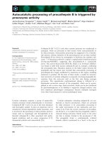
Fig. 1. Analysis of procathepsin B activity on Z-Arg-Arg-NH-MEC
with zymography (bottom) and native PAGE (top) at pH 7.4: (1)
procathepsin B; (2) cathepsin B; and (3) cathepsin B, previously
inactivated by a 2 h incubation at pH 7.6 and 37 °C. Further details
are provided in the Experimental procedures.
J. Rozman Pungerc
ˇ
ar et al. Autocatalytic processing of procathepsin B
FEBS Journal 276 (2009) 660–668 ª 2008 The Authors Journal compilation ª 2008 FEBS 661
collagen type I, which was efficiently hydrolyzed by
mature cathepsin B (data not shown). This is in agree-
ment with the general idea that procathepsin B and
other procathepsins cannot autocatalytically process at
neutral pH due to the inhibitory role of the propep-
tide, although the active site is already formed and
capable of hydrolyzing the substrates.
Autocatalytic processing of procathepsin B
is delayed in the presence of small molecule
inhibitors
To further understand the initial steps of procathep-
sin B autocatalytic processing, we attempted to inhibit
procathepsin B processing by addition of E-64, a
broad spectrum inhibitor of cysteine proteases. The
inhibitor concentrations were varied over a range that
was 5–20% of the molar concentration of procathep-
sin B. Because processing of procathepsin B is typically
45–50% efficient, a higher inhibitor concentration
would completely abolish any catalytic activity of the
enzyme, thereby preventing detection of cathepsin B
activity. All processing curves were sigmoid, showing a
bimolecular process with negligible procathepsin B
activity compared to the activity of the mature cathe-
psin B (Fig. 2). As demonstrated, autocatalytic pro-
cessing of procathepsin B was significantly delayed in
the presence of E-64, suggesting that E-64 primarily
inhibited the mature enzyme. However, from this
experiment, it was not possible to conclude whether
E-64 could bind also to procathepsin B. Thus, to
address this question, E-64 was replaced with the radio-
actively labelled analogue DCG-04 (
125
I-DCG-04) [23].
The major advantage of this inhibitor is the possibility
of detecting the radioactively labelled proteins by auto-
radiography. Samples of procathepsin B and cathep-
sin B were incubated in the presence of
125
I-DCG-04 at
pH 5.8 because processing was not expected to occur at
this pH [14]. As shown in Fig. 3B (lower panel), both
the proform and the mature form of cathepsin B were
found to bind
125
I-DCG-04, suggesting that both spe-
cies are catalytically active. However, labelling of the
zymogen was much weaker, suggesting a substantially
slower binding of the probe to the zymogen compared
to the mature enzyme.
To confirm the specific nature of interaction between
DCG-04 and cathepsin B species, the enzyme samples
were incubated with E-64 prior to labelling with DCG-
04. E-64 at a concentration of 5 lm completely abol-
ished binding of
125
I-DCG-04 to both cathepsin B
species (Fig. 3, lanes 2 and 5), confirming the specific
binding of the activity-based probe to the enzyme. In
an additional experiment, the inactive procathepsin B
Cys29Ser mutant did not label with the probe, thereby
excluding nonspecific binding of the probe to the
enzyme (Fig. 3, lanes 7–9). This is in agreement with
specific labelling of cathepsin B and procathepsin B as
the two active cathepsin species (Fig. 3, lanes 1 and 4).
In the last control experiment, preheated cathepsin B
samples incubated with
125
I-DCG-04 did not label with
the probe, consistent with its binding being specific
(Fig. 3, lanes 3, 6 and 9).
150010005000
100
80
60
40
20
0
Time (min)
Activity
Fig. 2. Autocatalytic processing of 0.17 lM procathepsin B in the
presence of 0 (s), 1.7 (d), 8.5 (h), 17 (
) and 34 (D)nM E-64 at
pH 4.5 and 37 °C. Aliquots were taken from the reaction mixtures
and added to 10 l
M Z-Arg-Arg-NH-MEC substrate solution. Fluores-
cence of the released 7-amino-4-methylcoumarin was followed con-
tinuously with a spectrofluorimeter at the excitation and emission
wavelengths of 370 nm and 460 nm, respectively. Further details
are provided in the Experimental procedures.
25
35
3
4
5
678 9
21
kDa
Coomassie
staining
Autoradiography
Fig. 3. Labelling of procathepsin B by
125
I-DCG-O4. Five micro-
grams of recombinant protein (pCatB, procathepsin B; CatB,
cathepsin B; pCatB C29S, catalytic procathepsin B mutant) were
diluted into acetate buffer (pH 5.6) and incubated in the absence or
presence of 5 l
M E-64 (E-64) for 40 min at 25 °C followed by the
addition of
125
I-DCG-04. In the control experiment, procathepsin B
was pre-heated to 95 °C for 5 min (P.H.). Samples were resolved
by SDS ⁄ PAGE (10–20% gradient gel). Gels were subsequently
stained with Coomassie brilliant blue R250 (upper panel) or analy-
sed by autoradiography (lower panel). Lanes: 1, pCatB; 2, pCatB +
E-64; 3, pCatB P.H.; 4, CatB; 5, CatB + E-64; 6, CatB P.H.; 7,
pCatB C29S; 8, pCatB C29S + E-64; 9, pCatB C29S P.H.
Autocatalytic processing of procathepsin B J. Rozman Pungerc
ˇ
ar et al.
662 FEBS Journal 276 (2009) 660–668 ª 2008 The Authors Journal compilation ª 2008 FEBS
Identification of cleavage sites during
procathepsin B autocatalytic processing
After demonstrating that the zymogen can exhibit
catalytic activity, we next aimed to validate the zymo-
gen activity on other substrates. Therefore, we
performed a mutation analysis of the cleavage region
between the propeptide and the mature enzyme around
Met56-Phe57, which is a conserved cleavage site during
processing [13,24]. All the mutants (Table 1) except the
C29S variant contain a common R54N replacement in
the putative P3 position, which was designed on the
basis of E-64 binding to cathepsin B, where the posi-
tively charged agmatine group, structurally related to
arginine, binds into the S3 substrate binding site [25].
The other mutations were focused on the P1 Met56
residue and ⁄ or on the P1¢–P4¢ residues (Phe57Thr58-
Glu59Asp60). Although the deletion mutants were
expected to increase tension in the flexible C-terminal
propeptide region and thus prevent cleavage in this
segment, the other mutants were expected to prevent
or delay cleavage due to diminished affinity [26].
Initially, processing of procathepsin B mutants was
analysed by SDS ⁄ PAGE. Proenzymes were clearly
present on the gel as 36 kDa bands (data not shown).
After a 3 h incubation of procathepsin B mutants in
the presence or absence of dextran sulfate prior to
electrophoresis, 29 kDa bands corresponding to
mature cathepsin B were observed (data not shown).
The cleavage sites were determined by N-terminal
sequencing of the mature enzymes after processing
(Table 1). Most of the mutants were cleaved after
Met56 (Ala56), with some additional cleavages occur-
ring in the mutated regions with several Ala residues.
However, introducing Pro in the P1 or P1¢ position
abolished cleavage at Met56 and resulted in alternative
cleavages upstream and ⁄ or downstream from the origi-
nal cleavage site, thereby preventing the formation of
a noncleavable procathepsin B mutant.
Next, we evaluated the activity of the mature forms
resulting from the processing of procathepsin B
mutants. All these forms of cathepsin B with different
N-terminal extensions exhibited similar activity against
Z-Arg-Arg-NH-MEC (not shown), in agreement with
the idea that the neo N-terminus of mature cathep-
sin B is not important for its catalytic activity. Finally,
the processing rates of the procathepsin B mutants
were compared. To ensure equal starting concentra-
tions, the procathepsin B variants were subjected to
processing in the presence of dextran sulfate to com-
plete the process reasonably quickly (approximately
1 h) and to prevent possible inactivation. Mature
cathepsin B generated was then active-site titrated by
E-64 directly in the processing mixture to determine
the processing efficiency. The processing rates of pro-
cathepsin B mutants and native procathepsin B (equal
concentrations) were then determined in the presence
and absence of dextran sulfate (Table 1). The R54N
procathepsin B variant, which served as a basis for all
other mutations, was processed at a rate almost three-
fold lower than the wild-type procathepsin B, support-
ing the proposed important role of Arg54 in substrate
recognition. Most of the other mutants were processed
somewhat faster than the R54N variant. The excep-
tions were the T58ADED and E59A ⁄ D60A mutants,
which were processed approximately five-fold faster
than the wild-type zymogen, and the F57A and
F57A ⁄ T58A ⁄ E59A ⁄ D60A mutants, which were pro-
cessed approximately two-fold slower. Surprisingly, the
F57P mutant was processed substantially faster than
the F57A mutant, probably due to different cleavage
sites, which could result from stepwise processing.
Because Quraishi and Storer [21] detected a process-
ing intermediate starting with L41, R40A and
K39A ⁄ R40A mutants on the wild-type background
were generated. However, the processing of these
mutants, which appear to have a role in GAG binding,
was up to two-fold faster than the processing of the
wild-type variant (t
1 ⁄ 2
= 28 versus 55 min, respec-
tively) [27]. This suggests that the Arg40-Leu41 cleav-
age may not be essential for processing because Arg is
the preferred residue in the S1 position of cysteine
cathepsins [26].
Discussion
Zymogen activation is one of the crucial steps in regu-
lating the activity of proteases [28,29]. Although there
have been a number of attempts to explain the mecha-
nism of autocatalytic activation of cysteine cathepsins
[1,30], none have succeeded in explaining the initial
activity of the proteases, which was observed at the
very beginning of processing [14,18–20,27]. In addition,
it has been suggested that processing may proceed
through several intermediate steps, although their
importance for the actual processing was not evaluated
[21]. The results obtained in the present study demon-
strate that the initial activity observed during process-
ing belongs to the activity of the cathepsin B zymogen,
as detected by a small synthetic substrate and affinity
labelling by the activity-based probe
125
I-DCG-04. As
seen in the crystal structure of the cathepsin zymogens
[7–10], the propeptide binds in the active site in a
direction opposite to that of the substrate, thereby pre-
venting substrate hydrolysis. The data thus suggest
that substrate hydrolysis can be explained by the
J. Rozman Pungerc
ˇ
ar et al. Autocatalytic processing of procathepsin B
FEBS Journal 276 (2009) 660–668 ª 2008 The Authors Journal compilation ª 2008 FEBS 663
Table 1. Processing of procathepsin B variants. Estimates of processing half-times of procathepsin B variants in the absence (–DS) and presence (+DS) of 25 lgÆmL
)1
dextran sulfate,
obtained by a discontinuous method, are given together with the respective SEM. The proenzyme concentration was 0.37 l
M in all the experiments. The cleavage sites determined by
N-terminal amino acid sequencing after autocatalytic processing of proenzyme variants in the absence of DS are marked with arrows. Partial propeptide sequence (residues 46 to 62 of
the propeptide) is given in the first line. Further details are given in the Experimental procedures.
Autocatalytic processing of procathepsin B J. Rozman Pungerc
ˇ
ar et al.
664 FEBS Journal 276 (2009) 660–668 ª 2008 The Authors Journal compilation ª 2008 FEBS
flexibility of the propeptide, which is presumably
greatly increased at acidic pH. This is supported by
in vitro studies of the interaction between the propep-
tide and mature enzymes, which demonstrated a sub-
stantially weaker affinity of the propeptides at acidic
than at neutral pH [15–17].
The major outcome of the mutagenesis studies was
that cathepsin B is not a very specific enzyme and is
capable of cleaving procathepsin B at different sites,
which is in agreement with the general broad specific-
ity of the cathepsins [26]. Although the preferential
cleavage site appears to be at the Met56-Phe57 bond,
mutating Met56 or Phe57 to Pro leads to new N-termi-
nal variants (Table 1). This prevented us from making
a catalytically active, nonprocessed or partially pro-
cessed zymogen, suggesting that the same probably
holds true for processing of other cysteine cathepsins.
On the basis of the results obtained in the present
study, as well as those of previous studies
[14,17,21,27], a common mechanism for the autocata-
lytic processing of papain-like cysteine endoproteases
is proposed. Initially, the pH change facilitates propep-
tide movement from its normal position within the
active-site cleft in the zymogen, thereby converting the
latter into an active form. This appears to be a
dynamic equilibrium, which is shifted towards the
inactive form at neutral pH and towards the active
form at acidic pH, consistent with the inability of pro-
cathepsin B to cleave a macromolecular proteinaceous
substrate at neutral pH. Moreover, this conformational
change, which is the only unimolecular step of the
mechanism, is not accompanied by any larger struc-
tural changes, such as unfolding of the ‘hook’ domain,
as demonstrated previously using the catalytic Cys29-
Ser procathepsin B mutant [14].
When two procathepsin B molecules come into close
contact, one active zymogen molecule cleaves the pro-
peptide from the second molecule. It is very likely that
propeptide removal occurs in at least two consecutive
steps, with the first one comprising the ‘hook’ removal,
as Quraishi and Storer [21] detected several intermedi-
ate forms starting downstream of the ‘hook’ region
(Leu41 and Cys43 from the propeptide). These short-
ened zymogen forms, with presumably higher enzy-
matic activity, facilitate the removal of the rest of the
propeptide from the interacting procathepsin B mole-
cules. Fully active mature cathepsin B molecules then
enter the cycle and process the majority of the intact or
partially processed zymogen molecules. It is possible
that, at least initially, intermediate forms and intact
zymogens are also cleaved by activated intact and par-
tially processed zymogens. This is in agreement with
the findings of a study [31] demonstrating that the trun-
cated procathepsin B zymogens, resulting from a gene
lacking exons 2 and 3 and with a propeptide shortened
by 34 residues, possess substantial catalytic activity.
Glycosaminoglycans, which can facilitate autocatalytic
activation of cysteine cathepsins, were shown to induce
a conformational change in procathepsin B upon bind-
ing, resulting in propeptide removal from the active site
cleft and conversion of the zymogen into a better
substrate for mature cathepsin B [27]. Moreover, such
procathepsin B processing was observed during a puri-
fication step on heparin Sepharose, even at pH 7.6,
demonstrating their extreme efficiency (data not
shown). In addition to glycosaminoglycans, other
charged surfaces were found to enable autocatalytic
processing at neutral pH because the processing of
procathepsin B during filtration through microcon
cellulose membrane at pH 7.6 was also observed (data
not shown). The molecular mechanism of cathepsin
activation induced by pH lowering and ⁄ or by glycosa-
minoglycans is probably similar in both cases, with the
only difference being that glycosaminoglycans and
other negatively charged surfaces are much more
efficient and can facilitate processing also at a higher
pH. Therefore, it is proposed that this unimolecular
conformational change has a dual role: first, it converts
the zymogen into an active form and, second, it con-
verts the zymogen into a better substrate, although the
latter may be more applicable to glycosaminoglycans
[27].
In vivo processing of cysteine cathepsins is probably
more complex. The relative insensitivity of procathep-
sin B processing to mutations in the linker region sug-
gests that cathepsins are well adapted to the cellular
environment, and explains why they can be activated
by multiple proteases [1,30,32]. All these different
pathways of activation may thus account for the pres-
ence of active cathepsin or procathepsin species outside
lysosomes, which, under normal conditions, are held
under the control of endogenous inhibitors, such as
cystatins and serpins [33]. However, the existence of
extralysosomal and extracellular cathepsins in disease
is not only linked to the secretion of various cathepsin
forms from lysosomes and subsequent processing at
the membranes, but also likely results from differential
trafficking and synthesis because different splice vari-
ants of cathepsins are found primarily in cancer
[3,5,31]. Moreover, the fact that cathepsin zymogens
are very resistant towards pH-induced inactivation,
combined with their ability to be readily activated even
under unfavourable conditions, poses a persistent
threat to the system, which cannot be so easily elimi-
nated because zymogens are resistant to inhibition by
endogenous inhibitors.
J. Rozman Pungerc
ˇ
ar et al. Autocatalytic processing of procathepsin B
FEBS Journal 276 (2009) 660–668 ª 2008 The Authors Journal compilation ª 2008 FEBS 665
In conclusion, procathepsin B was found to be an
active species, suggesting that autocatalytic activation
of cysteine cathepsins is a multi-step process, starting
with a unimolecular conformational change of the
zymogen, which unmasks the active site and, in the
presence of negatively charged molecules ⁄ surfaces, also
converts the zymogen into a better substrate. This is
followed by the bimolecular proteolytic removal of the
propeptide, which can be accomplished in one or more
steps. Such active cathepsin species could have impor-
tant roles in physiology, including the development of
several diseases such as cancer and arthritis.
Experimental procedures
Materials
Restriction enzymes were obtained from MBI Fermentas
(Burlington, Canada) and New England Biolabs (Steve-
nage, UK); T4 DNA ligase was obtained from Roche
(Basel, Switzerland); polynucleotide T4 kinase was obtained
from MBI Fermentas; and Vent DNA polymerase was
obtained from New England Biolabs. Oligonucleotides were
obtained from MWG-Biotech (Ebersberg, Germany).
Z-Arg-Arg-NH-MEC was obtained from Bachem (Buben-
dorf, Switzerland); E-64 was obtained from the Peptide
Research Institute (Osaka, Japan); and dextran sulfate was
obtained from Sigma (St Louis, MO, USA). DCG-04 was
prepared as described previously [23].
Procathepsin B and its mutants were synthesized in
E. coli and purified as described previously [12]. The recom-
binant proteins were nonglycosylated as a consequence of
the expression system. However, all the potential glycosyla-
tion sites are located on the surface of the protein pointing
towards the solvent and thus do not interefere with glycos-
aminoglycan binding, autocatalytic activation of the zymo-
gen or activity of the mature enzyme [9,13,27]. All proteins
were verified by SDS ⁄ PAGE and N-terminal aminoacid
sequence analysis. Protein concentrations were determined
from absorption spectra according to Pace et al. [34]. The
active proenzyme concentrations were determined by acti-
vation and active-site titration of the resulting enzyme with
E-64 [35].
Site-directed mutagenesis
Site-directed mutagenesis was performed using PCR as
described by Michael [36]. The plasmid and outer primer
oligonucleotides used were constructed by Kuhelj et al. [12].
The mutagenic oligonucleotides (5¢-CCACCCCAGAACGT
TATGTTTACCG-3¢ and 5¢-GCTCCTCCTGGGCCTT-3¢)
were used to introduce the R54N and C29S substitutions,
respectively (where the C29S mutation substituted active-
site Cys29 on the mature part of the enzyme to a serine
residue). Additional mutants were prepared using a vector
with cDNA for pcatB(R54N) as a template and the
following mutagenic oligonucleotides: 5¢-CCAGAACGTTA
TGTTTGCACTGAAGCTGCCTGC-3¢ (for the T58ADED
mutant: R54N, T58A, deletion of E59 and D60); 5¢-GAAC
GTTATGTTTACCGCAGCTCTGAAGCTGCCTGC-3¢
(for the ED59AA mutant: R54N, E59A, D60A); 5¢-CCAG
AACGTTATGTTTGCAGCTGCACTGAAGCTGCCTGC-3¢
(for the TED58AAA mutant: R54N, T58A, E59A, D60A);
5¢-CCCAGAACGTTATGGCTGCAGCTGCACTGAAGC
TGCCTG-3¢ (for the FTED57AAAA mutant: R54N,
F57A, T58A, E59A, D60A); 5¢-CCAGAACGTTATGGC
TACCGAGGACCTGAAGC-3¢ (for the F57A mutant:
R54N, F57A); 5¢-CCAGAACGTT(GC)CGTTTACCGA
GG-3¢ (a degenerate primer for M56A and M56P mutants;
R54N, M56A and R54N, M56P, respectively); and 5¢-GAA
CGTTATGCCGACCGAGGACC-3¢ (for F57P mutant:
R54N, F57P). The second set of mutants were prepared
using the vector with cDNA for pcatB (R54N, T58A,
deletion of E59 and D60) as a template and mutagenic
primers: 5¢-CCAGAACGTTCCGTTTGCACTGAA-3¢ (for
T58ADED_M56P mutant: R54N, M56P, T58A, deletion of
E59 and D60) and 5¢-GAACGTTATGCCGGCACTGA
AGCT-3¢ (for T58ADED_F57P mutant: R54N, F57P,
T58A, deletion of E59 and D60). Mutagenic oligonucleo-
tides were phosphorylated by T4 polynucleotide kinase
prior to the mutagenesis reaction. Each PCR mixture
(100 lL) contained 500 ng of a plasmid template, 50 pmol
of each of the three oligonucleotides (the two outer and a
mutagenic one), 20 nmol of each of the four deoxynucleo-
side triphosphates, Taq DNA ligase buffer, 5 U of Vent
DNA polymerase and 5 U of Taq DNA ligase. After 35
cycles of PCR amplification (94 °C for 60 s; 50 °C for 60 s;
65 °C for 240 s), the PCR products were cleaned by the
QIAEX II extraction kit (Qiagen, Valencia, CA, USA) and
cloning was carried out as described previously [12].
Propeptide numbering is used throughout, unless stated
otherwise.
Kinetic measurements
Processing of procathepsin B and its mutants was examined
at 37 °C and pH 4.5 (0.1 m acetate buffer, containing 1 mm
EDTA and 5 mm dithiothreitol) as described by Rozman
et al. [14]. Proenzyme (0.17–1.33 lm) was incubated in
1 mL of the processing buffer. Aliquots of 5, 10 or 20 lL
were taken from the reaction mixtures at appropriate times
and added to 2.495–2.48 mL of 10 lm Z-Arg-Arg-NH-
MEC substrate solution in 0.1 m phosphate buffer (pH 6.0)
containing 1 mm EDTA and 0.1% (w ⁄ v) polyethylene gly-
col 6000 (Serva, Wichita Falls, TX, USA). Fluorescence of
the released 7-amino-4-methylcoumarin was followed con-
tinuously with a C-61 spectrofluorimeter (Photon Technol-
ogy International, Birmingham, NJ, USA) at the excitation
Autocatalytic processing of procathepsin B J. Rozman Pungerc
ˇ
ar et al.
666 FEBS Journal 276 (2009) 660–668 ª 2008 The Authors Journal compilation ª 2008 FEBS
and emission wavelengths of 370 and 460 nm, respectively.
When specified, processing was accelerated by the addition
of dextran sulfate (25 lgÆmL
)1
) or decelerated by the addi-
tion of E-64 in the processing buffer. The final concentra-
tion of procathepsin B variants in the processing buffer was
0.37 lm throughout.
Detection of
125
I-DCG-04-labelled proteins
Proteins (1.7 lg) were incubated in 50 mm sodium acetate
(pH 5.8) containing 5 mm dithiothreitol, 150 mm NaCl and
1mm EDTA in the presence or absence of 5 lm E-64 for
40 min at 25 °C, followed by the addition of small amounts
of radioactive probe
125
I-DCG-04 and an additional 40 min
of incubation under the same conditions. In a control
experiment, protein sample was incubated for 5 min at
95 °C prior to the addition of
125
I-DCG-04. The samples
were then separated by SDS ⁄ PAGE and stained with Coo-
massie brilliant blue R250 or visualized by autoradiography
using a Typhoon Trio (GE Healthcare, Milwaukee, WI,
USA) as described previously [23].
N-terminal amino acid analysis
Procathepsin B variants (1.0–3.15 lg) were incubated in the
processing buffer at 37 °C for 3 h. The products were sepa-
rated by SDS ⁄ PAGE under reducing conditions on 12.5%
gels and electroblotted to poly(vinylidene difluoride) mem-
brane (Bio-Rad, Hercules, CA, USA). The protein bands
were subjected to Edman degradation on an Procise 492A
protein sequencer (Applied Biosystems, Foster City, CA,
USA).
Native polyacrylamide gel electrophoresis and
zymography
Native PAGE was performed on a 7% gel at pH 7.4 as
described by McLellan [37]. After electrophoresis, the gel
was incubated for 5 min in 0.1 m phosphate buffer (pH 6.0)
containing 10 mm dithiothreitol, 1 mm EDTA and 0.1%
(w ⁄ v) polyethylene glycol 6000, and covered by 5 mL of
40 lm substrate Z-Arg-Arg-NH-MEC in the same buffer.
Fluorescence of the released product was monitored under
an UV lamp. The gel was stained subsequently with
Coomassie brilliant blue R250.
Acknowledgements
We thank Adrijana Leonardi for N-terminal amino
acid sequencing and Professor Roger H. Pain for criti-
cal reading of the manuscript. The work was sup-
ported by grants (P0140 and J1-6488) to B.T. and V.T.
from the Slovenian Ministry of Higher Education,
Science and Technology and by the Human Frontier
Science Project Grant RGP0024 ⁄ 2006-C to B.T. and
M.B. The work was further supported by National
Institutes of Health National Technology Center for
Networks and Pathways grant U54 RR02084 to M.B.
and Sandler Family Supporting Foundation grant to
M.S.
References
1 Turk B, Turk D & Turk V (2000) Lysosomal cysteine
proteases: more than scavengers. Biochim Biophys Acta
1477, 98–111.
2 Turk V, Turk B & Turk D (2001) Lysosomal cysteine
proteases: facts and opportunities. EMBO J 20, 4629–
4633.
3 Vasiljeva O, Reinheckel T, Peters C, Turk D, Turk V &
Turk B (2007) Emerging roles of cysteine cathepsins in
disease and their potential as drug targets. Curr Pharm
Des 13, 387–403.
4 Kos J & Lah TT (1998) Cysteine proteinases and their
endogenous inhibitors: target proteins for prognosis,
diagnosis and therapy in cancer (review). Oncol Rep 5,
1349–1361.
5 Mohamed MM & Sloane BF (2006) Cysteine cathep-
sins: multifunctional enzymes in cancer. Nat Rev Cancer
6, 764–775.
6 Baici A, Lang A, Horler D, Kissling R & Merlin C
(1995) Cathepsin B in osteoarthritis: cytochemical and
histochemical analysis of human femoral head cartilage.
Ann Rheum Dis 54 , 289–297.
7 Coulombe R, Grochulski P, Sivaraman J, Menard R,
Mort JS & Cygler M (1996) Structure of human procat-
hepsin L reveals the molecular basis of inhibition by the
prosegment. EMBO J 15, 5492–5503.
8 Cygler M, Sivaraman J, Grochulski P, Coulombe R,
Storer AC & Mort JS (1996) Structure of rat procathep-
sin B: model for inhibition of cysteine protease activity
by the proregion. Structure 4, 405–416.
9 Podobnik M, Kuhelj R, Turk V & Turk D (1997) Crys-
tal structure of the wild-type human procathepsin B at
2.5 A resolution reveals the native active site of a
papain-like cysteine protease zymogen. J Mol Biol 271,
774–788.
10 Turk D, Podobnik M, Kuhelj R, Dolinar M & Turk V
(1996) Crystal structures of human procathepsin B at
3.2 and 3.3 Angstroms resolution reveal an interaction
motif between a papain-like cysteine protease and its
propeptide. FEBS Lett 384, 211–214.
11 Riedl SJ, Renatus M, Schwarzenbacher R, Zhou Q,
Sun C, Fesik SW, Liddington RC & Salvesen GS
(2001) Structural basis for the inhibition of caspase-3
by XIAP. Cell 104, 791–800.
12 Kuhelj R, Dolinar M, Pungerc
ˇ
ar J & Turk V (1995)
The preparation of catalytically active human cathepsin
J. Rozman Pungerc
ˇ
ar et al. Autocatalytic processing of procathepsin B
FEBS Journal 276 (2009) 660–668 ª 2008 The Authors Journal compilation ª 2008 FEBS 667
B from its precursor expressed in Escherichia coli in the
form of inclusion bodies. Eur J Biochem 229, 533–539.
13 Mach L, Mort JS & Glossl J (1994) Maturation of
human procathepsin B. Proenzyme activation and
proteolytic processing of the precursor to the mature
proteinase, in vitro, are primarily unimolecular
processes. J Biol Chem 269, 13030–13035.
14 Rozman J, Stojan J, Kuhelj R, Turk V & Turk B
(1999) Autocatalytic processing of recombinant human
procathepsin B is a bimolecular process. FEBS Lett
459, 358–362.
15 Carmona E, Dufour E, Plouffe C, Takebe S, Mason P,
Mort JS & Menard R (1996) Potency and selectivity of
the cathepsin L propeptide as an inhibitor of cysteine
proteases. Biochemistry 35, 8149–8157.
16 Fox T, de Miguel E, Mort JS & Storer AC (1992)
Potent slow-binding inhibition of cathepsin B by its
propeptide. Biochemistry 31, 12571–12576.
17 Quraishi O, Na
¨
gler DK, Fox T, Sivaraman J, Cygler
M, Mort JS & Storer AC (1999) The occluding loop in
cathepsin B defines the pH dependence of inhibition by
its propeptide. Biochemistry 38, 5017–5023.
18 Baker KC, Taylor MA, Cummings NJ, Tunon MA,
Worboys KA & Connerton IF (1996) Autocatalytic
processing of pro-papaya proteinase IV is prevented by
crowding of the active-site cleft. Protein Eng 9, 525–
529.
19 Mason RW, Gal S & Gottesman MM (1987) The iden-
tification of the major excreted protein (MEP) from a
transformed mouse fibroblast cell line as a catalytically
active precursor form of cathepsin L. Biochem J 248,
449–454.
20 McQueney MS, Amegadzie BY, D’Alessio K, Hanning
CR, McLaughlin MM, McNulty D, Carr SA, Ijames C,
Kurdyla J & Jones CS (1997) Autocatalytic activation
of human cathepsin K. J Biol Chem 272, 13955–13960.
21 Quraishi O & Storer AC (2001) Identification of inter-
nal autoproteolytic cleavage sites within the proseg-
ments of recombinant procathepsin B and procathepsin
S. Contribution of a plausible unimolecular autoproteo-
lytic event for the processing of zymogens belonging to
the papain family. J Biol Chem 276, 8118–8124.
22 Turk B, Dolenc I, Z
ˇ
erovnik E, Turk D, Gubens
ˇ
ek F &
Turk V (1994) Human cathepsin B is a metastable
enzyme stabilized by specific ionic interactions associ-
ated with the active site. Biochemistry 33, 14800–14806.
23 Greenbaum D, Medzihradszky KF, Burlingame A &
Bogyo M (2000) Epoxide electrophiles as activity-depen-
dent cysteine protease profiling and discovery tools.
Chem Biol 7, 569–581.
24 Rowan AD, Mason P, Mach L & Mort JS (1992) Rat
procathepsin B. Proteolytic processing to the mature
form in vitro. J Biol Chem 267, 15993–15999.
25 Turk D, Podobnik M, Popovic
ˇ
T, Katunuma N, Bode
W, Huber R & Turk V (1995) Crystal structure of
cathepsin B inhibited with CA030 at 2.0-A resolution: a
basis for the design of specific epoxysuccinyl inhibitors.
Biochemistry 34, 4791–4797.
26 Turk D, Gunc
ˇ
ar G, Podobnik M & Turk B (1998)
Revised definition of substrate binding sites of papain-
like cysteine proteases. Biol Chem 379, 137–147.
27 Caglic
ˇ
D, Pungerc
ˇ
ar JR, Pejler G, Turk V & Turk B
(2007) Glycosaminoglycans facilitate procathepsin B
activation through disruption of propeptide-mature
enzyme interactions. J Biol Chem 282, 33076–33085.
28 Neurath H (1984) Evolution of proteolytic enzymes.
Science 224, 350–357.
29 Turk B (2006) Targeting proteases: successes, failures
and future prospects. Nat Rev Drug Discov 5, 785–799.
30 Bromme D, Nallaseth FS & Turk B (2004) Production
and activation of recombinant papain-like cysteine
proteases. Methods 32, 199–206.
31 Mehtani S, Gong Q, Panella J, Subbiah S, Peffley DM
& Frankfater A (1998) In vivo expression of an alterna-
tively spliced human tumor message that encodes a
truncated form of cathepsin B. Subcellular distribution
of the truncated enzyme in COS cells. J Biol Chem 273,
13236–13244.
32 Dalet-Fumeron V, Boudjennah L & Pagano M (1996)
Competition between plasminogen and procathepsin B
as a probe to demonstrate the in vitro activation of pro-
cathepsin B by the tissue plasminogen activator. Arch
Biochem Biophys 335, 351–357.
33 Turk B, Turk D & Salvesen GS (2002) Regulating cys-
teine protease activity: essential role of protease inhibi-
tors as guardians and regulators. Curr Pharm Des 8,
1623–1637.
34 Pace CN, Vajdos F, Fee L, Grimsley G & Gray T
(1995) How to measure and predict the molar absorp-
tion coefficient of a protein. Protein Sci 4, 2411–2423.
35 Turk B, Krizˇ aj I, Kralj B, Dolenc I, Popovic
ˇ
T, Bieth
JG & Turk V (1993) Bovine stefin C, a new member of
the stefin family. J Biol Chem 268, 7323–7329.
36 Michael SF (1994) Mutagenesis by incorporation of a
phosphorylated oligo during PCR amplification.
Biotechniques 16, 410–412.
37 McLellan T (1982) Electrophoresis buffers for
polyacrylamide gels at various pH. Anal Biochem 126,
94–99.
Autocatalytic processing of procathepsin B J. Rozman Pungerc
ˇ
ar et al.
668 FEBS Journal 276 (2009) 660–668 ª 2008 The Authors Journal compilation ª 2008 FEBS