Báo cáo Y học: Assignment of molecular properties of a superactive coagulation factor VIIa variant to individual amino acid changes potx
Bạn đang xem bản rút gọn của tài liệu. Xem và tải ngay bản đầy đủ của tài liệu tại đây (170.91 KB, 6 trang )
Assignment of molecular properties of a superactive coagulation
factor VIIa variant to individual amino acid changes
Egon Persson
1
and Ole H. Olsen
2
1
Haemostasis Biology and
2
Medicinal Chemistry Research IV, Novo Nordisk A/S, Ma
˚
løv, Denmark
The most active factor VIIa (FVIIa) variants identified to
date carry concurrent substitutions at positions 158, 296 and
298 with the intention of generating a thrombin-mimicking
motif, optionally combined with additional replacements
within the protease domain [Persson, E., Kjalke, M. &
Olsen, O. H. (2001) Proc. Natl Acad. Sci. USA 98, 13583–
13588]. Here we have characterized variants of FVIIa
mutated at one or two of these positions to assess the relative
importance of the individual replacements. The E296V and
M298Q mutations gave an increased intrinsic amidolytic
activity (about two- and 3.5-fold, respectively) compared
with wild-type FVIIa. An additive effect was observed upon
their combination, resulting in the amidolytic activity of
E296V/M298Q-FVIIa being close to that of the triple
mutant. The level of amidolytic activity of a variant was
correlated with the rate of inhibition by antithrombin (AT).
Compared with wild-type FVIIa, the Ca
2+
dependence of
the intrinsic amidolytic activity was significantly attenuated
upon introduction of the E296V mutation, but the effect was
most pronounced in the triple mutant. Enhancement of the
proteolytic activity requires substitution of Gln for Met298.
The simultaneous presence of the V158D, E296V and
M298Q mutations gives the highest intrinsic activity and is
essential to achieve a dramatically higher relative increase in
the proteolytic activity than that in the amidolytic activity.
The N-terminal Ile153 is most efficiently buried in V158D/
E296V/M298Q-FVIIa, but is less available for chemical
modification also in the presence of the E296V or M298Q
mutation alone. In summary, E296V and M298Q enhance
the amidolytic activity and facilitate salt bridge formation
between the N-terminus and Asp343, E296V reduces the
Ca
2+
dependence, M298Q is required for increased factor X
(FX) activation, and the simultaneous presence of the
V158D, E296V and M298Q mutations gives the most pro-
found effect on all these parameters.
Keywords: factor VIIa variant; factor X activation; intrinsic
activity; superactivity; zymogenicity.
Coagulation factor VIIa (FVIIa), in contrast to other,
homologous serine proteases, possesses an active confor-
mation that is energetically unfavourable. The consequence
is a far from optimal enzymatic activity of free FVIIa, which
is dramatically enhanced upon binding to the cognate,
membrane-bound cofactor tissue factor (TF) or to its
extracellular, soluble portion (sTF) [1]. In the natural
environment, the zymogenicity of free FVIIa ensures timely
triggering and appropriate location of FVIIa haemostatic
activity upon vascular lesion and concomitant TF exposure.
The three-dimensional structure of the protease domain
of free FVIIa is, apart from certain loop regions, virtually
identical to that of thrombin and other constitutively active
and homologous serine proteases [2,3]. In addition, the
structural differences between free [3–5] and TF-bound
FVIIa [6,7] are subtle; thus the details in molecular
architecture that restrict the activity of free FVIIa remain
elusive. However, the high degree of similarity may be due
to the presence of an active site inhibitor in the structure of
the free FVIIa. The crystal (or solution) structure of
noninhibited FVIIa is presumably needed to reveal the
structural differences between ÔlatentÕ (zymogen-like) and
ÔactiveÕ FVIIa. However, information possibly pertaining to
the latent conformation of free FVIIa has been obtained
from the crystal structure of zymogen FVII [8]. This
structure suggests that relative b strand movements and a
hydrogen bond involving Glu296{154} (chymotrypsinogen
numbering is given in curly brackets to facilitate compar-
isons with homologous enzymes) regulate the activity state
of FVIIa.
Recent advances in our understanding of the mechanisms
regulating the activity of FVIIa have pinpointed side chains
that function as zymogenicity determinants in the free
enzyme. Replacements of these amino acid residues have
resulted in FVIIa molecules with improved intrinsic
(TF-independent) catalytic efficiency [9–11]. The relatively
high intrinsic activity of some of these FVIIa variants
suggests that the zymogen-like conformation of free factor
VIIa is dictated by a limited number of key amino acid
residues. We have previously shown that one of these
superactive FVIIa variants, containing the mutations
V158{21}D, E296{154}V and M298{156}Q, exhibits several
properties resembling TF-bound rather than free FVIIa [9].
Apart from increased intrinsic enzymatic activity and
inhibitor susceptibility as compared with wild-type FVIIa,
this mutant has a diminished requirement for calcium ions
and a more deeply buried protease domain N-terminus
Correspondence to E. Persson, Haemostasis Biology, Novo Nordisk
A/S, Novo Nordisk Park, DK-2760 Ma
˚
løv, Denmark.
Fax: + 45 44434417, Tel.: + 45 44434351,
E-mail:
Abbreviations: FVIIa, coagulation factor VIIa; FX, coagulation factor
X; sTF, soluble tissue factor (residues 1–219); TF, tissue factor;
AT, antithrombin (III).
(Received 20 June 2002, revised 2 October 2002,
accepted 22 October 2002)
Eur. J. Biochem. 269, 5950–5955 (2002) Ó FEBS 2002 doi:10.1046/j.1432-1033.2002.03323.x
(Ile153{16}) indicating salt bridge formation of this residue
with Asp343 {194}. To pinpoint the underlying mutation(s)
responsible for these molecular and functional properties of
the triple mutant, and thereby gain more knowledge about
the zymogenicity determinants of FVIIa, mutants with
substitutions at one or two of positions 158{21}, 296{154}
and 298{156} were expressed and characterized.
MATERIALS AND METHODS
Proteins, chemical reagents and standard methods
Wild-type FVIIa [12] and sTF [13] were prepared according
to published procedures. The concentrations of FVIIa and
sTF were determined by ELISA and spectrophotometry,
respectively, as described [9]. SDS/PAGE was run on
8–25% gradient gels using the PhastSystem (Amersham
Pharmacia) and followed by silver staining to check the
purity of the FVIIa mutants and to verify their conversion
to the activated, two-chain form. Factor X (FX) and factor
Xa were from Enzyme Research Laboratories (South Bend,
IN, USA) and antithrombin (AT) from Hematologic
Technologies (Essex Junction, VT, USA). Unfractionated
heparin was from Leo Pharmaceutical Products (Ballerup,
Denmark), potassium cyanate (KOCN) from Merck and
the chromogenic substrates S-2288 (
D
-Ile-Pro-Arg-p-nitro-
anilide) and S-2765 (benzyloxycarbonyl-
D
-Arg-Gly-Arg-
p-nitroanilide) from Chromogenix (Mo
¨
lndal, Sweden).
Mutagenesis and preparation of FVIIa mutants
The FVII expression plasmid pLN174 [14] was used as the
template for site-directed mutagenesis using the Quik-
Change kit (Stratagene, La Jolla, CA, USA). The primer
(only sense primer is given) used to introduce the E296V
mutation, with base substitution in italic and the affected
codon underlined, was GCC ACG GCC CTG
GTGCTC
ATG GTC CTC. The primers used to introduce the V158D,
E296V/M298Q and M298Q mutations have been described
[9]. Plasmid preparation, baby hamster kidney cell trans-
fection and selection, and the expression, purification and
autoactivation of FVII mutants were carried out as
described [9,15]. The presence of none but the desired
mutation(s) was verified by sequencing the portion of the
cDNA encoding residues 80–406, encompassing the
second epidermal growth factor-like and serine protease
domains, on a MegaBACE 1000 (Amersham Pharmacia
Biotech).
Activity and inhibition assays
The enzymatic activity and inhibition rates of the FVIIa
variants were measured as described [9] using an assay
buffer containing 50 m
M
Hepes, 0.1
M
NaCl, 5 m
M
CaCl
2
and 1 mgÆmL
)1
bovine serum albumin (pH 7.4). Briefly, the
amidolytic activity was measured by mixing 180 lL wild-
type or mutant FVIIa alone (final concentration 100 n
M
)or
180 lL FVIIa together with sTF (final concentrations
10 n
M
FVIIa and 50 n
M
sTF) with 20 lL10m
M
S-2288 at
25 °C. The measurement with free FVIIa was also per-
formed in an assay buffer where CaCl
2
was replaced by
EDTA. The ability of the FVIIa variants to activate FX (the
proteolytic activity) was studied by incubating 10 n
M
(M298Q-, V158D/M298Q-, E296V/M298Q- and V158D/
E296V/M298Q-FVIIa) or 50 n
M
(wild-type, V158D-,
E296V-, and V158D/E296V-FVIIa) FVIIa variant alone
or 5 n
M
FVIIa variant plus 100 n
M
sTF with various
concentrations of FX (0.1–4.8 l
M
)for20minatambient
temperature (22 ± 1 °C). Buffer containing S-2765 was
then added to give a chromogenic substrate concentration
of 0.5 m
M
, whereafter the factor Xa-catalyzed hydrolysis
was measured for 2 min. The inherent activity of the FX
substrateandoftheFVIIa/sTFmixtureweresubtracted
and the net amount of factor Xa generated was derived
from a standard curve. The kinetic parameters of FX
activation were calculated using
GRAFIT
4.06 (Erithacus
Software, Ltd). The rates of inhibition by AT/heparin and
potassium cyanate were studied according to published
methods [9] by incubation of the FVIIa variants (100 n
M
with AT and 1 l
M
with cyanate) with the inhibitor for 15
and 60 min, respectively, followed by measurements of the
residual amidolytic activity. Bovine serum albumin was
omitted from the buffer during the incubation with cyanate.
RESULTS
Enzymatic activity of FVIIa variants
Two forms of enzymatic activity are analyzed. The amido-
lytic activity is measured to assess the functional status of
the active site, whereas the proteolytic activity reflects both
this and exosite alterations resulting in further increased
macromolecular substrate turnover. In the absence of TF,
V158D/E296V/M298Q-FVIIa has seven- to eight-fold
higher intrinsic amidolytic activity compared with wild-type
FVIIa as measured by the rate of hydrolysis of the
chromogenic substrate S-2288 [9]. When the mutations
were introduced individually into FVIIa, V158D had no
significant effect on the amidolytic activity, whereas E296V
and M298Q yielded approximately two- and 3.5-fold
enhancement, respectively (Table 1). The result with
M298Q-FVIIa agrees with earlier reports [9,10]. The double
mutant E296V/M298Q-FVIIa had an amidolytic activity
six times higher than that of wild-type FVIIa and close to
that of V158D/E296V/M298Q-FVIIa. In addition, V158D/
E296V-FVIIa had significantly lower amidolytic than
E296V-FVIIa and V158D/M298Q-FVIIa had similar or
slightly lower activity than M298Q-FVIIa. This shows that
the simultaneous presence of the E296V and M298Q
mutations suffices to achieve an amidolytic activity similar
Table 1. Enzymatic activity of free FVIIa variants. All values are
means ± SD (n ¼ 3). The amidolytic activity is given as the ratio
between the activity of mutant and wild-type FVIIa.
FVIIa variant
Amidolytic activity
(mutant/wt)
FX activation
(k
cat
, · 10
)3
s
)1
)
Wild-type 0.088 ± 0.006
V158D 1.0 ± 0.1 0.069 ± 0.008
E296V 2.1 ± 0.3 0.083 ± 0.010
M298Q 3.4 ± 0.4 0.62 ± 0.04
V158D/E296V 0.6 ± 0.1 0.081 ± 0.009
V158D/M298Q 3.0 ± 0.5 0.60 ± 0.05
E296V/M298Q 6.0 ± 0.5 0.47 ± 0.07
V158D/E296V/M298Q 7.6 ± 0.5 2.2 ± 0.3
Ó FEBS 2002 Dissection of a superactive FVIIa variant (Eur. J. Biochem. 269) 5951
to that of the triple mutant and that the substitution of Asp
for Val at position 158 has an insignificant or negative
impact on the amidolytic activity. In complex with sTF,
none of the investigated mutants had an amidolytic activity
significantly different from that of wild-type FVIIa (not
shown).
The amidolytic activity of free FVIIa has been shown to
be at least about 10 times higher in the presence of 5 m
M
Ca
2+
than in the absence of the metal ion [16,17]. As
expected, in the absence of Ca
2+
wild-type FVIIa exhibited
an amidolytic activity corresponding to about one tenth of
its activity in the presence of 5 m
M
Ca
2+
. This was also the
case for V158D-FVIIa, M298Q-FVIIa and V158D/M298Q-
FVIIa. In contrast, all FVIIa variants containing the E296V
mutation retained a larger fraction of their activity when
Ca
2+
was omitted from the assay buffer; E296V-FVIIa
(retained 24% of the activity), V158D/E296V-FVIIa (29%),
E296V/M298Q-FVIIa (39%) and, in particular, V158D/
E296V/M298Q-FVIIa (64%). This shows that the substitu-
tion of Val for Glu296{154}, which contacts the acidic Ca
2+
binding loop in the protease domain, attenuates the Ca
2+
dependence of FVIIa and confirms that this replacement is
responsible for the diminished Ca
2+
requirement observed
for V158D/E296V/M298Q-FVIIa [9]. The additional sub-
stitution of Gln for Met298{156}, especially in combination
with the replacement of Val158{21} by Asp, further
attenuates the Ca
2+
dependence.
We have previously shown that, in the absence of TF, the
k
cat
values for FX activation by M298Q-FVIIa and V158D/
E296V/M298Q-FVIIa were increased 5.5- and 28-fold
compared with that of wild-type FVIIa, respectively [9]. In
agreement with these results, the new batches of the two
variants displayed seven- and 25-fold higher values, respect-
ively (Table 1). E296V/M298Q-FVIIa and V158D/M298Q-
FVIIa activated FX five to seven times more rapidly than
did wild-type FVIIa, rates similar to that seen with M298Q-
FVIIa. This indicates that the introduction of the V158D or
E296V mutation on the M298Q-FVIIa background does
not contribute to an increased proteolytic activity. Indeed,
an increased rate of FX activation was only observed for the
FVIIa variants containing the M298Q mutation, and
V158D-FVIIa, E296V-FVIIa and V158D/E296V-FVIIa
exhibited a rate of catalysis of FX activation similar to or
slightly below that of wild-type FVIIa. It is noteworthy that
FX activation occurs much faster when catalyzed by
V158D/E296V/M298Q-FVIIa than when catalyzed by
V158D/E296V-FVIIa, V158D/M298Q-FVIIa or E296V/
M298Q-FVIIa. This suggests that the triad composed of
residues 158{21}, 296{154} and 298{156} works as a unit
regulating macromolecular substrate processing by free
FVIIa. All FVIIa variants (including the wild-type enzyme)
gave K
m
values for FX between 2.2 and 2.8 l
M
indicating
that the mutations did not affect substrate affinity to a
detectable extent (data not shown). When bound to sTF,
none of the studied FVIIa variants exhibited an increased
ability to activate FX as compared with wild-type FVIIa
(not shown).
Inhibition of FVIIa variants by antithrombin
and potassium cyanate
The susceptibility of the FVIIa variants to inhibition by
two mechanistically different agents, antithrombin (active
site-directed) and potassium cyanate (N-terminal carbamy-
lation), was investigated. The rate of inhibition by anti-
thrombin reflects the reactivity of the active site and has
previously been found to nicely correlate to the level of
amidolytic activity of FVIIa variants [9]. The results herein
show that the new variants also obey this rule, with a strong
relationship between amidolytic activity enhancement and
increased inhibition rate (Table 2). The inhibition resulting
from potassium cyanate-mediated, N-terminal carbamyla-
tion reflects the degree of exposure of the protease domain
N-terminus. When compared with that of wild-type FVIIa,
the susceptibility to carbamylation was found to be
strikingly reduced for V158D/E296V/M298Q-FVIIa (and
reduced to some extent also for M298Q-FVIIa) indicative of
a more buried N-terminal amino group [9]. A majority
of the present FVIIa variants exhibits an intermediate level
of protection from carbamylation (Table 2). E296V-FVIIa
retains about half of its activity after incubation with
potassium cyanate, which is slightly more than wild-type
FVIIa. V158D/E296V-FVIIa, V158D/M298Q-FVIIa and
E296V/M298Q-FVIIa all retain about 60% of their activity,
which is similar to the residual activity of M298Q-FVIIa but
considerably less than that of V158D/E296V/M298Q-
FVIIa. This indicates that no single mutation is particularly
efficient in terms of promoting the insertion of the
N-terminus and, importantly, demonstrates that all three
mutations are needed for stable burial of the N-terminus,
most likely through salt bridge formation with
Asp343{194}).
DISCUSSION
V158D/E296V/M298Q-FVIIa has been found to possess
unique properties that differ dramatically from those of
wild-type FVIIa (Fig. 1). This includes an increased intrinsic
activity, a reduced activity dependence on Ca
2+
and a
buried protease domain N-terminus [9]. The characteriza-
tion of FVIIa variants containing one or two of the
mutations at positions 158{21}, 296{154} and 298{156} has
enabled the identification of the amino acid changes mainly
responsible for the unique profile of the triple mutant.
M298Q is the single mutation that enhances the amidolytic
activity to the largest extent. The E296V mutation, on the
other hand, appears to be responsible for the decreased
calcium ion dependence of the amidolytic activity, with the
other two mutations functioning as modulators of this
Table 2. Inhibitor susceptibility of free FVIIa variants. The residual
activity (%) after incubation with the inhibitor for 15 min (anti-
thrombin) and 60 min (KNCO) is given (means ± SD, n ¼ 3).
FVIIa variant
Inhibitor
Antithrombin KNCO
Wild-type 71 ± 4 38 ± 4
V158D 75 ± 6 44 ± 7
E296V 67 ± 6 48 ± 6
M298Q 48 ± 4 66 ± 5
V158D/E296V 74 ± 8 60 ± 6
V158D/M298Q 41 ± 5 58 ± 8
E296V/M298Q 24 ± 4 64 ± 3
V158D/E296V/M298Q 19 ± 2 87 ± 4
5952 E. Persson and O. H. Olsen (Eur. J. Biochem. 269) Ó FEBS 2002
property in the presence of valine at position 296. It is
possible that the increased activity of FVIIa after Ca
2+
binding to the acidic 210–220 {70–80} loop [18] is induced,
at least to some extent, by a conformational change
resulting from charge neutralization. The altered local
charge distribution upon replacement of Glu296 by Val
might in part mimic this effect and contribute to an
increased activity and reduce the positive effect of Ca
2+
binding (Fig. 1A). The relative intrinsic proteolytic activity,
i.e. the degree of enhanced catalysis of FX activation,
follows a pattern slightly different from that of the
amidolytic activity. The M298Q mutation, in contrast to
V158D and E296V, enhances the proteolytic activity and is
indeed present in all members of this family of FVIIa
variants with increased proteolytic activity. Moreover,
V158D/E296V/M298Q-FVIIa is far superior to the other
variants, indicating that the three mutations work together
in a concerted fashion to dramatically boost the proteolytic
activity. The fact that FX itself binds Ca
2+
precludes direct
studies of the influence of Ca
2+
on the proteolytic activity.
An intriguing property of V158D/E296V/M298Q-FVIIa is
the nonparallel increase in amidolytic and proteolytic
activity compared with wild-type FVIIa. Such a behaviour,
but less pronounced, is also observed for M298Q- and
V158D/M298Q-FVIIa. This demonstrates that the three
mutations together, and to some extent the M298Q
mutation alone, somehow result in an additional facilitation
of macromolecular substrate processing on top of the
activity increase detected with a low-molecular-mass,
chromogenic substrate. The V158D and E296V mutations
need to be present simultaneously to achieve a proteolytic
activity higher than that observed with M298Q-FVIIa. This
indicates that local net charge and charge distribution are
critical, presumably in order to allow for a stable local
conformation to involve in an exosite interaction with FX.
A recent study has clearly shown that the charge on residue
158 is pivotal for enhanced FX activation [19]. The relative
rate of hydrolysis of peptides of various lengths (from
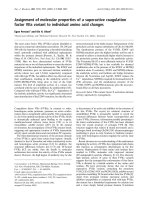
Fig. 1. Activation pocket region of FVIIa. (A) The carbon atoms of the N-terminal residues 153{16} to 158{21} are shown in green, those of b strand
B2 in the active FVIIa conformation (residues 296{154} to 305{163}) in gray and in the zymogen or inactive conformation (residues 296{154} to
302{160}) in magenta, and the Ca
2+
binding loop (residues 210{70} to 220{80}) is shown as a gray ribbon with the Ca
2+
ion represented by a
magenta sphere. The residues in position 158{21} and in positions 296{154} and 298{156} in the active B2 conformation are those found in V158D/
E296V/M298Q-FVIIa. The water molecule interacting with the backbone carbonyl of Ile153{16}, the backbone amides of residues 155{18} and
156{19} and the side chain of Gln298{156} is shown as a red sphere. (B and C) Detail of the activation pocket in V158D/E296V/M298Q-FVIIa and
wild-type FVIIa, respectively. The structure is from the FVIIa-TF complex (6, PDB entry code 1dan), except for the zymogen conformation of
strand B2 which is from the structure of FVII (8, PDB entry code 1jbu). The drawings were made using
QUANTA
2000 (Accelrys Inc.).
Ó FEBS 2002 Dissection of a superactive FVIIa variant (Eur. J. Biochem. 269) 5953
P4-P1¢ to P4-P7¢ of FX) by V158D/E296V/M298Q-FVIIa
as compared with that of wild-type FVIIa was constant,
indicative of that the three mutations do not simply increase
the accessibility of the substrate binding cleft to longer
substrates (E. Persson, A. M. Hansen, K. Madsen and O.
H. Olsen, unpublished observation). However, the peptides
may not correctly mimic the corresponding sequences when
part of FX. Finally, the high proteolytic activity of V158D/
E296V/M298Q-FVIIa appears to be accompanied by a
stabilized salt bridge between Ile153{16} and Asp343{194}.
Crystallographic data and molecular dynamics simula-
tions of FVII suggest that the purpose of the activation to
FVIIa is to maturate and open the substrate binding site, in
particular the S1 pocket, whereas an appropriate catalytic
triad geometry appears to be preformed in the zymogen
[8,20]. However, even after conversion to FVIIa the
conformational equilibrium appears to be shifted toward
an enzymatically latent form. Thus, the role of TF, apart
from localizing FVIIa to the site of vascular injury,
optimally positioning the active site [21] and contributing
to an extended, specificity-determining, factor IX/X binding
surface [22–24], is to stabilize the active FVIIa conforma-
tion. Strong evidence supports that Met306 in FVIIa is the
starting point for the TF-mediated effect on the FVIIa
conformation leading to allosteric stimulation of the
enzymatic activity [6,15,25,26]. Recently, site-directed muta-
genesis on FVIIa has been able to mimic the effect of TF
binding, at least in part, resulting in FVIIa molecules with
enhanced intrinsic activity [9–11,19]. Two published hypo-
theses accommodate an instrumental role of the activation
region in the regulation of the activity of free FVIIa, the first
dealing with the structural requirements for FVIIa to be in
an active conformation [8], whereas the other tries to explain
the effects of the activity-enhancing mutations [9]. They are
complementary and contribute to our understanding of
how the activity-enhancing mutations in this region of
FVIIa might exert their influence on the enzymatic activity.
Replacement of Met298{156} by Gln could prevent relative
b strand movement and stabilize strand B2 in a position
compatible with an active FVIIa conformation (Fig. 1A).
This is accomplished by introducing an extra hydrogen
bond to the water molecule that interacts only with residues
155{18} and 156{19} in wild-type FVIIa (Fig. 1B,C).
Substitution of Asp for Val at position 158{21} would not
be expected to affect the activity of FVIIa unless Gln is
simultaneously present at position 298{156} to allow the
establishment of a hydrogen bond between the two
introduced side chains that in turn stabilizes the inserted
N-terminus (Fig. 1B). An effect of the V158D mutation
becomes evident only after simultaneous replacement of
Glu296 by Val, presumably due to electrostatic repulsion
between Asp at position 158 and Glu at position 296. In
addition, according to the structure of zymogen FVII [8],
the change at position 296{154} (Glu to Val) eliminates
hydrogen bonds between the Glu side chain and the
backbone carbonyls of residues 158{21} and 159{22}. This
would remove an obstacle for the formation of a salt bridge
between the N-terminal residue 153{16} and Asp343{194}
as well as for the b strand reregistration that appears to be
required for FVIIa to attain its active conformation.
Together, the three mutations result in a highly active
FVIIa molecule that is more comfortable in the ÔactiveÕ b
strand registration and with a buried N-terminus.
Moreover, the mentioned b strand B2 and the preceding
loop contain Glu296{154} and Arg290{147} which have
been shown to be important for FX activation [25,27]. This
might explain why an ordering of this region selectively
increases the proteolytic activity more than the amidolytic
activity. In accordance with this, displacement of this region
by a peptide exosite inhibitor causes a larger effect on the
proteolytic activity of FVIIa than on its amidolytic activity
[5].
ACKNOWLEDGEMENT
We thank Anette Østergaard and Helle Bak for excellent technical
assistance.
REFERENCES
1. Pedersen, A.H., Nordfang, O., Norris, F., Wiberg, F.C., Chris-
tensen, P.M., Moeller, K.B., Meidahl-Pedersen, J., Beck, T.C.,
Norris, K., Hedner, U. & Kisiel, W. (1990) Recombinant human
extrinsic pathway inhibitor. Production, isolation, and character-
ization of its inhibitory activity on tissue factor-initiated coagu-
lation reactions. J. Biol. Chem. 265, 16876–16793.
2. Bode, W., Brandstetter, H., Mather, T. & Stubbs, M. (1997)
Comparative analysis of haemostatic proteinases: structural
aspects of thrombin, factor Xa, factor IXa and protein C. Thromb.
Haemostasis 78, 501–511.
3. Pike, A.C.W., Brzozowski, A.M., Roberts, S.M., Olsen, O.H. &
Persson, E. (1999) Structure of human factor VIIa and its
implications for the triggering of blood coagulation. Proc. Natl
Acad. Sci. USA 96, 8925–8930.
4. Kemball-Cook, G., Johnson, D.J.D., Tuddenham, E.G.D. &
Harlos, K. (1999) Crystal structure of active site-inhibited
human coagulation factor VIIa (des-Gla). J. Struct. Biol. 127,213–
223.
5. Dennis, M.S., Eigenbrot, C., Skelton, N.J., Ultsch, M.H., Santell,
L., Dwyer, M.A., O’Connell, M.P. & Lazarus, R.A. (2000)
Peptide exosite inhibitors of factor VIIa as anticoagulants. Nature
404, 465–470.
6. Banner, D.W., D’Arcy, A., Che
`
ne, C., Winkler, F.K., Guha, A.,
Konigsberg, W.H., Nemerson, Y. & Kirchhofer, D. (1996) The
crystal structure of the complex of blood coagulation factor VIIa
with soluble tissue factor. Nature 380, 41–46.
7. Zhang, E., St. Charles, R. & Tulinsky, A. (1999) Structure of
extracellular tissue factor complexed with factor VIIa inhibited
with a BPTI mutant. J. Mol. Biol. 285, 2089–2104.
8. Eigenbrot, C., Kirchhofer, D., Dennis, M.S., Santell, L., Lazarus,
R.A., Stamos, J. & Ultsch, M.H. (2001) The factor VII zymogen
structure reveals reregistration of b strands during activation.
Structure 9, 627–636.
9. Persson, E., Kjalke, M. & Olsen, O.H. (2001) Rational design of
coagulation factor VIIa variants with substantially increased
intrinsic activity. Proc. Natl Acad. Sci. USA 98, 13583–13588.
10. Petrovan, R.J. & Ruf, W. (2001) Residue Met
156
contributes to the
labile enzyme conformation of coagulation factor VIIa. J. Biol.
Chem. 276, 6616–6620.
11. Persson, E., Bak, H. & Olsen, O.H. (2001) Substitution of valine
for leucine 305 in factor VIIa increases the intrinsic enzymatic
activity. J. Biol. Chem. 276, 29195–29199.
12. Thim, L., Bjoern, S., Christensen, M., Nicolaisen, E.M., Lund-
Hansen,T.,Pedersen,A.H.&Hedner,U.(1988)Aminoacid
sequence and posttranslational modifications of human factor
VIIa from plasma and transfected baby hamster kidney cells.
Biochemistry 27, 7785–7793.
13. Freskga
˚
rd, P O., Olsen, O.H. & Persson, E. (1996) Struc-
tural changes in factor VIIa induced by Ca
2+
and tissue factor
5954 E. Persson and O. H. Olsen (Eur. J. Biochem. 269) Ó FEBS 2002
studied using circular dichroism spectroscopy. Protein Sci. 5,
1531–1540.
14. Persson, E. & Nielsen, L.S. (1996) Site-directed mutagenesis but
not c-carboxylation of Glu-35 in factor VIIa affects the association
with tissue factor. FEBS Lett. 385, 241–243.
15. Persson, E., Nielsen, L.S. & Olsen, O.H. (2001) Substitution of
aspartic acid for methionine-306 in factor VIIa abolishes the
allosteric linkage between the active site and the binding interface
with tissue factor. Biochemistry 40, 3251–3256.
16. Butenas, S., Lawson, J.H., Kalafatis, M. & Mann, K.G. (1994)
Cooperative interaction of divalent metal ions, substrate, and
tissue factor with factor VIIa. Biochemistry 33, 3449–3456.
17. Persson, E. & Petersen, L.C. (1995) Structurally and functionally
distinct Ca
2+
binding sites in the c-carboxyglutamic acid-con-
taining domain of factor VIIa. Eur. J. Biochem. 234, 293–300.
18. Wildgoose, P., Foster, D., Schiødt, J., Wiberg, F.C., Birktoft,
J.J. & Petersen, L.C. (1993) Identification of a calcium binding site
in the protease domain of human blood coagulation factor VII:
Evidence for its role in factor VII–tissue factor interaction.
Biochemistry 32, 114–119.
19. Petrovan, R.J. & Ruf, W. (2002) Role of zymogenicity-determin-
ing residues of coagulation factor VII/VIIa in cofactor interaction
and macromolecular substrate recognition. Biochemistry 41, 9302–
9309.
20. Perera, L., Darden, T.A. & Pedersen, L.G. (2002) Predicted
solution structure of zymogen human coagulation FVII. J. Com-
put. Chem. 23, 35–47.
21. McCallum, C.D., Hapak, R.C., Neuenschwander, P.F., Morris-
sey, J.H. & Johnson, A.E. (1996) The location of the active site of
blood coagulation factor VIIa above the membrane surface and its
reorientation upon association with tissue factor. A fluorescence
energy transfer study. J. Biol. Chem. 271, 28168–28175.
22. Huang, Q., Neuenschwander, P.F., Rezaie, A.R. & Morrissey,
J.H. (1996) Substrate recognition by tissue factor-factor VIIa.
Evidence for interaction of residues Lys
165
and Lys
166
of tissue
factor with the 4-carboxyglutamate-rich domain of factor X.
J. Biol. Chem. 271, 21752–21757.
23. Kirchhofer, D., Lipari, M.T., Moran, P., Eigenbrot, C. & Kelley,
R.F. (2000) The tissue factor region that interacts with substrates
factor IX and factor X. Biochemistry 39, 7380–7387.
24. Zhong, D., Bajaj, M.S., Schmidt, A.E. & Bajaj, S.P. (2002) The
N-terminal epidermal growth factor-like domain in factor IX and
factor X represents an important recognition motif for binding to
tissue factor. J. Biol. Chem. 277, 3622–3631.
25. Dickinson, C.D., Kelly, C.R. & Ruf, W. (1996) Identification of
surface residues mediating tissue factor binding and catalytic
function of the serine protease factor VIIa. Proc. Natl Acad. Sci.
USA 93, 14379–14384.
26. Dickinson, C.D. & Ruf, W. (1997) Active site modification of
factor VIIa affects interactions of the protease domain with tissue
factor. J. Biol. Chem. 272, 19875–19879.
27. Ruf, W. (1994) Factor VIIa residue Arg
290
is required for efficient
activation of the macromolecular substrate factor X. Biochemistry
33, 11631–11636.
Ó FEBS 2002 Dissection of a superactive FVIIa variant (Eur. J. Biochem. 269) 5955