- Trang chủ >>
- Khoa Học Tự Nhiên >>
- Vật lý
Temperature dependence of morphology and diameter of silicon nanowires synthesized by laser ablation
Bạn đang xem bản rút gọn của tài liệu. Xem và tải ngay bản đầy đủ của tài liệu tại đây (243.49 KB, 5 trang )
Temperature dependence of morphology and diameter
of silicon nanowires synthesized by laser ablation
Y.Q. Chen, K. Zhang, B. Miao, B. Wang, J.G. Hou
*
Structure Research Laboratory, Center for Physical Sciences, University of Science and Technology of China, Hefei 230026, China
Received 15 March 2002
Abstract
The silicon nanowires (SiNWs) with different diameters and morphologies were synthesized by laser ablation of a
target containing metals over a temperature range 910–1120 °C. The octopus-shaped wires of larger diameters were
formed in lower temperature zone (910–960 °C), while SiNWs and silicon nanoparticle chains of smaller diameters in
higher temperature zone (960–1120 °C). The distribution of the morphology and diameter of SiNWs as a function of
growth temperature differs from that reported by thermal evaporation of SiO powders. The study shows that the
morphology and diameter of SiNWs synthesized by laser ablation not only correlate closely with the growth
temperature of SiNWs, but also with the nature of a catalyst. By change of nucleation temperature and critical
nucleus size of nucleus droplets in vapor–liquid–solid (VLS) growth process, a catalyst can change relationships
between the morphology, diameter, and growth temperature of SiNWs. Ó 2002 Elsevier Science B.V. All rights
reserved.
1. Introduction
One-dimensional nanostructured materials,
such as nanotubes [1,2], nanowires [3–8], and
nanobelts [9], are a burgeoning and intriguing re-
search area both for their fundamental scientific
issues in meso-physics phenomena and for poten-
tial nano-device application. Silicon is one of the
most important electronic materials and holds
considerable technological promise for device ap-
plications. Therefore much attention has been paid
recently to the investigation of SiNWs. Until now,
a variety of techniques on synthesizing SiNWs
have been developed, including lithography and
etching techniques [10,11], scanning tunneling mi-
croscopy [12], thermal evaporization [13], and laser
ablation [3]. However, controlling the morphology
and size of as-grown SiNWs is still a challenging
issue. The recent studies [14,15] on SiNWs syn-
thesized by thermal evaporation of SiO powders
showed that the temperature of substrate for col-
lecting SiNWs played a dominant role in control-
ling the diameter of SiNWs and the formation of
various kinds of silicon nanowire-related mor-
phologies. It was demonstrated that the diameters
of SiNWs decreased with the descending growth
temperature and the morphology of SiNWs was
different in different deposition temperature zone.
7 June 2002
Chemical Physics Letters 358 (2002) 396–400
www.elsevier.com/locate/cplett
*
Corresponding author. Fax: +86-551-360-2803.
E-mail address: (J.G. Hou).
0009-2614/02/$ - see front matter Ó 2002 Elsevier Science B.V. All rights reserved.
PII: S 0 0 0 9 - 2 6 14 (02 )00671-1
Laser ablation synthesis of nanowires is quite
popular, by which long, uniform-sized, and single-
crystal SiNWs can be readily fabricated in bulk
quantities [16]. To our knowledge, there is no re-
port to date on the temperature dependence of the
morphology of SiNWs synthesized by laser abla-
tion. In this Letter, we present the results on this
project. Our results show that the morphology and
diameter of SiNWs synthesized by laser ablation
not only correlate closely with the growth tem-
perature of SiNWs, but also with the nature of a
catalyst.
2. Experimental
The experimental apparatus used for the pre-
sent work is similar to the one described previously
[3,17]. An alumina tube was mounted inside a
horizontal tube furnace. A target was made by
compressing Si powders (purity 99.99%) with 5
mol% Zr (purity P 92%; impurities, Mg, Fe, Ge,
Ca, Cl). The target was placed at the center inside
the furnace. A strip-like Si substrate (68 mm in
length and 20 mm in width) was placed at the
outlet end, near a cooling copper finger for col-
lecting the deposited products. There existed a
temperature gradient from center to the gas outlet
end of the furnace. A PtRh–Pt thermocouple was
used to measure the temperature distribution in
the alumina tube. After the tube had been evacu-
ated to 0.01 Torr by a mechanical vacuum pump,
5% H
2
–Ar gas mixture, as a carrier gas, was in-
troduced and kept flowing at a flow rate of 50
sccm. The pressure in the tube was controlled at
300 Torr. Then the furnace was heated to 1200 °C
at the central region. After the temperature and
pressure in the tube had been stabilized, pulsed
laser beam from an Nd:YAG laser (wavelength
532 nm, pulse width 7–8 nm, frequency 10 Hz,
average power 1.7 W) ablated the target for 1 h.
When ablation was over, the fluffy as-deposited
products with different colors and appearances
were found on the surface of the Si substrate. The
morphologies and electron diffraction patterns of
the as-deposited products were investigated by
transmission electron microscopy (TEM). The
chemical composition was analyzed by an energy
dispersive X-ray spectrometry (EDS) attached to
JEOL-2010 high-resolution TEM.
3. Results and discussion
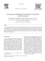
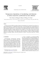
Fig. 1 shows the typical bright-field TEM im-
ages of the products, which are, respectively, cor-
responding to the different growth temperature
zones, A, B1, B2, and C, on the Si substrate
(shown in Fig. 2). The appearances and colors of
as-deposited products on the Si substrate are dis-
tinctly different, just by observing with the naked
eyes. A thin dark yellow gauze-like product was
formed in zone A, a thick dark yellow fluffy
product in Zone B, and a thin light yellow powder-
like product in zone C. As can be seen in Fig. 1,
both morphology and diameter of as-grown
SiNWs are different in different growth tempera-
ture zones. In zone A (1080–1120 °C), the nano-
wires had a uniform diameter of about 35 nm. The
presence of nanoparticles at the tips of the nano-
wires implies that growth mechanism of the
nanowires in this zone is vapor–liquid–solid (VLS)
growth [18]. Energy dispersive spectroscopy (EDS)
analysis indicated that the nanoparticles at the tips
of the nanowires only contained silicon and oxy-
gen. Within the detection limit of EDS measure-
ments (% 0:5%), no evidence of existence of Zr or
any other elements was detectable on the tip. Even
though the metal silicide was not found, it is be-
lieved that the function of these Si tips of the
nanowires is analogous to that of the metal silicide
catalyst in VLS growth process [15,16]. Namely,
melt Si nanoparticles may act as a nucleus for the
nanowires. In zone B (960–1080 °C), a large
quality of what is called silicon nanoparticle chains
were formed (Figs. 1b,c). Clearly, the diameter of
the silicon nanoparticle chain decreases with re-
ducing the growth temperature. Lee et al. [16] have
intensively studied the growth mechanism of the
silicon nanoparticle chains. They proposed that
nucleation and growth occurring alternatively re-
sulted in the formation of chains of silicon nano-
particles. The formation of the kinks of silicon
nanoparticle chains resulted from a change of
growth direction of the SiNWs. We examined the
kinks by using energy dispersive spectroscopy and
Y.Q. Chen et al. / Chemical Physics Letters 358 (2002) 396–400 397
confirmed that the kinks only contained silicon
and oxygen.
It should be noted that, from zone A to zone
B, the temperature dependence of the morphol-
ogy and diameter of the SiNWs is similar to that
by thermal evaporation, reported recently by
Peng et al. [14]. It can be seen that the diameter
of SiNWs decreases remarkably with a decrease
in growth temperature. They proposed that
the variation of the diameter resulted from the
Fig. 2. Schematic diagram of the different growth temperature zones on the silicon substrate.
Fig. 1. TEM images of the typical morphologies of SiNWs grown in: (a) zone A (1120–1080 °C); (b) zone B
1
(1080–1020 °C); (c) zone
B
2
(1020–960 °C); and (d) zone C (960–910 °C). Inset is electron diffraction patterns of the SiNWs.
398 Y.Q. Chen et al. / Chemical Physics Letters 358 (2002) 396–400
variation of the diameter of the droplet nucleated
at different temperature. It is recognized that the
melting point of nanoparticles decreases with the
reduction of their size. When metal silicide
nanoparticles of different sizes in the carrier gas
were present above the Si substrate, the larger
nanoparticles with higher melting points con-
densed on the higher temperature position of the
substrate, and the smaller nanoparticles with
lower melting points on the lower temperature
position of the substrate.
According to the above discussion, nanostruc-
tured morphology with smaller size should have
been anticipated in zone C (910–960 °C). However,
in addition to some aggregative fine particles, we
observed that a large quantity of silicon wires of
rather larger diameters (100–150 nm), called oc-
topus-shaped silicon nanowires, were formed in
zone C, a relatively lower temperature zone (Fig.
1d). It is interesting that the octopus-shaped sili-
con nanowires synthesized by thermal evaporation
of SiO powders were found at higher growth
temperature of more than 1230 °C [15]. Focusing
on the octopus-shaped structure, it can be seen
that two or more branches share the same tip,
which suggests that the tip might act as the nu-
cleation site for two or more branches when the
diameter of tip was large enough. Moreover, a
bifurcation phenomenon of the silicon nanowires
was also observed, which may be attributed to
renucleation of the crystal silicon in growth pro-
cess. On the other hand, the diameter of each
branch of the octopus-shaped nanowires decreases
gradually as the distance from the tip increases.
TEM investigations revealed that there existed two
kinds of structures in the entire length of the
branch. One was a crystal silicon core sheathed
with a thick amorphous outer layer of silicon ox-
ide, which originated from the tip and terminated
at reaching a certain length. The other, the rest of
the branch, was a complete amorphous silicon
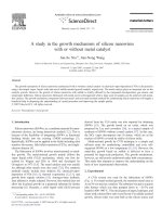
oxide. Fig. 3 is the HRTEM image of the interface
(arrow in Fig. 1d) of two kinds of structures along
the axis of the branch. It can be seen that the
growth direction of the crystal silicon was h 111i,
which is consistent with the fact that the growth
direction of SiNWs synthesized by metal catalyzed
VLS growth is predominantly h111i [3,14]. The
peculiar feature of the branch suggests that there
may exist a competitive growth between crystal
silicon core and outer layer of silicon oxide. When
the forming rate of outer layer of silicon oxide
exceeded the growth rate of crystal silicon, outer
layer of silicon oxide will surround the crystal sil-
icon. As a result, the growth of crystal silicon
ceased and silicon oxide of outer layer coalesced
together and extended.
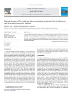
In order to investigate the reason why the
SiNWs of such larger diameters were deposited in
zone C, we analyzed the chemical composition of
the tip of the octopus-shaped wires in the zone C
by EDS. EDS analysis showed that the tip con-
tained Si, Mg, Ge, and O (oxygen came from the
outer layer of the tip), as shown in Fig. 4. From
Fig. 3. HRTEM image of the interface between the crystal
silicon and amorphous silicon oxides,along the axis of the
branch of the octopus-shaped wires.The arrow shown in Fig. 1d
indicates the interface.
Fig. 4. EDS spectrum taken from the tip of the octopus-shaped
SiNWs.
Y.Q. Chen et al. / Chemical Physics Letters 358 (2002) 396–400 399
Mg–Si, Si–Ge, and Mg–Ge binary phase diagrams
[19,20], it is evident that addition of Mg and Ge
into Si can reduce the melting point of the silicon
solid solution. Moreover, the melting points of
nanoparticles are usually lower than the corre-
sponding bulk material. All of these factors col-
lectively could effectively reduce the melting points
of the tips of larger diameter in zone C. This then
explains why these octopus-shaped wires could be
deposited in zone C, a relatively lower temperature
zone. Furthermore, it is known that the diameters
of the nanowires formed in VLS process have re-
lation to the critical diameter dc of the liquid
droplets nucleated in VLS process. Droplets larger
than d
c
will become stable nuclei, whereas droplets
smaller than d
c
will disappear gradually. The crit-
ical nucleus size can be expressed as r
Ã
¼
ðÀ2cÞ=DF
v
; where c is the specific interfacial free
energy of the condensate–vapor interface and DF
v
is the bulk free energy change per unit volume
[21,22]. We assume that the addition of Mg and Ge
into Si nanoparticles would increase interfacial
free energy c. Therefore r
Ã
becomes larger, which
means that the droplets with larger diameters
(> r
Ã
) can grow to form nanowires. This may be
the reason why the diameters of nanowires in zone
C are larger than those in zone A or zone B.
4. Conclusions
In summary, the diameter and morphology of
SiNWs synthesized by laser ablation not only
correlate closely with growth temperature, but also
with the nature of a catalyst. The nature of a
catalyst has a direct influence upon the nucleation
temperature and critical nucleus size of the drop-
lets in VLS growth process. The addition of Mg
and Ge into Si tips gave rise to the deposition of
octopus-shaped SiNWs of larger diameters in the
lower temperature zone. Furthermore, it is sug-
gested in our research that there is a corresponding
correlation between morphology and diameter of
nanowires. Nanowires of larger diameter (100–150
nm) were inclined to be octopus-shaped, while
nanowires of smaller diameter (10–15 nm) were
inclined to be nanoparticle-chain-shaped.
Acknowledgements
This work was supported by the NSF of China
(59972036, 10074059, and 19904012) and the
ICQS of Chinese Academy of Sciences.
References
[1] S. Iijima, Nature 354 (1991) 917.
[2] N.G. Chopra, R.J. Luyken, K. Cherry, V.H. Crespi,
M.L. Cohen, S.G. Louie, A. Zettl, Science 269 (1995) 966.
[3] A.M. Morales, C.M. Lieber, Science 269 (1998) 208.
[4] X.F. Duan, X.F. Wang, C.M. Lieber, Appl. Phys. Lett. 76
(2000) 1116.
[5] H.Y. Peng, X.T. Zhou, N. Wang, Y.F. Zheng, L.S. Liao,
W.S. Shi, C.S. Lee, S.T. Lee, Chem. Phys. Lett. 327 (2000)
263.
[6] W.S. Shi, Y.F. Zheng, N. Wang, C.S. Lee, S.T. Lee, Adv.
Mater. 13 (2000) 591.
[7] M.H. Huang, Y. Wu, H. Feick, N. Tran, E. Weber, P. Yang,
Adv. Mater. 13 (2001) 113.
[8] Y. Wu, B. Messer, P. Yang, Adv. Mater. 13 (2001) 1487.
[9] Z.W. Pan, Z.R. Dai, Z.L. Wang, Science 291 (2001) 1947.
[10] H.I. Liu, N.I. Maluf, R.F.W. Pease, J. Vac. Sci. Technol. B
10 (1992) 2846.
[11] H. Namatsu, S. Horiguchi, M. Nagase, K. Kurihara,
J. Vac. Sci. Technol. B 15 (1997) 1688.
[12] T. Ono, H. Saitoh, M. Esashi, Appl. Phys. Lett. 70 (1997)
1852.
[13] D.P. Yu, Z.G. Bai, Y. Ding, Q.L. Hang, H.Z. Zhang,
J.J. Wang, Y.H. Zou, W. Qian, G.C. Xiong, H.T. Zhou,
S.Q. Feng, Appl. Phys. Lett. 72 (1998) 3458.
[14] H.Y. Peng, Z.W. Pan, L. Xu, X.H. Fan, N. Wang,
C.S. Lee, S.T. Lee, Adv. Mater. 13 (2001) 317.
[15] Z.W. Pan, Z.R. Dai, L. Xu, S.T. Lee, Z.L. Wang, J. Phys.
Chem. B. 105 (2001) 2507.
[16] S.T. Lee, N. Wang, Y.F. Zhang, Y.H. Tang, MRS Bull.
August (1999) 36.
[17] T. Guo, P. Nikolaev, A. Thess, D.T. Colbert, R.E. Smalley,
Chem. Phys. Lett. 243 (1995) 49.
[18] R.S. Wagner, W.C. Ellis, Appl. Phys. Lett. 4 (1964) 89.
[19] G.V. Raynor, The Physical Metallurgy of Magnesium and
its Alloys, Pergamon, London, 1959.
[20] H. St
€
oohr, W. Klemm, Z. Anorg. Chem. 241 (1939) 305.
[21] J.W. Mullin, Crystallisation, Butterworths, London, 1972.
[22] X.F. Fan, L. Xu, C.P. Li, Y.F. Zheng, C.S. Lee, S.T. Lee,
Chem. Phys. Lett. 334 (2001) 231.
400 Y.Q. Chen et al. / Chemical Physics Letters 358 (2002) 396–400