- Trang chủ >>
- Khoa Học Tự Nhiên >>
- Vật lý
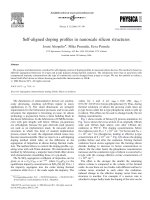
Self aligned silicon quantum wires on ag(1 1 0)
Bạn đang xem bản rút gọn của tài liệu. Xem và tải ngay bản đầy đủ của tài liệu tại đây (423.96 KB, 7 trang )
Surface Science Letters
Self-aligned silicon quantum wires on Ag(1 1 0)
C. Leandri
a
, G. Le Lay
a
, B. Aufray
a,
*
, C. Girardeaux
b
, J. Avila
c,d
,
M.E. Da
´
vila
c
, M.C. Asensio
c,d
, C. Ottaviani
e
, A. Cricenti
e
a
CRMCN-CNRS, Campus de Luminy, Case 913, 13288 Marseille Cedex 9, France
b
L2MP, Campus de Saint Je
´
ro
ˆ
me, 13397 Marseille Cedex 20, France
c
Instituto de Ciencia de Materiales de Madrid (CSIC), 28049 Cantoblanco, Madrid, Spain
d
LURE, Ba
ˆ
t. 209 D, Universite
´
Paris-Sud, BP 34, 91898 Orsay, France
e
Instituto di Struttura della Materia, CNR, Via Fosso del Cavaliere, 00133 Rome, Italy
Received 9 September 2004; accepted for publication 21 October 2004
Available online 13 December 2004
Abstract
Upon deposition of silicon onto the (1 1 0) surface of a silver crystal we have grown massively parallel one-dimen-
sional Si nanowires. They are imaged in scanning tunnelling microscopy as straight, high aspect ratio, nanostructures,
all with the same characteristic width of 16 A
˚
, perfectly aligned along the atomic troughs of the bare surface. Low
energy electron diffraction confirms the massively parallel assembly of these self-organized nanowires. Photoemission
reveals striking quantized states dispersing only along the length of the nanowires, and extremely sharp, two-compo-
nents, Si 2p core levels. This demonstrates that in the large ensemble each individual nanowire is a well-defined quan-
tum object comprising only two distinct silicon atomic environments. We suggest that this self-assembled array of
highly perfect Si nanowires provides a simple, atomically precise, novel template that may impact a wide range of
applications.
Ó 2004 Elsevier B.V. All rights reserved.
Keywords: Silver; Silicon; Self-assembly; Nanowires; Scanning tunneling microscropy; Photoelectron spectroscopy
In the quest for electronics on the nanoscale,
one-dimensional (1D) quantum structures are ex-
pected to play a key role [1,2]. Systems that might
act as nanowires (NWs) are of major importance,
but are rather difficult to prepare experimentally
[3]. Such NWs bear great potential to exhibit exo-
tic and attractive physical phenomena [4]. In re-
cent years, several self-organized quantum wire
arrays have been grown upon depositing metals
on semiconductor [3–9] or on metallic surfaces
exhibiting regularly spaced steps [10,11]. Self-orga-
nized formation of quasi-one-dimensional surface
0039-6028/$ - see front matter Ó 2004 Elsevier B.V. All rights reserved.
doi:10.1016/j.susc.2004.10.052
*
Corresponding author. Fax: +33 0 4 91 82 91 97.
E-mail address: (B. Aufray).
Surface Science 574 (2005) L9–L15
www.elsevier.com/locate/susc
oxide domains on Cu(1 1 0) leading to 1D confine-
ment of a Shoc kley surface state has been also ob-
served by Bertel and Lehmann [12]. On wide band
gap b-SiC(100) substrates, the spontaneous forma-
tion of stable atomic lines, i.e., carbon lines with C
atoms in sp
3
configuration on C- terminated sur-
faces as well as silicon lines at the phase transition
between Si-rich and Si-terminated surfaces has
also been observed [13,14]. Given the central role
of silicon in microelectron ics and the potential
occurrence of quantum size effects in silicon-based
devices [15], silicon NWs have attracted consider-
able interest [16,17]. However, with respect to pro-
cedures used, producing Si NWs with controlled
sizes is far from being trivial and aligning them
in a well-ordered fashion, a crucial issue, is
another problem.
We have succeeded in growing a massively paral-
lel assembly of straight silicon NWs on a clean,
nominally flat (misorientation $ 0.1°) (11 0) silver
surface. All NWs have the same orientation and
characteristic narrow width of 1.6nm and are
two-atom thick; they reach eventually hundreds of
nanometers in length. Strikingly, this ensemble
displays quantized electronic states with a 1D dis-
persion in valence band photoemission, while
high-resolution core level spectroscopy demon-
strates that all individual NWs within the assembly
have an identical and highly perfect atomic structure
which comprises two and only two distinct silicon
environments. Hence, this nanowire array provides
a novel, simple and atomically precise macroscopic
template that may impact, not only future electron-
ics, but and also a wide range of fields [18].
1D metal chains or stripes on silicon surfaces
have attracted considerable interest because of en-
hanced many-body interactions leading possibly to
an exotic state described by the Luttinger liquid
framework, or, typically, to metal-insulator transi-
tions [7,19–21]. However, conversely, only very
few studies concern the reverse silicon-on-metal
systems. Two investigations concern gold and cop-
per noble metal substrates [22,23]. In the last case,
short atomic silicon chains, albeit presenting many
defects, and displaying no localized electronic
states, could be grown on top of an initial 2D sur-
face alloy by depositing silicon onto clean Cu(11 0)
surfaces.
We have deposited silicon in situ under ultra-
high vacuum (UHV) (typical silicon coverage
$0.25 monolayer (ML) in silver (1 1 0) surface
atom density) from a direct-current heated piece
of silicon wafer (flashed at $1250 °C), controlling
the evaporation flux with a quartz monitor and
the deposition at room temperature (RT) by Auger
electron spectroscopy. To limit any possible inter-
mixing we have chosen a silver (1 1 0) substrate,
since numerous works have demonstrated the
atomic abruptness of the silver-silicon interface
compared to the diffusiveness of the gold-silicon
one and the reactivity of the Cu–Si one, which
forms silicides [24]. The clean, nominally flat
(1 1 0) surface (misorientation 0.1°) was prepared
by standard, repeated cycles of Ar+ bombard-
ments and annealing.
As imaged in scanning tunnelling microscopy
(STM) at RT in Fig. 1(a), thin silicon NWs,
reaching up to about 30 nm in length, are formed
at the early stages of the deposition at RT, appar-
ently from the self-assembly of nanodots, which
appear as their swiftly diffusing building blocks.
The density of the nanowires is typically $1.4 ·
10
12
cm
À2
at RT; as will be seen later it can be re-
duced upon mild annealing. All these NWs are
perfectly aligned along the [À11 0] direction of
the Ag(1 1 0) surface, showing rounded protru-
sions (Fig. 1(b)), equally spaced every second sil-
ver atomic distance (2a
2
= 0.577 nm); some of
them appear too large to represent single atoms
(the atomic diameters of Si and Ag in the bulk
crystals are 0.288 and 0.235 nm respectively).
The 2a
2
periodicity indicates that the NWs are
not simply composed of Si [À1 1 0] rows with a
‘‘bulk-like’’ inter-atomic distance; in such a case
a4a
2
periodicity would be expected, given the
excellent match between four Ag atomic distances
and three silicon ones along [À1 1 0]. Indeed, the
negligible misfit permits the perfect epitaxial
growth of silver (1 1 1) crystallites on the Si(1 11)
surface with common [1 1 0] directions [25]. The
NWs have the same definite width of 1.6 nm,
which corresponds to four silver atomic distances
(4a
1
) along the Ag[0 0 1] direction, and a maxi-
mum apparent height of $0.2 nm (Fig. 1(c)); their
mutual separations vary between 1.5 and 15 nm.
The NWs are markedly asymmetric along their
L10 C. Leandri et al. / Surface Science 574 (2005) L9–L15
SUR
FACE SCIENCE
LETTERS
widths, as shown by the height profile, which
eventually indicates that their atomic structure
may not be trivial, although we can not exclude
tip convolution effects. Upon mild annealing at
230 °C for about 10 min they further markedl y
elongate, keeping the same narrow width, well be-
yond 100nm, as shown in Fig. 2; in this case their
density is reduced typically by a factor $7. Just
from the STM images we can not give a reliable
atomic model of the NWs. However, we surmise
that their very narrow width is due to a strong
epitaxial strain, consequently the actual geometry
might resemble one of the metallic bulk silicon
phases obtained at high pressures, e.g., the b-tin
like phase or rather the simple hexagonal phase
[26]. If true, this would point to a possible super-
conductivity of the NWs.
As seen in Fig. 3(a), LEED patterns display, in
addition to the integer order spots of the unrecon-
structed Ag(1 1 0) surface, thin streaks elongated
along the [1 0 0]* reciprocal direction, either con-
necting these spots or situated in half-order posi-
tion along the orthogonal [À 1 1 0]* direction. In
excellent agreement with the STM images, these
patterns corroborate, at the macroscopic scale,
the order within the NWs with a 2a
2
periodicity
along their lengths, the narrow width of the silicon
NWs, and a lack of periodicity in the perpendicu-
lar direction, reflecting their varia ble separations.
Since the NWs differ only in length, these un-
equal separations are no obstacle to probe the
macroscopic electronic response using ad vanced
synchrotron radiation photoemission (PES) meth-
ods. We have performed high-resolution (HR)
angle-integrated (AI) measurements at RT of the
valence bands (VBs) and of the Si 2p core-levels
(CLs). A typical Si 2p spectrum is shown in Fig.
3(b) togeth er with its synthesis with two, spin–orbit
splitted, components, as obvious on the raw data,
using standard fitting procedures [27]. These two
components, separated by 0.24 eV, are remarkably
narrow, with respectively 0.17 and 0.20eV Full
Widths at Half Maximum comparing favourably
with the narrowest FWHMs of Si 2p bulk lines ob-
tained for Sb covered Si(11 1) samples [27]. This
proves the perfect atomic order within the NWs
and the existence of just two non-equivalent silicon
environments. Given the $0.2 nm maximum height
of the NWs we can surmise that one of them may
correspond to Si atoms (Si
1
) in direct contact with
the Ag surface (hence, at the lowest BE because of
the most effective metallic screening) and the other
to Si atoms (Si
2
) bonded to the (Si
1
) ones, although
we can not exclude the possibility that peculiar Ag
Fig. 1. Topographic images of $0.25 monolayer of Si deposited on Ag(1 1 0) at room temperature: (a) 42 · 42 nm
2
overview with Si
nanowires and nanodots, (b) 12.1 · 12.1nm
2
zoom revealing the atomic rows of the bare substrate along the [À110] direction and the
profile of the nanowires, (c) height profile along the black line in (b). Imaging conditions: À1.7 V sample bias and 1.1nA tunnel current
in (a) and (b). Note that the width of the NWs (1.6nm) can serve as distance marker while their length direction points to the [À110].
C. Leandri et al. / Surface Science 574 (2005) L9–L15 L11
SUR
FAC
E
SCI
ENCE
LET
TERS
atom rows participate also in the structural organi-
zation of the NWs. With photoelectron diffraction
experiments on each component, one could
determine precisely the two different local Si envi-
ronments, possibly solve the complete atomic
structure of the NWs, and, along with detailed sim-
ulations, interpret the protrusions seen in the STM
images. Since the atomi c structure of the thinnest
silicon wires is a matter of intense theoretical re-
search this structural determination might have a
decisive impact [17,28,29].
The best fits shown in Fig. 3(b) were obtained
upon including an asymmetry parameter of 0.09,
higher than that reported for a pristine sil ver Ag
3d CL [30]. This is direct evidence of the metallic-
ity of the Si NWs (all spectra taken at various pho-
ton energies, incidence and detection angles are
markedly asymmetric). This metallicity is consis-
tent with the fact that the density of states at the
Fermi energy increases compared to that of the ini-
tial silver surface (Fig. 3(c)), as well as with scan-
ning tunnelling spectroscopy (STS) measurements
(not shown here) performed on individual NWs:
the I(V) spectra (tunnelling current versus sam-
ple-to-tip voltage) do not significantly deviate
from those performed on the pristine Ag surface.
This metallic character could be a proximity effect
due metal-induced gap states, or be analogous to
the 2D surface alloy initially formed by Si on
Cu(1 1 0), or rather be the consequence of the
stabilisation of a high-pressure silicon phase, as
mentioned above [31,23,26].
The most striking result is the presence of new,
discrete, elect ronic states, compared to the feature-
less sp valence band of the pristine Ag(1 1 0) sur-
face. A maximum number of four new states
were detected; they are clear ly noticed in the mea-
surement geometry of Fig. 3(c). To precise their
Fig. 2. Si nanowires (image size: 45 · 100nm
2
) before (a), and after (b), annealing at 230 °C. The diffusing companion nanodots
disappear after complete incorporation for longer annealing times. Imaging conditions: À1.7 V sample bias and 1.14 nA tunnel current
in (a) and À0.4 V and 0.7 nA in (b).
L12 C. Leandri et al. / Surface Science 574 (2005) L9–L15
SUR
FACE SCIENCE
LETTERS
nature, we further performed a detailed angle-re-
solved (AR) photoemission study. These states,
which do not exist on pristine silver (no confusion
with the bare Ag(1 1 0)
Y Schockley-type surface
state is possible [32]), do not disperse at normal
emission as a function of the photon energy. This
proves that they are associated with the Si NWs. In
the measurement geometry of Fig. 4, that is, along
the direction of the NWs, the two deepest new
states, previously detected in AI-PES, are observed
at $2.4 and $3.1 eV BE at 51° off normal emis-
sion. We emphasize that the photon energy, the
polarization direction, as well as the collection
angle, with respect to the wires strongly influence
the detection of the new states. Besides a strongly
dispersive Ag bulk sp band, the analysis of repre-
sentative spectra taken along the NWs after anneal-
ing $230 °C(Fig. 4(a)), reveals that these two deep
levels disperse markedly, by $0.4eV. The disper-
sion relations of these two new states are plotted
in Fig. 4(b). We stress that no dispersion at all
was noticed in the direction orthogonal to the
NWs; hence the dispersion is purely one-dimen-
sional, as already shown in Ref. [11] . Such behav-
iour can be expected for quantum well levels due
to confinement within the NWs, i.e., the electronic
wave is quantum mechanically confined in two
directions: along the normal to the surface, as well
as perpendicular to the NWs, while the electronic
movement is not restricted along the [À1 1 0] direc-
tion, leading to pronounced 1D dispersion along
C–X in k-space.
Fig. 3. Low energy electron diffraction and angle integrated photoemission on a macroscopic surface area covered with h $ 0.25
monolayer of silicon at room temperature. (a) LEED pattern taken at 43 eV primary energy. (b) Si 2p core level spectrum (dots) and its
synthesis (solid line overlapping the data points) with two asymmetric components (bottom curves). The spectrum was recorded at
normal incidence at hm = 140eV photon energy with the hemispherical photoelectron analyser axis (16° acceptance angle) aligned at 45°
from the normal to the surface. The fitting parameters are a spin–orbit splitting of 605meV, a Lorentzian FWHM of 40meV, Gaussian
FWHMs of respectively 135 and 185 meV, an asymmetry parameter of 0.09. (c) Normal incidence valence band spectra (hm = 79eV)
limited to the sp region for the initial pristine Ag(1 1 0) surface (bottom curve) and for the same surface as in (a) and (b) (top curve) The
detector axis is at 45° from the surface normal in the incidence plane, parallel to the direction of the nanowires. The zero of energy is
taken at the Fermi level and the relative intensities of the two spectra take into proper account all measurement conditions. The total
energy resolution is $40 meV.
C. Leandri et al. / Surface Science 574 (2005) L9–L15 L13
SUR
FAC
E
SCI
ENCE
LET
TERS
To conclude, we stre ss that the quantized, sili-
con NWs that we have grown and characterize d
under UHV might be stabilized by atomic hydro-
gen termination, which could make them semicon-
ducting [16] or oxidized, which could make them
insulating. Indeed, the growth can be further pur-
sued. We have done such tests, which show that
extremely long, larger and much thicker crystalline
nanowires, also perfectly aligned along the [À110]
direction can be produced [33]. We plan to use
these NWs as nucleation objects for further
growth by Chemical Vapour Deposition. We can
also envisage to cover these Si NWs by overlayers
and even the embedment of these nanostructures
inside a silver matrix upon epitaxial regrowth of
silver overlayers, which is very easy on Si surfaces.
Indeed, one can foresee the impact of such mas-
sively parallel arrays of one-dimensional silicon
metallic, semiconducting or insulating nanostruc-
tures, from narrow, ultra-thin, nanowires to larger
and thicker ones, in future electronics. Another
particularly exciting potentiality is for aligning
large molecules, like C
60
, organic ones, nanotubes
and polymers and interfacing with biological
systems.
Acknowledgment
The original LEED-AES and STM work
started at the CRMC2-CNRS in Marseille as part
of the Thesis work of Christel Leandri; we espe-
cially thank Dr. H. Oughaddou for help in the
measurements and many discussions. The expert
technical assistance of A. Ranguis, J.Y. Hoarau
and J.P. Dussaulcy is greatly acknowledged. We
thank Dr. P. de Padova for stimulating discus-
sions. The angle-integrated photoemission experi-
ments were carried out at the VUV beamline of
the Italian synchrotron radiation facility ELET-
TRA, in Trieste; we are grateful to the entire staff
of the beamline for help during the measurements.
The angle-resolved photoemission measurements
were carried out at the Spanish–French SU8
beamline of the LURE, the French synchrotron
radiation facility in Orsay; we warmly thank
M.A. Valbuena for help during the measurements.
Fig. 4. Angle-resolved photoemission valence band spectra and
dispersion relations of the deep lying quantum levels (QW
a
and
QW
b
) from the silicon nanowires. (a) a series of spectra at
different collection angles after annealing at $230°C. (b)
Dispersion relations of the two quantum well states: the
binding energies of each state versus k
||
, the momentum of the
photoelectron parallel to the surface along the corresponding
C–X direction of the second and third (1 1 0) surface Brillouin
zones. The colour code reflects the intensities of the different
features. Experimental conditions: h $ 0.25 silicon ML; typical
resolutions of 1° and 50meV; hm = 75 eV; binding energies are
referenced to the Fermi level; light was incident at 45° from the
surface normal, the plane of incidence is parallel to the [À110]
direction of the wires the polar angles of detection in the
incidence plane are indicated; tick marks point to the positions
of the quantized states.
L14 C. Leandri et al. / Surface Science 574 (2005) L9–L15
SUR
FACE SCIENCE
LETTERS
Support from the ELETTRA and LURE staffs is
greatly acknowledged.
References
[1] D. Appell, Nature 419 (2002) 553.
[2] Y. Cui, C. Lieber, Science 291 (2001) 851.
[3] D.R. Bowler, Phys. Rev. B 62 (2000) 7237.
[4] O. Gurlu, O.A.O. Adam, H.J.W. Zandvliet, B. Poelsema,
Appl. Phys. Lett. 83 (2003) 4610.
[5] I.K. Robinson, P.A. Bennet, F.J. Himpsel, Phys. Rev.
Lett. 88 (2002) 096104.
[6] H.W. Yeom, J. Electron. Spectrosc. Rel. Phenom. 114–116
(2001) 283.
[7] P. Segovia, D. Purdie, M. Hengsberger, Y. Baer, Nature
402 (1999) 504.
[8] H.S. Yoon, S.J. Park, J.E. Lee, C.N. Whang, I W. Lyo,
Phys. Rev. Lett. 92 (2004) 096801.
[9] D. Sanchez-Portal, S. Riikonen, R.M. Martin, Phys. Rev.
Lett. 93 (2004) 146803.
[10] F. Picaud, C. Ramseyem, C. Girardet, P. Jensen, Phys.
Rev. B 61 (2004) 16154.
[11] J.E. Ortega, A. Mugarza, V. Repain, S. Rousset, V. Perez-
Dieste, A. Mascaraque, Phys. Rev. B 65 (2002) 165413.
[12] E. Bertel, J. Lehmann, Phys. Rev. Lett. 80 (1988) 1497;
K. Berge, A. Gerlach, G. Meister, A. Goldmann, E. Bertel,
Phys. Rev. B 70 (2004) 155303.
[13] V. Derycke, et al., Phys. Rev. Lett. 81 (1998) 5868.
[14] P. Soukiassian, et al., Phys. Rev. Lett. 79 (1997) 907.
[15] A.T. Tilke, et al., Phys. Rev. B 68 (2003) 075311.
[16] D.D.D. Ma, et al., Science 299 (2003) 1874.
[17] Y. Zhao, B.I. Yakobson, Phys. Rev. Lett. 91 (2003)
035501.
[18] V. Derycke, et al., Nature Mater. 2 (2003) 253.
[19] C. Kumpf, et al., Phys. Rev. Lett. 85 (2000) 4916.
[20] J.M. Luttinger, J. Math. Phys. 4 (1963) 1154.
[21] T. Uchihashi, U. Ramsperger, Appl. Phys. Lett. 80 (2002)
4169.
[22] A. Franciosi, et al., Phys. Rev. B 32 (1985) 6917.
[23] C. Polop, et al., Phys. Rev. B 63 (2001) 115414.
[24] G. Le Lay, et al., Surf. Sci. 307–309 (1994) 280, and
references therein.
[25] G. Le Lay, Surf. Sci. 132 (1983) 169.
[26] P. Focher, et al., Europhys. Lett. 26 (1994) 343.
[27] A. Cricenti, et al., Phys. Rev. B 62 (2000) 9931.
[28] B.X. Li, et al., Phys. Rev. B 65 (2002) 125305.
[29] P. Sen, et al., Phys. Rev. B 65 (2002) 235433.
[30] G. Le Lay, et al., Europhys. Lett. 45 (1998) 65.
[31] U. Landman, et al., Phys. Rev. Lett. 85 (2000) 1958.
[32] A. Gerlach, et al., Surf. Sci. 443 (1999) 221.
[33] C. Leandri et al., in preparation.
C. Leandri et al. / Surface Science 574 (2005) L9–L15 L15
SUR
FAC
E
SCI
ENCE
LET
TERS