Báo cáo khóa học: Structural characterization of the human Nogo-A functional domains Solution structure of Nogo-40, a Nogo-66 receptor antagonist enhancing injured spinal cord regeneration ppt
Bạn đang xem bản rút gọn của tài liệu. Xem và tải ngay bản đầy đủ của tài liệu tại đây (575.86 KB, 11 trang )
Structural characterization of the human Nogo-A functional domains
Solution structure of Nogo-40, a Nogo-66 receptor antagonist enhancing injured
spinal cord regeneration
Minfen Li
1
, Jiahai Shi
1
, Zheng Wei
1
, Felicia Y. H. Teng
2
, Bor Luen Tang
2
and Jianxing Song
1,2
1
Department of Biological Sciences and
2
Department of Biochemistry, National University of Singapore, Singapore
The recent discovery of the Nogo family of myelin inhibitors
and the Nogo-66 receptor opens up a very promising avenue
for the development of therapeutic agents for treating spinal
cord injury. Nogo-A, the largest member of the Nogo fam-
ily, is a multido main protein containing at le ast two regions
responsible for inhibiting central ner vous system (CNS)
regeneration. So far, no structural information is available
for Nogo-A or any of its structural domains. We have sub-
cloned and expressed two Nogo-A fragments, namely the
182 residue Nogo-A(567–748) and the 66 residue Nogo-66 in
Escherichia coli. CD and NMR characterization indicated
that Nogo-A(567–748) was only partially structured while
Nogo-66 was highly insoluble. Nogo-40, a truncated form
of Nogo-66, has been previously shown to be a Nogo-66
receptor antagonist that is able to enhance CNS neuronal
regeneration. Detailed NMR examinations revealed that a
Nogo-40 peptide had intrinsic helix-forming propensity,
even in an aqueous environment. The NMR structure of
Nogo-40 was therefore d etermine d in the presenc e of the
helix-stabilizing solvent trifluoroethanol. The solution
structure of Nogo-40 revealed two well-defined helices linked
by an unstructured loop, representing the first structure of
Nogo-66 receptor binding ligands. Our results provide the
first structural insights into Nogo-A functional domains and
may have implications in further d esigns of peptide mimetics
that would enhance CNS neuronal regeneration.
Keywords: CNS neuronal regeneration; NMR spectroscopy;
Nogo-40; NogoA; spinal cord injury.
Survivors of severe central nervous system (CNS) i njury
often suffer f rom permanent disability. Previously, it was
thought that the inability of CNS neurons to regenerate was
due to the absence of growth-promoting factors in CNS
neurons. However, recent discoveries c hallenge this dogma.
It has been shown that the failure of CNS neuronal
regeneration re sults to a large extent from the existence of
inhibitory molecules of axon outgrowth in adult CNS
myelin [1]. So far, three proteins have been identified that
cause inhibitory effects on CNS neuronal r egeneration,
namely Nogo [2–4], myelin-associated glycoprotein [5] and
oligodendrocyte myelin glycoprotein [6]. All three molecules
appear to exert t heir inhibitory action through t he initial
binding of the Nogo-66 receptor (NgR) [3], first identified as
a high affinity neuronal r eceptor for Nogo-A [7]. NgR
binding leads to subsequent activation of signaling path-
ways that possibly involve Rho activation, and the induc-
tion of growth cone collapse [8]. These discoveries raise a
promising possibility to enhance axonal growth by disrupt-
ing the interaction between NgR and its ligands.
Of the three myelin-associated molecules above, the
CNS-enriched Nogo belonging to t he reticulon protein
family has received intense attention recently. Nogo has
several splicing variants, among which N ogo-A is the
largest, composed of 1192 a mino acids (Fig. 1). Recent
studies have demonstr ated that NogoA is a multidomain
protein containing several discrete regions with growth
inhibitory functions [4,9–11]. Two major inhibitory regions
have been identified. The first is a stretch in the middle of
the Nogo-A molecule (residues 544–725 for m ouse and
residues 5 67–748 f or huma n Nogo-A proteins) that
restricts neurite outgrowth and cell spreading and induces
growth cone co llapse. The second is the e xtracellular 6 6
amino acid loop called Nogo-66 that is also capable of
inhibiting neurite g rowth and inducing growth c one
collapse [4,9–11]. The Nogo-66 domain has been shown
to be anchored on the oligodendrocyte surface and binds
to the neuronal g lycophosphatidylinositol-linked NgR, via
its leucine-rich r epeat containing do main. The binding of
Nogo-66 to N gR is competitively inhibited by a peptide
consisting of the N-terminal 40 residues of Nogo-66,
named Nogo-40 [12,13]. This Nogo-40 peptide has been
experimentally demonstrated to be a strikingly e ffective
NgR antagonist capable of enhancing recovery from spinal
cord injury [13].
In contrast to the extensive functional studies on Nogo-
A, thus far no structural information has been available for
any region of the Nogo-A protein. In the present study, w e
cloned and expressed the two functional regions of human
Nogo-A and performed structural characterization by CD
Correspondence to J. Song, Department of Biochemistry, National
University of Singapore; 10 Kent Ridge Crescent, Singapore 119260.
Fax: +65 6779 2486, Tel.: +65 6874 1013,
E-mail:
Abbreviations: CNS, central nervous system; IPTG, isopropyl thio-
b-
D
-galactoside; NgR, Nogo-66 receptor; rmsd, root mean square
deviation; TFE, trifluoroethanol.
(Received 10 June 2004, revised 6 July 2004, accepted 12 July 2004)
Eur. J. Biochem. 271, 3512–3522 (2004) Ó FEBS 2004 doi:10.1111/j.1432-1033.2004.04286.x
and NMR spectroscopy. While Nogo-66 is highly insoluble,
the 182 residue fragment was found to be partially
structured and could be further induced to form a helical
structure with the introduction of 4 m
M
Zn
2+
.We
conducted f urther NMR studies on two truncated forms
of Nogo-66: Nogo-40 and Nogo-24. Although Nogo-40
and Nogo-24 appeared to be unstructured i n aqueous
buffer, a detailed N MR analysis revealed that these have
intrinsic helix-forming propensity. This observation,
together with results from secondary structure predictions,
offered the rationale to study the structure of Nogo-40 after
its intrinsic helix-forming propensity is stabilized by the
introduction of the helix-stabilizing solvent trifluoroethanol.
We report here the structure of Nogo-40, a Nogo-66
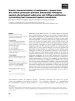
Fig. 1. Schematic representation of the doma in organization o f the hum an Nogo-A protein. (A) T he do main o rganizat ion of human Nogo-A s howing
the N -terminal stretch region Nogo-A(567–748) and the extracellular 66 amino a cid loop Nogo-66 w ith growth cone collapsing f unctions. The black
boxes indicate transmembrane domains. (B) The amino acid sequence of Nogo-40, a Nogo-66 receptor antagonist that has been demonstrated to
enhance CNS neuronal regeneration. (C) The amino acid se quence of the N-terminal 24 residues of Nogo-40.
Fig. 2. Expression and purification of Nogo-A(567–748) and Nogo-66. (A) Coomasie Brilliant Blue stained SDS/PAGE gel showing the expression
and affinity-purification of the human Nogo-A(567–748) protein. Lane 1, total cell extract before isopropyl thio-b-
D
-galactoside (IPTG) induction;
lane 2, total cell e xtract after 0.5 m
M
IPTG in duction at 20 °C o vernight ; l ane 3, supernatant of the cell lysate after h igh speed centrifugation; lane 4,
pellet of the c ell lysate after high s peed centrifugation; lane 5, N i-agarose beads with bound Nogo-A(567–748); lane 6, protein molecular mass
markers; lane 7, affinity-purified Nogo-A(567–748) protein; lane 8, protein molecular mass marke rs. (B) Coomasie Brilliant Blue stained SDS/
PAGE gel showing the expression an d affinity-purificatio n of the Nogo-66 protein under denaturing conditions. Lane 1, total cell e xtract before
IPTG induction; lane 2, total cell extract after 0.5 m
M
IPTG induction at 20 °C overnight; lane 3, Ni-agarose beads with bound Nogo-66; lane 4,
elution 1 u nder d enaturing c ondition s (in th e prese nce of 8
M
urea); lane 5, elutio n 2 u nder d enaturin g con ditions (in the presence of 8
M
urea); lane
6, elution 3 under denaturing conditions (in the presence of 8
M
urea); lane 7, elution 4 under denaturing conditions (in the presence of 8
M
urea);
lane 8, protein molecular mass markers.
Ó FEBS 2004 NMR characterization of the Nogo-A functional domains (Eur. J. Biochem. 271) 3513
receptor antagonist, determined by NMR spectroscopy.
The obtained results may contribute to further understand-
ing of Nogo-A function and aiding in future designs of
NgR antagonists.
Experimental procedures
Cloning and expression of the Nogo-A fragments
The Nogo-A cDNA (designated KIAA 0886) was obtained
from the Kazusa DNA Research Institute (Kazusa-
Kamatari, K isarazu, Chiba, Japan). A DNA fragment
encoding a 182 residue Nogo-A fragment from residues
567–748 (designated as Nogo-A(567–748); Fig. 1) was
generated b y P CR w ith a pair of p rime rs: 5 ¢-CG
CGCGCGCGGATCCACTGGTACAAAGATTGCT-3¢
(forward) a nd 5¢-CGCGCGCGCCTCGAGCTAAAAT
AAGTCAACTGGTTC-3¢ (reverse). A DNA fragment
encoding human Nogo-66 corresponding to residues
1055–1120 of Nogo-A (Fig. 1) was likewise obtained by
PCR. The PCR fragment encoding Nogo-A(567–748) was
subsequently clo ned into BamHI/XhoI r estriction sites of
the expression vector pET 32a (Novagen). The fragment
encoding Nogo-66 was cloned into the NdeI/BamHI
restriction sites of pET-15b (Novagen). The DNA
sequences were confirmed by automated DNA sequencing.
The recombinant H is-tagged Nogo-A(567–748) and N ogo-
66 were expressed in Escherichia coli BL21 cells. B riefly, the
cells w ere cultured at 37 °C until D ¼ 0.6. Isopropyl thio-
b-
D
-galactoside was then added at a final concentration of
0.5 m
M
to induce the recombinant protein expression
overnight at 20 °C. The Nogo-A(567–748) protein was
purified by Ni
2+
-affinity chromatography under native
conditions, while the Nogo-66 protein was purified under
denaturing conditions because Nogo-66 was found in the
inclusion body.
For heteronuclear NMR experiments the Nogo-A(567–
748) and Nogo-66 proteins were prepared in
15
N-labeled
form using a similar expression protocol except t hat E. coli
BL21 cells were grown in minimal M9 media instead of rich
(2YT) media, with t he addition of [
15
NH
4
]
2
SO
4
for
15
N-labeling.
Peptide synthesis and purification
Nogo-40 peptide with a sequence of RIYKGVIQAIQ
KSDEGHPFRAYLESEVAISEELVQKYSNS(1–40) a nd
the Nogo-24 peptide consisting of the N-terminal 24
Fig. 3. CD and NMR characterization of Nogo-A(567–748). (A) Far-UV CD spectra of Nogo-A(567–748) collected at 20 °C in a phosphate buffer
at pH 6.5 (black line) and in a Tris/HCl buffer at pH 6.5 containing 4 m
M
zinc ion (grey line). (B) The
1
H-
15
N HSQC spectrum of Nogo-A(567–
748) collected at 20 °C in a phosphate b uffer at pH 6.5.
3514 M. Li et al.(Eur. J. Biochem. 271) Ó FEBS 2004
residues o f Nogo-40 were chemically synthesized using the
standard Fmoc method. The peptides were purified by
HPLC on a r everse-phase C
18
column (Vydac), and its
identity was verified by MALDI-TOF mass s pec trometry
and NMR resonance assignments.
Circular dichroism spectroscopy
CD experiments were performed on a Jasco J-810 spectro-
polarimeter equipped with a thermal controller. The far-UV
CD spect ra o f Nogo-A(567–748), N ogo-40 and Nogo-24
were collected at 20 °C at p eptide concentrations of
10–50 l
M
using 1 mm path length cuvettes with a 0.1 nm
spectral resolution. Data from five independent scans were
added and average d.
NMR experiments and structure calculation
NMR samples in aqueous buffer were p repared by dissol-
ving the Nogo-40 and Nogo-24 synthetic peptides in 50 m
M
phosphate buffer (pH 6.5) to a final concentration of
1m
M
. NMR samples for structure determination contained
1m
M
Nogo-40 in e ither (50 : 50, v/v) trifluoroethanol
(TFE)-d
3
/H
2
O or TFE-d
3
/D
2
O in the presence of 50 m
M
phosphate (final p H or pD 6.5). The deu terium lock
signal for the NMR spectrometers was provided by the
addition of 50 lLD
2
O.
NMR e xperiments including two-dimensional NOESY
[14], TOCSY [15], DQF-COSY and
1
H-
15
NHSQC[16]
were performed on a Bruker Avance-500 spectrometer
equipped with an actively shielded cryoprobe and pulse field
gradient units. A mixing time of 250 ms was used for
NOESY and 65 ms for TOCSY experiments. Spectral
processing and analysis w ere carried out using
XWINNMR
(Bruker),
NMRPIPE
[17] and
NMRVIEW
[18] software.
Sequence-specific assignments f or Nogo-40 were a chieved
through identification of spin systems in the TOCSY spectra
combined with sequential NOE connectivities in the
NOESY spectra [19].
For structural calculations, NOE connectivities were
collected from NOESY s pectra of Nogo-40 in TFE/H
2
O
or TFE/D
2
O mixtures. All N OE data wer e grouped i nto
four categories: strong, medium, weak and v ery weak,
corresponding to upper bound interproton distance
restraints of 3.0, 4.0, 5.0 and 6.0 A
˚
, respectively. The
sum of t he Van d er Waals r adii of 1.8 A
˚
was set to be
the lower distance bound. Due to resonance line
broadening, overlap or small
3
J
HNHa
, or all thre e, the
measurement of
3
J
HNHa
basedonaDQF-COSYspec-
trum was on the whole unsuccessful. Therefore, the
backbone dihedral angles were set to center at
)60 degrees for residues having both aN(i+3) NOEs
and large helical conformational shifts. The solution
structure of Nogo-40 was c alculated on a Linux-b ased
PC station using the simulated annealing protocol [20] in
the
CRYSTALLOGRAPHY
and
NMR
system [21]. The struc-
tures were analyzed by
INSIGHTII AND M OLMOL
graphic
softwares [22].
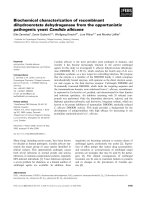
Fig. 4. NMR characterization of Nogo-24 NH-NH region of a NOESY spectrum of Nogo-24 (mixing time of 250 ms) acquired in an aqueous buffer
(50 m
M
phosphate buffer at pH of 6.5). The o bserved sequential NH-NH NOEs are labeled.
Ó FEBS 2004 NMR characterization of the Nogo-A functional domains (Eur. J. Biochem. 271) 3515
Results
Expression and structural characterization of
Nogo-A(567–748) and Nogo-66
Nogo-A(567–748) and Nogo-66 were successfully cloned
and expressed as His-tagged proteins. As shown in Fig. 2,
both recombinant proteins could b e a ffinity-purified by
affinity columns either under native c ondition for Nogo-
A(567–748) or under denaturing condition for Nogo-66.
Attempts to refold Nogo-66 by d ialysis and fast dilution
were unsuccessful, indicating that Nogo-66 is highly insol-
uble. On the other hand, the 182 residue Nogo-A(567–748)
was soluble and its molecular mass as determined by
MALDI-TOF MS m atched that predicted from the amino
acid sequence. Interestingly t he apparent molecular m ass of
Fig. 5. CD and NMR characte rization of Nogo-40. (A) Far-UV CD spectra of Nogo-40 collected at 20 °C in the presence of methanol at different
concentration s. Black, 50 m
M
phosphate b uffer (pH 6.5); pink, 20% TFE; green, 36%; cyan, 49%; dark violet, 60%; brown, 68%; dark green, 74%
and blue, 80%. (B) Far-UV CD spectra of Nogo-40 collected at 20 °C in the presence of TFE at different concentrations. black: 50 m
M
phosphate
buffer (pH 6.5 ); pink, 20% TFE solution; green, 36%; cyan, 49% and red, 60%. (C) NH-aliphatic region of a NOESY spectrum (mixing time of
250 ms) of Nogo-40 acquired in a 50 m
M
phosphate buffer (pH 6.5) at 15 °C. (D) NH-aliphatic region of a NO ESY spectrum of N ogo-40 (mixing
time of 250 ms) acquired i n a 50 : 5 0 (v/v) TFE/H
2
Omixtureat35°C.
3516 M. Li et al.(Eur. J. Biochem. 271) Ó FEBS 2004
Nogo-A(567–748) estimated by SDS/PAGE (Fig. 2A) was
about 37 kDa, much larger than that expected for a 182
residue protein. This anomalous behavior on SDS/PAGE
has been previously observed for cloned Nogo-A fragments
and w as attributed to the e xistence of a high number of
charged residues in Nogo-A [4,10].
The structural properties of Nogo-A(567–748) were first
investigated by CD spectroscopy. As shown in Fig. 3A, the
CD spectrum o f Nogo-A(567–748) in aqueous buffer had a
maximal negative peak at 202 nm and had no significant
positive signal a t 198 nm, indicating that the polypeptide
was not fully structured [23]. However, the existence of t he
maximal negative signal at around 202 nm, rather than
198 nm, together with the negative shoulder signal at
225 nm, indicated that the polypeptid e was also not
assuming a Ôrandom coilÕ structure. To explore whether
Nogo-A(567–748) h ad any specific interact ion with metal
ions, we utilized CD measurements to monitor conforma-
tional changes induced by the addition o f metal ions,
including Ca
2+
,Mg
2+
,Cu
2+
,Ni
2+
and Zn
2+
.OnlyZn
2+
was able to induce a significant conformational change in
the polypeptide. As shown in F ig. 3 A, the CD spectrum of
Nogo-A(567–748) with dual negative signals at 206 and
221 nm in the presence of 4 m
M
Zn
2+
resembles that for a
typical h elical protein. The results indicate that Zn
2+
could
specifically induce, to a significant degree, the polypeptide to
assume a helical conformation.
The structural properties of the Nogo-A(567–748) were
further assessed by the NMR HSQC experiment, which is
very sensitive to both secondary structures and tertiary
packings. As shown in Fig. 3B, the poor chemical disper-
sions of the spectrum a t both
1
Hand
15
N dimen sions
indicated that Nogo-A(567–748) did not have a tight side-
chain packing. In particular, the number of observed NMR
cross peaks was only a bout 35, much less than expected for
a 182 residue protein, thus indicating that slow conform-
ational exchanges existed over most regions of the protein.
Usually, slow conformational exchange w ould result in
significant line-broadening for HSQC peaks and make these
peaks undetectable. The manifested HSQC peaks in Fig. 3B
most likely resulted from the unstructured and flexible
regions of the N ogo-A(567–748), while the p eaks for the
regions undergoing slow conformational changes were
undetectable. The results above indicated that Nogo-
A(567–748) was partially structured, p robably with some
properties characteristic of m olten g lobule s tates [ 24–27].
Interestingly, upon addition of 4 m
M
Zn
2+
,nonewHSQC
peaks appeared but the intensities of the existing peaks
became s tronger (spectrum not shown). This observation
suggests that although the introduction of Zn
2+
was able to
Fig. 6. NMR spectral assignment of Nogo-40. The NH-aH region of a NOESY spectrum of Nogo-40 (mixing time of 250 ms) acquired in a 50 : 50
(v/v) TFE/H
2
O mixture at 35 °C with sequential assignments indicated. Several m edium-range NOEs defining h elical structures are labeled.
Ó FEBS 2004 NMR characterization of the Nogo-A functional domains (Eur. J. Biochem. 271) 3517
significantly enhance the helical structure of Nogo-A(567–
748) as detected by CD, it was not sufficient to make the
tertiary packing as tight as those f ound in a well-structured
protein.
CD and NMR characterization of Nogo-24 and Nogo-40
The purified Nogo-66 was found to be highly insoluble in
both aqueous buffer and a TFE/H
2
Omixture.Anattempt
to acquire a
1
H-
15
N HSQC spectrum of Nogo-66 was
unsuccessful. We therefore focused our NMR structure
determination on N ogo -40, which h as been shown p revi-
ously to be an excellent NgR a ntagonist by virtue of its
ability to interact with NgR without eliciting downstream
inhibitory signaling [10,11].
Secondary structure p rediction suggested that Nogo-40
had a strong propensity to form a helical structure (data not
shown). However, the preliminary CD and NMR study
indicated that Nogo-40 was largely unstructured in aqueous
buffers. As a r esult, it was not possible to assign the N MR
spectra of Nogo-40 under these conditions due to the severe
peak overlap. To gain insight into the intrinsic secondary
structure preference of Nogo-40 experimentally, we d issec-
ted Nogo-40 into two fragments, namely the N- and
C-terminal parts. While several attempts to synthesize the
C-terminal part of Nogo-40 f ailed, the peptide Nogo-24,
comprising the N-terminal 24 residues of Nogo-66, was
successfully produced. The sequential assignment o f Nogo-
24 was s uccessfully achieved and t he chemical shifts
determined (data not shown). The NOE assignment shown
Fig. 7. The s econdary structures of Nogo-24 and Nogo-40. (A) Ca proton conformational shifts of Nogo-24 (grey) and Nogo-40 (black). (B) The
NOE patterns of Nogo-40 u sed to define its secondary structure.
3518 M. Li et al.(Eur. J. Biochem. 271) Ó FEBS 2004
in Fig. 4 clearly indicates that sequential NH-NH NOE
connectivities exist over many residues of Nogo-24, strongly
indicating intrinsic helix-forming propensity in the Nogo-24
peptide, even in aqueous buffer. This observation, together
with the secondary structure predictions for Nogo-40,
prompted us to conduct further NMR studies of Nogo-40
in the presence of TFE and methanol, which is well-known
for its ability to stabilize intrinsic helixes.
Figure 5A shows t he CD spectra of Nogo-40 in aqueous
buffer and methanol/H
2
O mixtures. The CD spectrum of
Nogo-40 in the aqueous buffer has a negative peak at
198 nm, indicating that Nogo-40 had no stable confor-
mation in aqueous buffer [23]. Interestingly, with the
introduction of meth anol, the CD spectra of Nogo-40
undergo dramatic changes. The CD spectra of Nogo-40 in
the presence of methanol at a concentration of 74% or
above show one positive peak at 198 nm and two
negative peaks at 208 and 222 nm, r espectively. This
observation clearly indicates that Nogo-40 adopts a well-
formed helical conformation in the presence of 74% or
higher percentages of methanol. S imilarly, as shown in
Fig. 5B, TFE is also able to stabilize the helical conforma-
tion of Nogo-40. It appears that 50% TFE is sufficient to
stabilize a full helical conformation for the p eptide.
NMR s pectroscopy was further utilized to explore the
structural properties of Nogo-40. The very narrow reson-
ance dispersion of amide protons ( 0.7 p.p.m) and the lack
of side-chain packing with aromatic ring protons in aqueous
buffer (Fig. 5C) demonstrate that Nogo-40 in aqueous
buffer had no stable structure, which is consistent with the
CD results above. In contrast, the same NOESY region of
Nogo-40 in the 50 : 50 (v/v) TFE/H
2
O mixture (Fig. 5D)
shows a dramatically increased dispersion of amide protons
( 1.5 p.p.m) a nd extensive side-chain packing with aro-
matic ring protons, indicating that N ogo-40 adopts a well-
formed helical structure in the presence of 50% TFE.
NMR structure determination of Nogo-40
Based on the observations above, the structure determin-
ation of Nogo-40 by NMR spectroscopy was thus carried
out in a 50 : 50 (v/v) T FE/H
2
O m ixture. F igure 6
presents a NH-aH region o f NOESY spectrum of
Nogo-40 with sequential assignments labeled. The aH
conformational shifts (Fig. 7A) suggest that Nogo-40
contains two helical fragments, one at the N-terminal part
and the other over the C-terminus. The medium-range
NOE connectivities such as daN(i, i+2), daN(i, i+3),
daN(i, i+4) and dab(i, i+3) used f or ide ntification of
secondary structures, again support the observation that
two helical segments exist in Nogo-40 (Fig. 7B). It is also
noteworthy t hat the helical conformational s hifts already
existed for Nogo-24 in aqueous buffer (Fig. 7A), although
were less pronounced than those f or Nogo-40 in 50%
TFE.
Fifty Nogo-40 structures were calculated from the NMR
restraints detailed i n Table 1 with a simulated a nnealing
protocol implem ented by the Crystallography and NMR
system. O ut o f these, the 10 lowest-energy structures with a
distance violation of less than 0.3 A
˚
and a dihedral angle
violationoflessthan5° were selected for further analysis.
The structural statistics for the 10 selected structures are also
included in Table 1. The low values of distance and dihedral
angle energies i ndicate that all s elected structures satisfy the
experimental NMR c onstraints. Moreover, the covalent
geometry is well-respected as demonstrated by the low root
mean square deviation (rmsd) values for the bond lengths
(0.0019 A
˚
) and the valence a ngles (0.4°).
All 10 s tructures o f N ogo-40 contain t wo helices, one
over residues 7–12 and another over residues 26–37.
Superimposition of the 10 structures over either helix
(Fig. 8A,B) gives low rmsd v alues (Table 1), indicating that
both helices are well defined. However, due to the absence of
NOEs between N- and C-terminal helices, their relative
orientation cannot be determined. A more detailed exam-
ination of the 10 selected structures shows that there are two
populations among the 10 structures. Five of these struc-
tures, as r epresented in F ig. 8C, contain only two helices
(one from residues 7 to 12 and a nother f rom 2 6 t o 37).
However, another set of five structures, as represented in
Fig. 3D, has an additional helix over residues 20–24.
Indeed, conformational shifts shown in F ig. 7A and
medium-range NOEs in Fig. 7B indicate a helical confor-
mation over residues 20–24. Possibly due to the existence of
side-chain–side-chain NOEs among residues His17, Phe19,
Tyr22 and Leu23, the helix over residue 20–25 is distorted to
some extent and consequently became undetectable in five
of the 10 selected structures. Figure 8E shows a represen-
tation of the e lectrostatic potential associated with the
contact s urface of the N ogo-40 structure. The m ost
interesting observation here is that the N- and C-terminal
parts of Nogo-40 have opposite electrostatic potential
surfaces. More specifically, the N-terminal nine residues of
Table 1. NMR restraints used for structure calculation and structural
statistics for the 10 selected lowest-energy structures.
Restraints for structure determination
NOE distance constraints 198
Sequential 122
Medium range (|i-j| £ 4) 76
Statistics for structure calculation
Final energies (kcalÆmol
)1
)
E(total) 63.5 ± 6.6
E(bond) 2.3 ± 0.3
E(angle) 28.5 ± 1.9
E(improper) 4.2 ± 1.1
E(Van der Waals) 21.0 ± 2.5
E(NOE) 7.3 ± 2.5
Root mean square deviations
from idealized geometry
Bond (A
˚
) 0.002 ± 0.0001
Angle (degree) 0.400 ± 0.0135
Improper (degree) 0.285 ± 0.0350
NOE (A
˚
) 0.027 ± 0.0046
Average RMSD (A
˚
) from the
lowest-energy structure for
backbone/heavy atoms
Whole (2–39) 3.00/4.00
N-terminal helix (7–12) 0.22/1.11
C-terminal helix (26–37) 0.61/1.58
Additional helix (20–24) 0.78/1.69
Ó FEBS 2004 NMR characterization of the Nogo-A functional domains (Eur. J. Biochem. 271) 3519
Nogo-40 constitute a large positive surface (blue) while the
C-terminal residues make up a large negative surface (red).
Discussion
The discovery that the molecular interaction between Nogo-
66 and NgR poses inhibitory effects on the CNS neuronal
regeneration makes the Nogo-66–NgR interface an extre-
mely promising target for design of molecules to treat CNS
injuries. H owever, it has been extensively speculated that in
addition to the N ogo-66 loop, other regions of N ogo-A
might a lso play c ritical r oles in inhibiting CNS neuronal
regeneration [7–11]. Indeed, a recent study showed that
Nogo-A, the longest m ember of the Nogo transcripts
encoding for more than 1000 amino acid residues, has at
least two discrete regions with neuronal growth inhibitory
effects [4,11]. As no previous structural study has been
reported f or Nogo-A, w e carried out a detailed CD a nd
NMR investigation in an attempt to gain structural insights
into these two functional regions. Our results revealed that
although Nogo-A(567–748) is functionally active, it is only
partially structured either due to the loss of t he stabilizing
contacts provided by other parts of the Nogo-A protein or is
a member of so called natively unstructured proteins, which
only become w ell-structu red upon binding to their i nter-
acting partners or cognate receptors [28,29], or even both.
Interestingly, the observation t hat t he Zn
2+
was able t o
specifically induce the formation of h elical structures in
Fig. 8. Solution structure of Nogo-40. (A) The 10 lowest-energy structures superimposed over the N -terminal helix over residues 7–12. (B) The same
10 lowest-energy structures superimposed over the C-terminal helix over residues 26–37. (C) Ribbon representation of one conformational ensemble
of Nogo-40 structure with only two helices formed. (D) Ribbon representation of another conformational ensemble of Nogo-40 structure with an
additional helix over residues 20–24. (E) Representation of the electrostatic potential associated with the contact surface of the Nogo-40 solution
structure. Two distinctive surfaces are observed: the N-terminal surface is l argely positive (blue) while the C -terminal part is negative (red).
3520 M. Li et al.(Eur. J. Biochem. 271) Ó FEBS 2004
Nogo-A(567–748) m ight constitute an int eresting clue for
future functional studies of Nog o-A.
On the other hand, the recent identification of Nogo-40
as a potent NgR antagonist suggests a promising starting
point for the design of potential therapeutic agents to
enhance CNS neuronal regeneration. Knowledge o f the
three-dimensional s tructure of Nogo-40 i s n ecessary for
both understanding the endogenous Nogo-66–NgR inter-
action and for the rational design of other NgR-binding
antagonists. Although Nogo-40 is highly disordered in
aqueous buffer, close NMR examination indicates that it
has an intrinsic propensity to assume helical conformations.
This provides a k ey rationale for the use of TFE, which
represents a common practice in stabilizing the structure o f
a polypeptide with intrinsic helical propensity to enable their
further analysis [29].
The NMR structure of Nogo-40 reveals that the N- and
C-terminal segments of Nogo-40 have opposite electro-
static potential surfaces, thus providing an important
clue for understanding the Nogo-40–NgR interaction.
Recently, the deter mination of t he crystallographic s truc-
ture of the N gR ectodomain l ed to the speculation that
one potential Nogo-66 binding site on NgR has charac-
teristics of a negative cavity, consisting of residues Asp111,
Asp114, Ser113 and Asp138 [30,31]. As shown in Fig. 8E,
the C -terminal p art of Nogo-40 is highly negatively
charged, making it unlikely as a candidate for binding to
this acidic NgR cavity. On the other hand, it is highly
probable that t he N-terminal positive p art i s r esponsible
for its binding to the NgR negative cavity. This is in
complete agreement with previous findings that deletion of
the first five residues at the N-terminal end of Nogo-66
greatly diminished NgR binding, and deletion of the first
10 residues a bolished NgR b inding [10]. I t has also been
shown that residues 30–33 of Nogo-66 (con taining residues
Glu31 and Glu32) are important for NgR binding. Given
the fact t hat both N- and C-terminal residues of Nogo-40
were r equired for NgR binding, it would be logical to
speculate that the C-terminal part of Nogo-40 may bind to
a positively charged surface on NgR, which is not r evealed
by the current NgR structure. Alternatively, it is also
possible t hat this part of Nogo-40 may even bind to other
molecules s uch as the recently i dentified NgR coreceptor
p75
NTR
in the formation of a multicomponent complex.
In summary, our study represents the first structural
insights into the two functional regions of Nogo-A
critical for inhibiting CNS neuronal regeneration. The
results showed that the region consisting of Nogo-A(567–
748) is only partially structured but can be induced to
form a helical structure via interaction w ith Zn
2+
.
Furthermore, the d etermination of the Nogo-40 solution
structure offers a starting point for further understanding
the interaction between NgR and Nogo-40, and for
future designs of molecules to enhance CNS neuronal
regeneration using NMR methodology as demonstrated
previously [32–35].
Acknowledgements
This work is supported by the NMRC grant R183-000-092-214, the
BMRC grant R-183-000-097-305 and the BMRC Young Investigator
Award R-154-000-217-305 to J. Song and BMRC grant R-183-000-
098-305 to B .L. T ang. The authors acknowledge J . Le febvre for peptide
synthesis, H. Zhan g, Y.H. H an for acce ssing NMR spectrometer a nd
X.H. Wu at the Protein and Proteomics Center (PPC), National
University of Singapore for MALDI-TOF mass spectrometric analysis.
References
1. Woolf, C.J. & Bloechlinger, S. (2002) Neuroscience. It takes more
than two to N ogo. Science 297, 1132–1134.
2. Oertle, T. & Sc hwab, M .E. (2003) Nogo and its paRTNe rs. Trends
Cell Biol. 13, 187–194.
3. McGee, A.W. & Strittmatter, S.M. (2003) The Nogo-66 receptor:
focusing myelin inhibition of axon regeneratio n. Trends Neurosci.
26, 193–198.
4. Schwab, M.E. (2004) Nogo and axon regeneration. Curr. Opin.
Neurobiol. 14, 118–124.
5. Liu, B.P., Fournier, A., GrandPre, T. & Strittmatter, S.M. (2002)
Myelin-associated glycoprotein as a functional ligand for the
Nogo-66 receptor. Science 297, 1190–1193.
6. Wang, K.C., Koprivica, V., K im, J.A., Sivasankaran, R., Guo, Y.,
Neve, R.L. & He, Z. (2002) Oligodendrocyte-myelin glycoprotein
is a Nogo receptor ligand that inhibits neurite outgrowth. Nature
417, 941–944.
7. Fournier, A.E. & Strittmatter, S.M. (2001) Repulsive fa ctors
and axon regeneration in the CNS. Curr. Opin. Neurob iol. 11,89–
94.
8. Wang,K.C.,Kim,J.A.,Sivasankaran,R.,Segal,R.&He,Z.
(2002) P75 interacts with the Nogo receptor as a co-receptor for
Nogo, MAG and OMgp. Natur e 7, 74–78.
9. Chen,M.S.,Huber,A.B.,vanderHaar,M.E.,Frank,M.,Schnell,
L., Spillmann, A.A., Christ, F. & Schwab, M.E. (2000) Nogo-A is
a m yelin-associated neurite outgrowt h in hib itor and an ant igen for
monoclonal antibody IN-1. Nature 403, 434–439.
10. GrandPre, T., Nakamura, F., Vartanian, T. & Strittmatter, S.M.
(2000) Identification of the Nogo in hibitor of axon r egeneration as
a Reticulon protein. Nature 403, 439–444.
11. Oertle, T., van der Haar, M.E., Bandtlow, C .E., Robeva, A.,
Burfeind, P ., Bu ss, A., Huber, A.B., Simonen, M ., Schnell, L.,
Brosamle, C., Kaupmann, K., Vallon, R. & Schwab, M.E. (2003)
Nogo-A inhibits n eurite outgrowth and cell spreading with three
discrete regions. J. Neurosci. 23, 5393–5406.
12. GrandPre, T., Li, S. & Strittmatter, S.M. (2002) Nogo-66 receptor
antagonist peptide promotes a xonal r egeneration. Nature 41 7,
547–551.
13. Li, S. & Strittmatter, S.M. (2003) Delayed systemic Nogo-66
receptor antago nist prom otes recove ry from spinal cord injury.
J. Neurosci. 23, 4219–4227.
14.Jeener,J.,Meier,B.H.,Bachmann,P.&Ernst,R.R.(1979)
Investigation o f exchange p rocesses by two-dimensional NMR
spectroscopy. J. Chem. Phys. 71, 4546–4553.
15. Bax, A. & Davis, D.G . (1985) MLEV-17-based two-dimensional
homonuclear magnetization t ransfer spectroscopy. J. Mag n.
Reson. 65, 355–360.
16. Sattler, M., Schleucher, J. & Griesinger, C. (1999) Heteronuclear
multidimensional NMR experiments for the structure determi-
nation of p roteins in solution employing pul sed field gradients.
Prog. NMR Spectrosc. 34, 93–158.
17. Delaglio, F., Grzesiek, S., Vuis ter,G.W.,Zhu,G.,Pfeifer,J.&
Bax, A. (1995) NMRPipe: a multidimensional spectral processing
system based on UNIX pipes. J. Biomol. NMR 6, 277–293.
18. Johnson, B.A. & Blevins, R.A. (1994) NMRView: a computer
program for the visualization and analysis of NMR d ata. J. Bio-
mol. NMR 4, 603–614.
19. Wagner, G. & Wuthrich, K. (1982) Sequential resonance assign-
ments in protein 1H nuclear magnetic resonance spectra. Basic
pancreatic trypsin inhibitor. J. Mol. Biol. 155, 347–366.
Ó FEBS 2004 NMR characterization of the Nogo-A functional domains (Eur. J. Biochem. 271) 3521
20. Song, J., Gilquin, B., Jamin, N., Drakopoulou, E., Guenneugues,
M., Dauplais, M., Vita, C. & Menez, A. (1997) NMR solution
structure of a two-disulfide derivative of charybdotoxin: structural
evidence for c onservation of scorpion toxin alpha/beta motif and
its hydrophobic side chain p acking. Biochemistry 36, 3760–3766.
21. Brunger, A.T., Adams, P.D., Clore, G.M., Delano, W.L., Gros, P.,
Grosse-Kunstleve, R.W., Jiang, J., Kuszewski, J ., Nilges, M .,
Pannu, N.S. et al. (1998) C rystallography & NMR system: A new
software suite for macrom olecular structure determination. Acta.
Crystallogr. D54, 905–921.
22. Koradi,R.,Billeter,M.&Wu
¨
thrich, K. (1996) MOLMOL: a
program for display and analysis of macromolecular structures.
J. Mol. Graphics 14, 51–55.
23. Venyaminov, S.Y. & Yang, J.T. (1996) Determination of protein
secondary s tructure. I n Circ ular Dichroism and the C onformational
Analysis of Biomolecules (Fasman, G.D., ed.), pp. 69–107. Plenum
Press, New York.
24. Schulman, B.A., Kim, P.S., D obson, C.M. & Redfield, C. (1997)
A residue-specific NMR view of the non-cooperative unfolding of
a molten globule. Nat. Struct. Biol. 4, 630–634.
25. Song, J., Bai, P., Luo, L. & Peng, Z.Y. (1998) Contribution of
individual residu es t o formation of the native-like tertiary topol-
ogy in the alpha-lactalbumin molten glob ule. J. Mol. Biol. 280,
167–174.
26. Song,J.,Jamin,N.,Gilquin,B.,Vita,C.&Menez,A.(1999)A
gradual d isruption of tight side-chain packing: 2D, 1H-NMR
characterization of acid -induced unfolding of CHABII. Nat.
Struct. Biol. 6, 129–134.
27. Bai, P., Song, J., Luo, L. & Pe ng, Z.Y. (2001) A mode l of d ynamic
side-chain–side-chain interactions in the a lpha-lactalbumin m olten
globule. Prot ein Sci. 10, 55–62.
28. Wright, P.E. & Dyson, H.J. (1999) Intrinsically unstructured
proteins: re-assessing the protein structure-function paradigm.
J. Mol. Biol. 293, 3 21–331.
29.Song,J.,Chen,Z.,Xu,P.,Gingras,R.,Ng,A.,Leberer,E.,
Thomas, D.Y. & Ni, F. (2001) Molecu lar interactions of the Gb
binding domain of the Ste20p/PAK family of protein kinases. An
isolated but fully functional Gb binding domain from Ste20p is
only partially fold ed as s hown by heteronuclear NMR spectros-
copy. J. Biol. Chem. 276, 4 1205–41212.
30. He,X.L.,Bazan,J.F.,McDermott,G.,Park,J.B.,Wang,K.,
Tessier-Lavig ne, M., He, Z. & Garcia, K .C. (2003) St ructu re of the
Nogo receptor ectodomain: a recognition module implicated in
myelin inhibition. Neuron. 38, 177–185.
31. Barton, W.A., Liu, B.P., T zvetkova, D., Jeffrey, P.D., Fournier,
A.E., Sah, D., Cate, R., Strittmatter, S.M. & Nikolo v, D.B. ( 2003)
Structure and axon outgrowth inhibitor binding of the Nogo-66
receptor and related proteins. EMBO J. 22, 3291–3302.
32. Diercks, T., Coles, M. & Kessler, H. (2001) Applications of NMR
in drug discovery. Curr. Opin. Chem. Biol. 5, 285–291.
33. Pellecchia, M., Sem, D.S. & Wuthrich, K. (2002) NMR in drug
discovery. Nat. Rev . Drug Discov. 1, 211–219.
34. Song, J. & Ni, F. ( 1998) NMR for the d e sign o f f unctional
mimetics of prote in–protein i nteractions: o ne ke y is in the building
of bridges. Biochem. Cell Biol. 76, 1 77–188.
35. Homans, S.W. (2004) NMR spectroscopy tool s for structure-
aided drug design. Angew Chem. Int. Ed. E ngl. 43, 290–300.
Supplementary material
The following material is available from http://
www.blackwellpublishing.com/products/journals/suppmat/
EJB/EJB4286/EJB4286sm.htm
Tables S1 and S 2. Chemical shifts of Nogo-24 in a 25 mM
phosphate buffer (pH 6.8) at 298 K, and chemical shifts of
Nogo-40 in a 50/50 % (TFE/H2O) mixture at 308 K.
3522 M. Li et al.(Eur. J. Biochem. 271) Ó FEBS 2004