Báo cáo khoa học: Structural characterization of the lipopolysaccharide O-polysaccharide antigen produced by Flavobacterium columnare ATCC 43622 potx
Bạn đang xem bản rút gọn của tài liệu. Xem và tải ngay bản đầy đủ của tài liệu tại đây (200.72 KB, 7 trang )
Structural characterization of the lipopolysaccharide
O
-polysaccharide
antigen produced by
Flavobacterium columnare
ATCC 43622
Leann L. MacLean
1
, Malcolm B. Perry
1
, Elizabeth M. Crump
2
and William W. Kay
2
1
Institute for Biological Sciences, National Research Council, Ottawa, Ontario, Canada;
2
Department of Biochemistry and
Microbiology, University of Victoria, Victoria, British Columbia, Canada
The structure of the antigenic O-chain polysaccharide of
Flavobacterium columnare ATCC 43622, a Gram-negative
bacterium that causes columnaris disease in warm water fish,
was determined by high-field 1D and 2D NMR techniques,
MS, and chemical analyses. The O-chain was shown to be
an unbranched linear polymer of a trisaccharide repeat-
ing unit composed of 2-acetamido-2-deoxy-
D
-glucuronic
acid (
D
-GlcNAcA), 2-acetamidino-2,6-dideoxy-
L
-galactose
(
L
-FucNAm) and 2-acetamido-2,6-dideoxy-
D
-xylo-hexos-
4-ulose (
D
-Sug) (1 : 1 : 1), having the structure:
Keywords: Flavobacterium columnare; lipopolysaccharide;
NMR.
Flavobacterium columnare, formerly referred to as Flexi-
bacter columnaris or Cytophaga columnaris [1], is a Gram-
negative bacterium which causes columnaris disease [2] in
warm water fish, a disease that is the second leading cause of
mortality in pond raised catfish in the south-eastern United
States.
The virulence factors of F. columnare are relatively
unknown, but it has been suggested that, in pathogenesis,
adhesion of the bacterium may be related to its surface
polysaccharide constituents [3–6]. This investigation was
directed towards characterization of the lipopolysaccharide
(LPS) and putative capsule produced by the bacterium, as a
first step in identifying their possible role in pathogenesis in
fish. In addition, it was considered that characterization of
the LPS O-polysaccharide (O-PS) antigen would provide a
structural knowledge basis for the development of a specific
antibody diagnostic agent and possible target molecules for
a conjugate based vaccine.
Experimental procedures
Bacterial culture
F. columnare (ATCC 43622, NRCC 6160) was grown at
16 °C in a 52-L fermentor in medium of composition:
tryptone, 4 g; yeast extract, 0.4 g; MgSO
4,
0.5 g; CaCl
2
,
0.5 g; sodium acetate, 0.2 g; maltose, 10 gÆL
)1
;pHwas
adjusted to 7.00 with 0.1
M
NaOH. A 2.5-L inoculum
grown at 22 °C was used, with stirring at 200 r.p.m. and
dissolved oxygen at 20%. Cells were killed with 1% phenol
(final concentration, 2 h at 4 °C) in late exponential phase at
25.5hgrowth(A
600
¼ 3.34). After acidification with acetic
acid to pH 4 at 0 °C to break the gel-like constitution, the
suspended cells were harvested by centrifugation (yield
300 g wet paste).
Preparation of LPS and
O
-PS
F. columnare cells (300 g wet paste) were extracted for
15 min at 65 °C with vigorously stirred 50% (w/v)
aqueous phenol (1.2 L), and, after cooling (4 °C) and
low-speed centrifugation, the separated water and phenol
layers were collected by aspiration, and dialyzed against
running water until free from phenol. The lyophilized
dialyzed retentates were dissolved in sodium acetate
(0.02
M
, pH 7.0, 80 mL) and then treated sequentially
with RNase, DNase and proteinase K (37 °C, 2 h each).
The digests were cleared by low-speed centrifugation
(4000 g) and then subjected to ultracentrifugation
(105 000 g,12h,4°C). Only the phenol phase-soluble
product afforded a precipitated LPS gel. The gel was
½!4Þ-b-d-GlcpNAcA-ð1!4Þ-a-l-FucpNAm-ð1!3Þ-a-d-Sugp-ð1!
33
""
Ac Ac
Correspondence to M. B. Perry, Institute for Biological Sciences,
National Research Council, Ottawa, Canada K1A 0R6.
Fax: + 1 613 941 1327, Tel.: + 1 613 990 0837.
Abbreviations:
D
-Sug, 2-acetamido-2,6-dideoxy-
D
-xylo-hexos-4-ulose;
D
-GlcNAcA, 2-acetamido-2-deoxy-
D
-glucuronic acid;
L
-FucNAm,
2-acetamidino-2,6-dideoxy-
L
-galactose (2-acetamidino-2-deoxy-
L
-fucose); LPS, lipopolysaccharide; O-PS, O-polysaccharide;
CPS, capsular polysaccharide.
(Received 21 May 2003, revised 25 June 2003,
accepted 27 June 2003)
Eur. J. Biochem. 270, 3440–3446 (2003) Ó FEBS 2003 doi:10.1046/j.1432-1033.2003.03736.x
dissolved in water and lyophilized to yield LPS (1.68 g),
which was used in all further studies.
The addition of acetone (6 vol.) to the supernatant from
the above ultracentrifugate remaining after the collection of
LPS afforded a precipitate (94 mg), which, on Sephadex G-
50 column chromatography, yielded a void-volume elution
product (55 mg) tentatively identified as capsular polysac-
charide (CPS).
O
-PS
LPS (1.40 g) was delipidated by treatment with 1% (v/v)
acetic acid (100 mL) at 100 °C for 2 h and, after removal of
precipitated lipid A (170 mg), the lyophilized water-soluble
products were fractionated by Sephadex G-50 column
chromatography to yield an O-PS fraction (K
av
0.03–0.12,
390 mg) and a low-molecular-mass putative core oligosac-
charide fraction (K
av
0.75, 110 mg).
Chromatography and electrophoresis
Descending preparative paper chromatography was per-
formed on water-washed Whatman 3
MM
paper using
butanol/ethanol/water (4 : 1 : 5, by vol., top layer). Detec-
tion was with 2% ninhydrin in acetone, and mobilities are
quoted relative to
D
-glucosamine/HCl (R
GN
). GLC was
performed with an Agilent 6850 Series gas chromatograph
fitted with a flame ionization detector and a Phenomenex
Zebron capillary column ZB-50 (30 m · 0.25 mm · 25 lm)
using a temperature program 170 °C (delay 4 min) to
240 °Cat4°CÆmin
)1
. GLC/MS was performed under the
same conditions using a Hewlett–Packard 5985 GLC/MS
system and an ionization potential of 70 eV. Retention
times are quoted relative to hexa-O-acetyl-
D
-glucitol
(T
G
¼ 1.00).
Polysaccharide was separated by Sephadex G-50 column
(2 · 85 cm) chromatography using 0.05
M
pyridinium
acetate (pH 4.5) as the mobile phase, and the eluate was
continuously monitored using a Waters R403 differential
refractometer.
LPS samples (2 lg) were electrophoresed in 14% poly-
acrylamide in the presence of deoxycholate. Bands were
detected using the silver-staining directions of Tsai & Frasch
[7].
NMR spectroscopy
1
Hand
13
C NMR spectra were recorded on a Varian
Inova 400 spectrometer with samples in 99% D
2
Oat
55 °C, and internal acetone standard (2.225 p.p.m. for
1
H and 31.07 p.p.m. for
13
C) employing standard
COSY, TOCSY (mixing time 80 ms), NOESY (mixing
time 200 ms), heteronuclear single quantum correlation
(HSQC), and heteronuclear multiple-bond correlation
(gHMBC) (optimized for 5 Hz long-range coupling
constant).
Chemical procedures
Quantitative conversion in the O-PS of the acetamidino
function into an acetamido function was effected by
treatment of the O-PS with 5% aqueous triethylamine
(3 h, 70 °C) as previously described [8] to yield the modified
O-PS. Simultaneous reduction of the carboxy function of
the uronic residue C and the 4-keto function of residue B
was made by treatment of the native O-PS(47mg)inwater
(10 mL) with 1-(3-dimethylaminopropyl)-3-ethyl carbodi-
imide (150 mg) maintained at pH 4.7 over 4 h followed by
reduction at 0 °C by sodium borodeuteride (100 mg, 2 h),
followed by neutralization with acetic acid, dialysis against
distilled water, and recovery of the reduced O-PS (40 mg) in
the void-volume fraction from Sephadex G-50 gel-filtration
chromatography. A similar preparation of reduced O-PS
was made by reduction with NaBH
4
under the same
experimental conditions.
General methods
Hydrolyses were carried out in sealed tubes with either 6
M
HCl (100 °C, 3 h) or 2
M
trifluoroacetic acid (105 °C, 4 h),
and samples were concentrated to dryness in a stream of
nitrogen and examined directly or after derivatization.
Alditol acetates were prepared after reduction (NaBD
4
or
NaBH
4
) and acetylation (Ac
2
O) of isolated aldoses, as
previously described [8]. The absolute configuration of
derived 2-acetamido-2-deoxyhexoses was confirmed by
GLC analysis of their acetylated 2-(S)-butyl glycosides,
prepared under previously described conditions [9].
Optical rotations were measured at 20 °C in 10-cm
microtubes, using a Perkin-Elmer 243 polarimeter.
Results
Fermenter-grown cells of F. columnare were extracted by a
modified hot phenol/water method [10], and a S-type LPS,
found almost exclusively in the phenol phase of the cooled
extract, was obtained in 12% yield by ultracentrifugation of
the concentrated dialyzed extract. Deoxycholate/PAGE
analysis of the LPS gave a typical ladder-like banding
pattern in which the step separations suggested that the LPS
was composed of repeating trisaccharide units [11]. On
treatment with 6 vol. acetone, the ultracentrifugate afforded
a precipitate which, on Sephadex G-50 gel filtration, gave a
void-volume fraction ( 2% yield) of a glycan tentatively
identified as CPS. A lower-molecular-mass fraction (K
av
0.7,
180 mg) which gave a strong colorimetric (phenol/H
2
SO
4
)
reaction for carbohydrate contained glycopeptides in which
the oligosaccharide moieties had similar structure and
composition (results not reported) to those previously
found in glycoproteins produced by Flavobacterium
meningosepticum [12].
The LPS was delipidated by treatment with hot dilute
acetic acid and after removal of precipitated lipid A (8%),
the O-PS (86%) was collected in the void-volume fraction
obtained by Sephadex G-50 gel filtration of the water-
soluble products.
The O-PS had [a]
D
)90.1 ° (c 8.9, water) Anal. C, 44.61;
H, 6.18; N, 7.12% and ash, nil. GLC analysis of the
acetylated 2
M
trifluoroacetic acid (105 °C, 4 h) O-PS
hydrolysis products gave a low yield ( 2%) mixture of
mannose, galactose and
L
-glycero-
D
-manno-heptose. These
glycoses probably originate from a core oligosaccharide
component; however, no significant hydrolysis products
from the major O-PS component were detected.
Ó FEBS 2003 Flavobacterium columnare polysaccharide (Eur. J. Biochem. 270) 3441
The 1D
1
H-NMR spectrum of the O-PS showed inter
alia: three anomeric glycose H1 proton signals at 5.14 (J
1,2
2.2 Hz), 4.97 (J
1,2
3Hz)and4.70(J
1,2
8.8 Hz) p.p.m. with
J
1,2
couplings indicative of two a-linkage and one b-linkage,
respectively; two methyl signals at 1.21 and 1.17 p.p.m. (6H)
characteristic of two 6-deoxyhexose residues; an N-acyl
substituent at 2.25 p.p.m. (3H); and four signals (2.10–1.93
p.p.m.) characteristic of methyl signals of two N-acetyl and
two O-acetyl substituents.
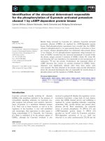
The
13
C-NMR spectrum of the O-PS (Fig. 1) showed
inter alia three anomeric signals at 102.6 (J
C-1,H-1
164 Hz),
97.1 (J
C-1,H-1
172 Hz) and 97.0 (J
C-1,H-1
180 Hz) p.p.m.
having J
C-1,H-1
coupling constants indicative of one b-link-
age and two a-linkages, respectively, together with a sharp
singlet at 93.9 p.p.m. subsequently identified as the C4
resonance of a 4-ketohexose residue. Also present were
two sharp singlets at 15.8 and 11.9 p.p.m. characteristic
of methyl shifts of 6-deoxyhexose residues, signals at
167.0 p.p.m. (C¼N) and 19.8 p.p.m. (CH
3
-C¼N) charac-
teristic of acetamidino groups, and ring carbon signals
at 55.3, 52.8 and 51.1 p.p.m. indicative of C-N-linked sub-
stituents, together with a total of four signals subsequently
assigned to methyl groups of two N-acetyl substituents (22.8
and 23.0 p.p.m.), and two O-acetyl substituents (21.1 and
20.9 p.p.m.). Five signals attributed to carbonyl substituents
were observed in the 175.7–173.6 p.p.m. region. The
preliminary data suggest that the O-PS is a polymer of
regular trisaccharide repeating units composed of three
aminoglycose residues.
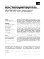
The chemical shift assignments in the
1
H-NMR and
13
C-NMR spectra and the characterization of the glycose
components in the O-PS were determined from the appli-
cation of COSY, TOCSY, NOESY and
1
H,
13
C-HSQC and
HMBC experiments (Table 1, Fig. 2). For the analysis,
Fig. 1.
13
C-NMR spectrum of F. columnare O-PS recorded at 55 °C (125 MHz).
Table 1.
1
H and
13
C NMR chemical shifts of the native LPS O-PS from F. columnare ATCC 43622. Spectra run in D
2
Oat55°C with internal
acetone reference (2.225 p.p.m. for
1
H and 31.07 p.p.m. for
13
C). Coupling constants (Hz) are given in parentheses. Tentative assignments for
residue A: N-H7 (8.83 p.p.m.) and N-H7
1
(8.57 p.p.m.) at 35 °C(10%D
2
O/90% H
2
O, v/v).
Glycose
residue
Chemical shift (p.p.m.)
H1/C1 H2/C2 H3/C3 H4/C4 H5/C5 H6/C6
A 5.14 (2.2) 4.28 (10.2) 5.16 (nr) 4.13 (2) 4.57 1.17
97.1 (172) 51.1 71.5 78.8 67.9 15.8
B 4.97 (3) 4.24 (3.2) 3.78 (nr) – 3.91 1.21
97.0 (180) 52.7 77.7 93.9 70.1 11.9
C 4.70 (8.8) 3.93 (10.0) 5.26 (9.8) 4.08 (10.0) 3.79 –
102.6 (164) 55.4 76.5 74.1 77.8 175.4
3442 L. L. MacLean et al.(Eur. J. Biochem. 270) Ó FEBS 2003
glycose residues were arbitrarily labeled A–C in order of
decreasing chemical shifts of the anomeric protons.
Glycose A was initially identified as a-FucpNAm. In a
COSY experiment, overlapping correlation peaks of H1A
to H2A and H2A to H3A were observed due to
O-acetylation at O3A causing an upfield shift of its
signal to 5.16 p.p.m. A correlation cross-peak for H4A to
H5A was not observed in the COSY spectrum because of
the small scalar coupling (H
4,5
2 Hz), but was evident
from TOCSY data linking H4A to the H6A methyl
signal at 1.20 p.p.m. (3H). A direct correlation of H2A to
C2A (51.1 p.p.m.) in HSQC and long-range HMBC and
correlations from the N-acyl proton (2.25 p.p.m.) to the
carbonyl signal at 167.0 p.p.m. were characteristic of an
acetamidino group, thus identifying A as an a-FucpNAm
residue, the a-configuration being assigned from consid-
eration of the anomeric proton and carbon coupling
constant data (J
Hl,2
2Hz,J
C-1,H-1
172 Hz).
From NMR data, residue C was assigned the b-gluco-
pyranose configuration from its observed large ring region
J
H,H
coupling constants for J
2,3
, J
3,4
and J
4,5
( 10 Hz), and
from its anomeric coupling constants, J
C-1,H-1
164 Hz, and
J
1,2
8.8 Hz. Thecorrelation of H2C tothe C2C at55.4 p.p.m.
is consistent with the presence of a C2 acetamido substituent,
and the lack of a proton at C6, considered in conjunction
with the long-range correlation of H5C to the carbonyl shift
at 175.4 p.p.m., seen in a HMBC experiment, is consistent
with the presence of a C6 carboxylic acid function and allows
C to be identified as a b-GlcpANAc residue.
Residue B was identified as an a-linked 2-acetamido-2,6-
dideoxyhexos-4-ulose residue from further NMR data. The
observed correlation from H2B to the corresponding C2B in
an HSQC experiment, the anomeric coupling constants J
1,2
of 3 Hz and J
C-1,H-1
of 180 Hz, considered in conjunction
with the fact that connectivities could only be followed from
H1B to H2B and H3B, and from the methyl resonance of
H6B to H5B with no evidence of connectivities to any
proton signals at C4B. The presence of a C4 keto group
function and the absence of a proton at C4B was further
supported from an observed long-range correlation between
H3B and the C4B carbon signal at 93.8 p.p.m. seen in
an HMBC experiment, thus identifying B as an a-linked
2-acetamido-2,6-dideoxy-xylo-hexos-4-ulose residue.
Further characterizations of the O-PS component
glycoses were made from chemical studies. Residue A was
identified as 2-acetamidino-2,6-dideoxy-
L
-galactose after its
conversion in the O-PS into its corresponding 2-acetamido
derivative by treatment with hot aqueous trimethylamine.
The quantitative transformation was verified from the 1D
1
H-NMR spectrum of the modified polymer in which a shift
of the characteristic carboxy resonance at 167 (Am) in the
native O-PS to 175.3 (Ac) p.p.m. in the modified O-PS was
observed. The HCl hydrolysate of the modified O-PS, in
contrast with the native O-PS, gave a single aminoglycose
product, which was isolated by preparative paper chroma-
tography and identified as 2-amino-2,6-dideoxy-
L
-galactose
HCl (R
GN
1.47) from its specific optical rotation {[a]
D
)81 °
(c0.2,water).Lit.[a]
D
–95° [13]}, the identity of its
1
H-
NMR spectrum with that of an authentic sample, and the
fact that its reduced (NaBD
4
) and acetylated product on
GLC/MS gave a single peak corresponding in retention
time (T
G
0.93) and mass spectrum to an authentic sample
of 1,3,4,5,-tetra-O-acetyl-2-acetamido-2,6-dideoxy-
D
-galact-
itol-[1-
2
H].
The hexuronic acid component C was identified as
2-acetamido-2-deoxy-
D
-glucuronic acid from the isolation
Fig. 2.
1
H-
13
CHSQCshiftcorrelationmap
of the spectral regions
1
H (1.0–5.5 p.p.m.)
and
13
C (10–104 p.p.m.) of the F. columnare
O-PS with resonances labeled for residues
A, B and C.
Ó FEBS 2003 Flavobacterium columnare polysaccharide (Eur. J. Biochem. 270) 3443
of 2-amino-2-deoxy-
D
-glucose-[6-
2
H
2
], from the hydrolysis
product of the reduced (NaBD
4
) carbodiimide-activated
O-PS. The latter glycose isolated by preparative paper
chromatography (R
GN
1.00) was identified by GLC/MS
of its reduced (NaBD
4
) acetylated derivative 1,3,4,5,6-
penta-O-acetyl-2-acetamido-2-deoxyglucitol-[1-
2
H, 6-
2
H
2
]
(T
G
1.22), and its
D
-configuration was confirmed from
the specific optical rotation of its hydrochloride derivative
{[a]
D
+67° (c0.3,water).Lit.[a]
D
+72°} and by GLC
analysis of its derived acetylated 2-(S)-butyl glycosides [11].
Residue B was identified as 2-acetamido-2,6-dideoxy-
D
-
xylo-hexos-4-ulose (D-Sug). The above preparative paper
chromatography of the hydrolysed reduced (NaBD
4
)
carbodiimide-activated O-PS also yielded two separated
aminoglycose fractions identified as a mixture of 2-amino-
2,6-dideoxy-
D
-(and
L
)galactose {R
GN
1.48; [a]
D
)4 ° (c 0.2,
water) [13]} and 2-amino-2,6-dideoxy-
D
-glucose {R
GN
1.83;
[a]
D
+52° (c 0.4, water); Lit. [a]
D
+50° [14]}, in
approximately equal yield. GLC/MS of their individual
reduced (NaBH
4
) and acetylated alditiol derivatives gave
single peaks corresponding in retention times to 1,3,4,5-
tetra-O-acetyl-2-acetamido-2,6-dideoxygalactitol (T
G
0.93)
and 1,3,4,5-tetra-O-acetyl-2-acteamido-2,6-dideoxyglucitol
(T
G
0.90) standards. The mass spectrum of each derivative
showed a fragmentation pattern with characteristic ions of
the C1–C2 fragment at m/z 144, 102, 84, and 60 showing
that C1 was not deuterium labeled. However, the expected
M+1) 60 ¼ 317 molecular-ion and the expected frag-
ment ions at m/z 261 (C2 to C6, 303–42, loss of ketene)
confirmed that deuterium labeling was present. The chro-
matographically isolated 2-amino-2,6-dideoxygalactose
fraction was a mixture of the
D
-and
L
-forms of the
aminoglycose, as evidenced from its optical rotation, and
from GLC analysis of its acetylated 2-(S)-butyl glycoside
derivatives. This finding is consistent with this fraction being
composed of a
L
-FucN component originating from the
O-PS residue A and the
D
-FucN from the reduced residue B.
The isolation of optically pure 2-amino-2,6-dideoxy-
D
-
glucose (D-QuiN), the major reduction product of residue
B, further confirms the
D
-configuration assigned to residue
B. Preparative paper chromatographic separation of the
hydrolysis products of NaBH
4
-reduced carbodiimide-
activated O-PS afforded the hydrochloride derivatives of
2-amino-2-deoxyglucose, 2-amino-2,6-dideoxyglucose, and
2-amino-2,6-dideoxygalactose, the
1
H-NMR spectra of
which were identical with those of authentic reference
glycoses, and further confirms their characterization. The
combined MS data and the isolation of the two aminoglyc-
oses with the respective
D
-galacto and
D
-gluco configura-
tions (epimers at C4) establishes that B is a 4-ketohexose
(or 4-acetal derivative) and, from its anomeric proton and
carbon chemical shift and coupling constant data, is present
in the a-
D
-hexopyranosyl configuration in the O-PS, and is a
2-acetamido-2,6-dideoxy-a-
D
-xylo-hexos-4-ulose residue.
The sequence of the glycose residues and their linkage
positions in the O-PSwereestablishedfrom1Dand2D
NOE and long-range multiple-bond
1
H-
13
C(HMBC)
correlations experiments. Interresidue NOEs were seen
from H1B to H4C andtoitsownH2B,fromH1A to
H2A and across the glycosidic bond to H3B,andalsofrom
H1C to H3C and H5C and across the ring to H4A.HMBC
experiment results were consistent with the proton NMR
data showing correlations between C1B (97.0 p.p.m.) to
H4C,fromC1C (102.6 p.p.m.) to H4A and from C1A
(97.1 p.p.m.) to H3B, thus defining the sequence and
linkage position in the O-PS repeating trisaccharide units
as fi4)-b-C-(1fi4)-a-A-(1fi3)-a-B-(1fi, leading to a basic
repeating unit with the structure:
Consistent with the above conclusion, the NMR analysis
of the native O-PS showed that the chemical shifts of the
linkage position carbon atoms C4A,C3B,andC4C
experience significant deshielding, further confirming the
linkage position assignments. As NMR data indicated the
presence of two O-acetyl substituents in the native O-PS,
they can only be located at the available O3 positions of
residues A and C. Partial de-O-acetylation of the O-PS with
dilute ammonium hydroxide (50 °C,1h)resultedinthe
hydrolytic removal of the acetyl substituent on residue A (a-
L-FucpNAm) and partial ( 20%) removal from residue C
(b-D-GlcpNAcA). The de-O-acetylation of A effected
deshielding of C3A (71.5–68.2 p.p.m.) and H3A (5.16–
4.04 p.p.m.), thus establishing the acetyl substituent loca-
tion at C3A in the native O-PS. The O-acetyl substitution on
residue C (b-D-GlcpNAcA) was indicated to be at position
C3C as these
1
Hand
13
C resonances experience similar
downfield shifts on de-O-acetylation. A consideration of the
experimental evidence thus leads to the full structure of the
F. columnare ATCC 43622 LPS native O-chain being an
unbranched polymer of a repeating trisaccharide having the
structure:
½C½A½B
½! 4Þ-b-d-GlcpNAcA-ð1!4Þ-a-l-FucpNAm-ð1!3Þ-a-d-Sugp-ð1!
½C½A½B
½!4Þ-b-d-GlcpNAcA-ð1!4Þ-a-l-FucpNAm-ð1!3Þ-a-d-Sugp-ð1!
33
""
Ac Ac
3444 L. L. MacLean et al.(Eur. J. Biochem. 270) Ó FEBS 2003
Discussion
In this investigation, it was shown by 1D and 2D NMR
analysis, MS, and chemical methods that the O-PS of the
LPS produced by F. columnare ATCC 43622 is a linear
unbranched polymer of trisaccharide units composed of
D-GlcNAcA, L-FucNAm and D-Sug having the structure
fi4)-b-D-GlcpNAcA-(1fi4)-a-L-FucpNAm-(1fi3)-a-D-
Sugp-(1fi, in which the linkage positions and the sequence
and pyranoside nature of the glycose residues were estab-
lished from NMR analyses. In the native O-PS the
D-GlcpNAcA and L-FucpNAm residues were both acetyl-
ated at their O3 positions.
It is interesting to note that O3-linked D-Sug was
found to be a component of the O-PS of the fish
pathogen Vibrio ordalii serotype O:2 [15], which is the
cause of vibriosis among feral and farmed fish and
shellfish. The only other reported bacterial source of this
glycose is the specific CPS of Streptococcus pneumoniae
type 5 [16]. However, in the latter polysaccharides, the
glycose is found in its b-
D
-configuration in contrast with
the a-
D
-configuration found in the F. columnare O-PS. In
agreement with previous studies, we also found that the
presence of this 4-ketoglycose in the polymeric structure
rendered the O-PS unstable under alkaline conditions
and even prolonged storage in aqueous solutions at
pH 7. A similar result was found in a study of forbeside
C, a saponin of Asterias forbesi [18], which also has a
component D-Sug residue.
After the precipitation of the LPS from the phenol phase
extract of F. columnare cells by ultracentrifugation, a low
yield of CPS material was obtained from the ultracentri-
fugate by acetone precipitation followed by Sephadex G-50
gel-filtration chromatography, yielding a lipid-free high-
molecular-mass void-volume fraction. On analysis, the
material proved to have the same structure as the homo-
logous LPS O-PS. This material could be considered to be a
putative capsule or simply free O-PS. The significance of the
O-PS and putative CPS in pathogenesis requires further
investigation. In the fish pathogens, Vibrio ordalii O:2 [15]
and Vibrio anguillarum O:2 [17], their respective LPS O-PS
components and CPSs shared the same respective homo-
logous structures, and the same constitution may pertain in
F. columnare.
Pathogenesis studies have shown a correlation between the
capacity of F. columnare to adhere to fish gill epithelium and
virulence [6,19,20]. However, the nature of the adhesins
involved have not been identified, but possible candidates are
LPS, capsule, fimbriae or other appendages of the bacterium,
a hypothesis requiring further investigation.
It is of note that the structure of the LPS O-antigen of
F. columnare differs structurally from the LPS O-antigen
of the fish pathogen Flavobacterium psychrophilium [21],
which is a linear polymer of a trisaccharide repeating unit
composed of
L
-rhamnose, 2-acetamido-2-deoxy-
L
-fucose,
and 2-N-acetyl-4-N-[(3S,5S)-3,5-dihydroxyhexanoyl]-
D
-
bacillosamine (1 : 1 : 1) [22].
Acknowledgements
This work was supported by funding from the Canadian Bacterial
Diseases Centres of Excellence Program. We thank Perry Fleming for
the large scale production of bacterial cells, and Dr E. Vinogradov for
helpful discussions.
References
1. Bernardet, J.F., Segers, P., Vancanneyt, M., Berthe, F., Kersters,
K. & Vandamme, P. (1996) Cutting a Gordian knot: emended
classification and description of the genus Flavobacterium,emen-
ded description of the Flavobacteriaceae, and proposal of Flavo-
bacterium hydatis nom. Nov (basonym, and Cytophaga aquatilis
Strohl and Tait 1978). Int. J. Syst. Bacteriol. 46, 128–148.
2.Durborow,R.M.,Thune,R.L.,Hawke,J.P.&Camus,A.C.
(1998) Columnaris Disease: a Bacterial Infection Caused by Flavo-
bacterium Columnare. Publication no. 479 Southern Regional.
Aquaculture Center, Stoneville, MI, USA.
3. Ofek, I. & Doyle, R.J. (1994) In Bacterial adhesion to cells and
tissues, pp. 1–16. Chapman & Hall, London.
4. Decostere, A., Haesebrouck, F., Van Driessche, E., Charlier, G. &
Ducatelle, R. (1999) Characterization of the adhesion of Flavo-
bacterium columnare (Flexibacter columnaris) to gill tissue. J. Fish
Dis. 22, 465–474.
5. Decostere, A., Haesebrouch, F. & Derriese, L.A. (1998) Char-
acterization of four Flavobacterium columnare (Flexibacter
columnaris) strains from tropical fish. Vet Microbiol. 62, 35–45.
6. Decostere, A., Haesebrouck, F., Charlier, G. & Ducatelle, R.
(1999) The association of Flavobacterium columnare strains of high
and low virulence with gill tissue of black mollies (Poecilia sphe-
nops). Vet. Microbiol. 67, 287–298.
7. Tsai, G.M. & Frasch, C.E. (1982) A sensitive silver stain for
detecting lipopolysaccharides in polyacrylamide gels. Anal. Bio-
chem. 119, 115–119.
8.Hermansson,K.,Perry,M.B.,Altman,E.,Brisson,J R.&
Garcia, M.M. (1993) Structural studies of the O-antigenic poly-
saccharide of Fusobacterium necrophorum. Eur. J. Biochem. 212,
801–809.
9. Leontein, K., Lindberg, B. & Lonngren, J. (1978) Assignment of
absolute configuration of sugars by g.1.c. of their acetylated gly-
cosides formed from chiral alcohols. Carbohydr. Res. 62, 359–362.
10. Johnson, K.G. & Perry, M.B. (1973) Improved techniques for the
preparation of bacterial lipopolysaccharides. Can. J. Microbiol.
22, 29–34.
11. Perry, M.B. & Babiuk, L.A. (1983) Structure of the polysaccharide
chains of Pasteurella haemolytica (serotype 4) lipopolysaccharide.
Biochem. Cell Biol. 62, 108–114.
12. Reinhold, B.B., Hauer, C.R., Plummer, T.H. & Reinhold, V.N.
(1995) Detailed structural analysis of a novel, specific O-linked
glycan from the prokaryote Flavobacterium meningosepticum.
J. Biol. Chem. 270, 13197–13203.
13. Perry, M.B. & Daoust, V. (1973) A synthesis of 2-Amino-2,6-di-
deoxy-D-(and L-)galactose (L-Fucosamine) and 2-Amino-2,6-di-
deoxy-D-(and L-)talose (Pneumosamine). Can. J. Chem. 51, 974–977.
14. Perry, M.B. & Daoust, V. (1973) A synthesis of 2-Amino-
2,6-dideoxy-L-mannose (L-Rhamnosamine) and 2-Amino-
2,6-dideoxy-L-glucose (L-Quinovosamine). Carbohydr. Res. 27,
460–463.
15. Sadovskaya, I., Brisson, J R., Khieu, N.H., Mutharia, L.M. &
Altman, E. (1998) Structural characterization of the lipopoly-
saccharide O-antigen and capsular polysaccharide of Vibrio ordalii
serotype O: 2. Eur. J. Biochem. 253, 319–327.
16. Jansson, P E., Lindberg, B. & Lindquist, U. (1985) Structural
studies of the polysaccharide from Streptococcus pneumoniae type
5. Carbohydr. Res. 140, 101–110.
17. Sadovskaya, I., Brisson, J R., Mutharia, L.M. & Altman, E.
(1996) Structural studies of the lipopolysaccharide O-antigen and
capsular polysaccharide of Vibrio anguillarum serotype O: 2.
Carbohydr. Res. 283, 111–127.
Ó FEBS 2003 Flavobacterium columnare polysaccharide (Eur. J. Biochem. 270) 3445
18. Findlay, J.A., Jaseja, M. & Brisson, J R. (1987) Forbeside C, a
saponin from Asterias forbesi. Complete structure by nuclear
magnetic resonance methods. Can. J. Chem. 65, 2605–2611.
19. Decostere, A. (2002) Flavobacterium columnare infection in fish:
the agent and its adhesion to the gill tissue. Verh. K. Acad.
Geneeskd. Belg. 64 (6), 421–430.
20. Decostere, A., Ducatelle, R. & Haesebrouck, F. (2002) Flavo-
bacterium columnare (Flexibacter columnaris) associated with
severe gill necrosis in koi carp (Cyprinus carpio L). Vet. Rec. 150
(22), 694–695.
21. Crump, E.M., Perry, M.B., Clouthier, S.C. & Kay, W.W. (2001)
Antigenic characterization of the fish pathogen Flavobacterium
psychrophilium. Appl. Environ. Microbiol. 67, 750–759.
22. MacLean, L.L., Vinogradov, E., Crump, E.M., Perry, M.B. &
Kay, W.W. (2001) The structure of the lipopolysaccharide
O-antigen produced by Flavobacterium psychrophilium (259–93).
Eur. J. Biochem. 268, 2710–2716.
3446 L. L. MacLean et al.(Eur. J. Biochem. 270) Ó FEBS 2003