mutagenesis and functional analysis of the pore forming toxin halt 1 from hydra magnipapillata
Bạn đang xem bản rút gọn của tài liệu. Xem và tải ngay bản đầy đủ của tài liệu tại đây (1.11 MB, 17 trang )
Toxins 2015, 7, 407-422; doi:10.3390/toxins7020407
OPEN ACCESS
toxins
ISSN 2072-6651
www.mdpi.com/journal/toxins
Article
Mutagenesis and Functional Analysis of the Pore-Forming
Toxin HALT-1 from Hydra magnipapillata
Yvonne Jing Mei Liew, Wai Tuck Soh, William Febry Jiemy and Jung Shan Hwang *
Faculty of Applied Sciences, UCSI University, No. 1, Jalan Menara Gading, UCSI Heights, Cheras,
56000 Kuala Lumpur, Malaysia; E-Mails: (Y.J.M.L.);
(W.T.S.); (W.F.J.)
* Author to whom correspondence should be addressed; E-Mail: ;
Tel.: +603-9101-8880 (ext. 3379); Fax: +603-9102-3606.
Academic Editor: John P. Berry
Received: 12 December 2014 / Accepted: 27 January 2015 / Published: 3 February 2015
Abstract: Actinoporins are small 18.5 kDa pore-forming toxins. A family of six actinoporin
genes has been identified in the genome of Hydra magnipapillata, and HALT-1
(Hydra actinoporin-like toxin-1) has been shown to have haemolytic activity. In this study,
we have used site-directed mutagenesis to investigate the role of amino acids in the
pore-forming N-terminal region and the conserved aromatic cluster required for cell
membrane binding. A total of 10 mutants of HALT-1 were constructed and tested for their
haemolytic and cytolytic activity on human erythrocytes and HeLa cells, respectively.
Insertion of 1–4 negatively charged residues in the N-terminal region of HALT-1 strongly
reduced haemolytic and cytolytic activity, suggesting that the length or charge of the
N-terminal region is critical for pore-forming activity. Moreover, substitution of amino acids
in the conserved aromatic cluster reduced haemolytic and cytolytic activity by more than
80%, suggesting that these aromatic amino acids are important for attachment to the lipid
membrane as shown for other actinoporins. The results suggest that HALT-1 and other
actinoporins share similar mechanisms of pore formation and that it is critical for HALT-1
to maintain an amphipathic helix at the N-terminus and an aromatic amino
acid-rich segment at the site of membrane binding.
Keywords: actinoporins; cytolysin; equinatoxin II; pore-forming protein; mutations; cnidarian
Toxins 2015, 7
408
1. Introduction
Actinoporins are a group of potent α-pore forming toxins (α-PFTs) that were first identified in sea
anemones [1]. They are low molecular weight proteins with 18.5 kDa and are able to destroy cells
containing sphingomyelin, a major component of plasma membrane lipids [2]. Equinatoxin II (EqtII)
from Actinia equina and sticholysin II (StII) from Stichodactyla helianthus are the two best-studied
α-PFTs. These α-PFTs were later referred to as actinoporins. The crystal structures of EqtII and StII
revealed that actinoporins are single-domain proteins with a compact β-sandwich composed of
12 β-strands aligned in two β-sheets flanked on each side by two short α-helices [3–5]. A cluster of
exposed aromatic amino acids including a phosphocholine (POC) binding site has been shown to be
functionally important for membrane binding. The aromatic amino acids provide initial contact between
the protein and the cell membrane while the POC binding site recognizes the headgroup of
sphingomyelin in the plasma membrane [6].
The N-terminal region comprising 30 residues appears to be the largest amphipathic part of
the protein, and this region is the only part of the protein that can undergo conformational changes,
whereby it detaches from the core of the protein, without disrupting the fold of the β-sandwich [3].
The flexibility and amphipathic character of this N-terminal region are crucial for the mechanism of
pore formation as this region is proposed to extend and penetrate into the plasma membrane to form the
pore [7]. Pore formation is a multi-step process. The first step is the recognition of sphingomyelin in
the lipid membrane. This was supported by the discovery of a POC binding site on the surface of StII
that specifically binds to the phosphocholine headgroup of sphingomyelin [5]. After binding to
sphingomyelin, the next step of pore formation is the translocation of the N-terminal region into the
lipid-water interface [8]. The flexibility and the amphipathic nature of this N-terminal region allow it to
be loosened from the protein and inserted into the lipid membrane [9]. Subsequently, three or four
actinoporins oligomerize in the plasma membrane and form an ion conductive pore, leading to the influx
of water into the cell [7,10].
Actinoporins were recently found in Hydra, a freshwater hydrozoan living in unpolluted lakes and
streams [11]. A subsequent study identified six HALT (Hydra actinoporin-like toxin) genes in the
genome of Hydra [12]. These authors also showed that HALT-1 had haemolytic activity similar to,
although weaker than, that of equinatoxin II (EqtII) from Actinia equina. To further analyze the
haemolytic activity of HALT proteins, we have used site-directed mutagenesis to mutate critical residues
in the conserved aromatic cluster required for membrane binding and in the amphipathic
N-terminal α-helix and tested their effect on pore formation. Mutation of residues in these domains
strongly reduced haemolytic and cytolytic activity, suggesting that HALT proteins form pores through
a similar mechanism to other actinoporins.
2. Results
2.1. Expression of HALT-1 and Its Mutants
Ten mutants of HALT-1 were generated by site-directed mutagenesis. Mutants 2E3, 2EE3, 2EED3
and 2EEDE3 altered the length of the N-terminal α-helix region by incorporating amino acid(s)
successively near the N-terminus. Mutants K76A, Y110A, W113A, A114W and Y129A altered amino
Toxins 2015, 7
409
acids in or near to the conserved aromatic cluster, which is involved in the interaction with cell membrane
(Figure 1 and Table 1). To assess whether these mutants have changes in their structures, mutant protein
stability was analyzed with SDM (Site-Directed Mutator) and I-Mutant 3.0. Out of these five mutants,
SMD’s prediction resulted in four neutral mutations and one stabilizing mutation (Table S1). On the
other hand, I-Mutant predicted that three mutants would have minor disturbances (∆∆Gs are greater than
−2.0 but less than −0.5) in the local structure while the other two would remain neutral (Table S2).
One mutant, 35∆C, was generated by deleting the only cysteine in HALT-1 (Figure 1 and Table 1).
All mutants were cloned into the expression vector pET28a and transformed into BL21(DE3). Figure 2
shows the SDS-PAGE images of mutant proteins with the expected protein band at approximately 25 kDa
(18.6 kD of HALT-1 mutant and 6.4 kD of vector peptide including 6 histidine tags). Eight out of ten
mutants were successfully expressed and purified in this study. Two mutants, A114W and 35∆C,
exhibited a low expression level and poor solubility. As a result, sufficient quantities could not be
purified for further testing.
****
EEDE
HALT-1
SticholysinII
EquinatoxinII
Bandaporin
1
1
1
1
*
ASGAALGVIAKVGVDAALQQIDDVW--KGKTVRYWKCAVENRSSKTLYALGTTQESGSMT
---ALAGTI-IAGASLTFQVLDKVLEELGKVSRKIAVGIDNESGGTWTALNAYFRSGTTD
-SADVAGAV-IDGASLSFDILKTVLEALGNVKRKIAVGVDNESGKTWTALNTYFRSGTSD
-SLAVAGAV-IEGGNLVMSVLDRILEAIGDVNRKIAIGVENQSGKSWTAMNTYFRSGTSD
58
56
58
58
HALT-1
SticholysinII
EquinatoxinII
Bandaporin
59
57
59
59
*
* **
TVFADIPPKSTGVF-VWEKSRG-AAKGAVGVVHYKY-GNKVLNIMASIPYDWNLYKAWAN
VILPEFVPNTKALLYSGRKDTGPVATGAVAAFAYYMSSGNTLGVMFSVPFDYNWYSNWWD
IVLPHKVPHGKALLYNGQKDRGPVATGAVGVLAYLMSDGNTLAVLFSVPYDYNWYSNWWN
VVLPHSVPSGKALLYDGQKTRGPVATGVVGVFAYAMSDGNTLAVMFSIPYDYNLYSNWWN
115
116
118
118
HALT-1
SticholysinII
EquinatoxinII
Bandaporin
116
117
119
119
*
VHLSDHKE-----SFSDLYKGKNGAKYPTRAGNWGEV-----DGTKFFLTEKSHAEFKVI
VKIYSGKRRADQGMYEDLYYG-NPYR---GDNGWHEKNLGYGLRMKGIMTSAGEAKMQIK
VRIYKGKRRADQRMYEELYYNLSPFR---GDNGWHTRNLGYGLKSRGFMNSSGHAILEIH
VKTYSGMKRADQSMYEDLYYHASPFK---GDNGWHSRNLGYGLKCRGFMNSSGAAKLEIH
165
170
175
175
HALT-1
SticholysinII
EquinatoxinII
Bandaporin
166
171
176
176
FSGISRVSKA
VSRA
168
173
179
179
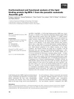
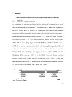
Figure 1. Ten mutations of HALT-1. The amino acid sequence of HALT-1 was aligned
with three different actinoporins: sticholysin II (Stichodactyla helianthus), equinatoxin II
(Actinia equina) and Bandaporin (Anthopleura asiatica). Signal peptides and propeptides for
all sequences are excluded in this alignment. Residues with the asterisk marked on top are
mutations from insertion, substitution or deletion. Magenta highlights the residues involving
in contact with the polar head group of the cell membrane, and light blue highlights the only
cysteine present in HALT-1. The
α-helix and
β-strand of HALT-1 are predicted by
PSIPRED and marked on the top of the sequence.
Toxins 2015, 7
410
Table 1. Mutations introduced in HALT-1 amino acid sequence.
Name
2E3
2EE3
2EED3
2EEDE3
35∆C
K76A
Y110A
W113A
A114W
Y129A
Type of mutation
Insertion
Insertion
Insertion
Insertion
Deletion
Substitution
Substitution
Substitution
Substitution
Substitution
Position
3
4
5
6
35
76
110
113
114
129
Mutation
E
EE
EED
EEDE
Removal of cysteine
K→A
Y→A
W→A
A→W
Y→A
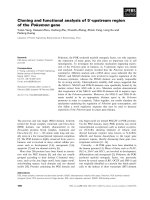
Figure 2. SDS-PAGE image of HALT-1 mutant proteins expressed in BL21(DE3).
Expression of mutants was regulated in the presence (induced) or absence (uninduced) of
1 mM IPTG. Lane 1 in all panels is the protein molecular marker. (A) lane 2, induced 2E3;
lane 3, uninduced 2E3; lane 4, induced 2EE3; lane 5, uninduced 2EE3; (B) lane 2, induced
2EED3; lane 3, uninduced 2EED3; lane 4, induced Y110A; lane 5, uninduced Y110A;
lane 6, induced K76A; lane 7, uninduced K76A; (C) lane 2, induced 2EEDE3; lane 3,
uninduced 2EEDE3; (D) lane 2, induced A114W; lane 3, uninduced A114W; lane 4, induced
35ΔC; lane 5, uninduced 35∆C; lane 6, induced W113A; lane 7, uninduced W113A; lane 8,
induced Y129A; uninduced Y129A.
Toxins 2015, 7
411
2.2. Haemolytic Activity of HALT-1 and Its Mutants
Recombinant HALT-1 was prepared in a serial concentrations of 5, 10, 15, 20, 25, and 30 μg/mL and
tested for haemolytic activity using human red blood cells. The results in Figures 3 and 4 show that more
than 50% of erythrocytes were completely lysed after 30 min of incubation with 15 μg/mL of
recombinant HALT-1, while 80% haemolysis occurred when 30 μg/mL were used. These results are
similar to those of Glasser et al. [12] and confirm that HALT-1 has haemolytic activity, but is 5–10 times
less active than equinatoxin II.
The mutants 2E3, 2EE3, 2EED3 and 2EEDE3, which lengthen the N-terminal α-helix, exhibited more
than 80% reduction in haemolytic activity compared to wild type HALT-1. At 30 μg/mL, both mutants
2E3 (one-amino acid insertion at the N-terminal region) and 2EE3 (two-amino acid insertion) only
exhibited ~18% and ~8% of haemolysis, respectively (Figure 3). Further amino acid insertion in mutants
2EED3 and 2EEDE3 (having 3 and 4 amino acid insertions) completely abolished haemolytic activity
(below 5%). Mutants with single alanine (A) substitutions in and near the conserved aromatic cluster
(K76A, Y110A, W113A and Y129A) caused an almost complete loss of haemolytic activity even at a
concentration as low as 5 μg/mL (Figure 4).
80
70
Haemolysis (%)
60
50
WT
2E3
40
2EE3
30
2EED3
2EEDE3
20
10
0
0
5
10
15
20
25
30
35
Protein concentra on (μg/mL)
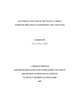
Figure 3. Comparison of wild type (WT) and mutants (2E3, 2EE3, 2EED3 and 2EEDE3)
haemolytic activities. A total of 4 amino acids (Glutamic acid-E, Glutamic acid-E, Aspartic
acid-D and Glutamic acid-E) were inserted in succession at the N-terminus. Mutants
generated were of significantly lower haemolytic activity as compared to the wild type.
Toxins 2015, 7
412
80
70
Haemolysis (%)
60
WT
50
Y110A
40
K76A
W113A
30
Y129A
20
10
0
0
5
10
15
20
Protein concentra on (μg/mL)
25
30
35
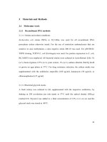
Figure 4. Comparison of wild type (WT) and mutants (Y110A, K76A, W113A and Y129A)
haemolytic activities. Three aromatic amino acids and one lysine were substituted
individually into alanine (A) and all mutants exhibited a significantly lower haemolytic
activity as compared to wild type.
2.3. Cytolytic Activity of HALT-1 and Its Mutants
The MTT assay was used to assess the cytolytic activity of recombinant HALT-1 and its mutants.
HeLa cells were incubated with recombinant HALT-1 at various concentrations (2, 5, 10, 15, 20,
25 μg/mL) for 24 h and the concentration of viable cells was measured by recording the changes in
absorbance. As shown in Figure 5, recombinant HALT-1 reduced the viability of HeLa cells in a
dose-dependent manner and the cells were completely killed by HALT-1 at a concentration of
20 μg/mL after 24 h of incubation. The IC50 value, which is defined as the concentration of recombinant
HALT-1 at which 50% of cells die, was found to be approximately 15 μg/mL. By comparison, the IC50
of EqtII on lung fibroblasts was reported to be 17 ng/mL after one hour of incubation [13]. The IC50
value for EqtII tested on two tumor cell lines, Ehrlich ascites carcinoma and leukemia cells was also
very low [14]. Thus, while HALT-1 is clearly cytotoxic, our results showed that it is less potent than
equinatoxin EqtII.
Figure 5 shows the cytolytic activity of HALT-1 mutants with a lengthened N-terminal α-helix (2E3,
2EE3, 2EED3 and 2EEDE3). The mutant proteins showed essentially no cytolytic activity (Figure 5).
The viability of HeLa cells was maintained at more than 80% when treated with mutant HALT-1
proteins. The IC50 of the wild type HALT-1 was 15 μg/mL under the same conditions. The mutants
K76A, Y110A, W113A and Y129A, which disrupt the conserved aromatic cluster involved in membrane
binding, also showed nearly no cytolytic activity at all concentrations tested (Figure 6).
Toxins 2015, 7
413
120
Cell Viability (%)
100
80
WT
2E3
60
2EE3
2EED3
40
2EEDE3
20
0
0
5
10
15
20
Protein concentra on (μg/mL)
25
30
Figure 5. Cytolytic activity of the wild type (WT) and mutants (2E3, 2EE3, 2EED3 and
2EEDE3). A total of 4 amino acids (Glutamic acid-E, Glutamic acid-E, Aspartic acid-D and
Glutamic acid-E) were inserted in succession at the N-terminus. Mutants generated displayed
significantly lower cytotoxic activity compared to the wild type.
120
Cell Viability (%)
100
80
WT
Y110A
60
K76A
W113A
40
Y129A
20
0
0
5
10
15
20
Protein concentra on (μg/mL)
25
30
Figure 6. Cytotoxic activity of the wild type (WT) and mutants (Y110A, K76A, W113A
and Y129A). Three aromatic amino acids and one lysine were substituted into alanine (A)
and all the mutants generated exhibited a significantly lower cytotoxic activity as compared
to the wild type.
Toxins 2015, 7
414
3. Discussion
The first 20 residues of HALT-1 protein constitute a signal peptide. Unlike other actinoporins such
as equinatoxins, the HALT-1 signal peptide is not followed by a propeptide and thus its active form is
likely not regulated by the endoproteolytic cleavage. Directly following the HALT-1 signal peptide is
the N-terminal region, which comprises about 30 amino acids. In this region, amino acids 14 to 25 are
predicted to form an α-helix (Figure 1). As all actinoporins are structurally and functionally conserved,
we assume that the N-terminal α-helix of HALT-1 exhibits an amphipathic nature and can be translocated
into the plasma membrane pore [12]. In EqtII, the corresponding α-helix encompasses residues 10 to 28
(Figure 1) and contains both negatively charged aspartic acid (residues 10 and 17) and glutamic acid
(residue 24) that greatly facilitates the transfer of its flexible N-terminus into the cation selective
pores [7]. Evidence has shown that an additional negative charge at the N-terminus of EqtII by sulfhydryl
modification improved the cationic selectivity and thus increased the conductance of the pore [15].
Nevertheless, our results show that extending the N-terminus of HALT-1 with 1–4 negatively charged
residues reduced haemolytic and cytolytic activity compared to the wild type (Figures 3 and 5).
This could be due to the increased length of the N-terminus or the increased negative charge.
Supporting the first idea are the fact that all actinoporins have approximately 30 amino acids forming
an amphipathic helix at the N-terminus and the fact that removal of five or ten residues from the
N-terminus of EqtII reduced haemolytic activity and presumably pore formation [16]. Similarly, the
addition of 1–4 amino acid(s) at the N-terminus of HALT-1 alters the length of the N-terminal
α-helix and might inhibit pore formation and diminish haemolytic and cytolytic activity. Alternatively,
changes in length of the N-terminus may disrupt the ability of the N-terminal α-helix to dissociate from
the β-sheet core of the actinoporin structure during pore formation. The requirement for structural
flexibility of the N-terminal α-helix was clearly demonstrated in a double cysteine mutant which
introduced a disulfide bond and locked the α-helix to the core β-sheet and prevented pore formation [6,9].
Apart from the size constraint, the amphipathic nature in the N-terminal region is another important
determinant for successful integration of this region into the plasma membrane. An electrophysiological
study on the planar lipid bilayer has suggested that when the N-terminal region of actinoporins is
translocated into the interfacial surface of plasma membrane, it is essential to have the negatively
charged amino acids facing the pore lumen, while the polar amino acids are positioned toward the
hydrophobic edge [7]. The N-terminal region of HALT-1 also contains negatively charged aspartic acids
at position 15, 22 and 23, although they are not located at the positions where the negatively charged
amino acids are found in other actinoporins. Based on our findings, the addition of one negatively
charged residue (E) reduced 80% of haemolytic activity while two or more negatively charged residues
(ED or EDE or EEDE) led to further reduction in activity. This indicates that excess negative charges at
the end of the N-terminus of HALT-1 were unfavorable for haemolytic and cytolytic activity.
Interestingly, although both sticholysins StnI and StnII have 93% sequence identity [17], the N-terminal
region of sticholysin StnI has two additional D and E residues compared to sticholysin StnII and exhibits
lower haemolytic activity [18–20]. Thus, similar to other actinoporins, HALT-1 requires the N-terminus,
including the amphipathic α-helix, to efficiently make pores in lipid membranes. However, we do not
rule out the possibility that extending the N-terminus of HALT-1 resulted in a change of the native
structure and thus the loss of protein function.
Toxins 2015, 7
415
Strands β6, β7 and helix α2 of actinoporins contain a conserved cluster of aromatic amino acids and
a POC (phosphocholine) binding site which are required for specific recognition of the phosphocholine
head group of spingomyelins in the cell membrane [21,22]. The aromatic amino acids, Tyr113, Trp116
and Tyr137 in EqtII and Tyr111, Trp114 and Tyr135 in StnII (Figure 1) have been identified as critical
for this binding [22,23]. In HALT-1, the corresponding amino acids are Tyr110 (in strand β8), Trp113
(in strand β8) and Tyr129 (in helix α2) (Figure 1) and they are expected to form a similar aromatic patch
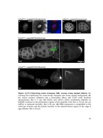
on the surface of the molecule as shown in the model in Figure 7. In fact, these three aromatic amino
acids are conserved in all actinoporin family members [24]. Substitution of these aromatic amino acids
by alanine in HALT-1 mutants Y110A, W113A and Y129A led to an almost complete loss of haemolytic
and cytolytic activity (Figures 4 and 6). The loss of activity observed in these three mutants may not be
due to structural instability, since mutations at the surface of a protein are usually not deleterious as
compared to mutations in the core, and both protein stability prediction tools indicated that the mutations
are either neutral or slightly destabilizing. Similar results have been obtained with EqtII. Replacement
of Trp116 with phenylalanine reduced the mutant membrane-binding significantly, leading to the loss
of haemolytic activity [22]. Replacement of Trp116 with alanine also reduced membrane binding and
haemolytic activity [6]. The importance of these aromatic amino acids became more evident when the
cocrystal structure of StnII and POC at 2.4 Å resolution revealed that POC bound to a cavity in which
its choline moiety interacts with the electron-rich aromatic ring of Tyr111 and Tyr135 [5]. A similar
POC-binding cavity is also predicted in HALT-1 whereby the aromatic rings from Tyr110, Trp113 and
Tyr129 project into the cavity (Figure 7).
A
B
Figure 7. Cont.
Toxins 2015, 7
416
C
Figure 7. Aromatic amino acid residues of HALT-1, EqtII and StnII. All residues are shown
as sticks in different colours. (A) Tyr110 (skyblue), Trp113 (magenta) and Tyr129 (yellow)
from HALT-1; (B) Tyr113, (skyblue), Trp116 (magenta) and Tyr137 (yellow) from EqtII;
(C) Tyr111 (skyblue), Trp114 (magenta) and Trp135 (yellow) from StnII. All these tyrosine
(Tyr) and tryptophan (Trp) are well conserved between HALT-1, EqtII and StnII and they
are predicted to form an aromatic patch on surface of the molecule which could be essential
for the insertion into water-lipid interface.
Finally, the K76A mutant also resulted in a decrease of haemolytic and cytolytic activity
(Figures 4 and 6). K76 is positioned at the end of strand β5 (Figure 1) and is a conserved residue also
found in EqtII (Lys77) and StnII (Lys75). Our result is in good agreement with the results of
Anderluh et al. [25] showing that lysine 77 of EqtII (Figure 1), when substituted with cysteine, led to
the decrease of haemolytic activity although the structure remained intact. Furthermore, the authors
claimed that the decrease of haemolytic activity was due to the loss of positive charge and thus the
inability of the K77C mutant to oligomerize on lipid membranes. Consistent with this interpretation, the
haemolytic activity was almost completely restored when a positive charge was reintroduced at position
77. Further supporting data were derived from the 2D crystallization of StnII with lipid monolayer [5].
In this high resolution model, it clearly showed that K75 (which lies in the loop between β5 and β6) of
StnII was in close proximity to lipidic interface, stabilizing the conformation of the toroidal pore. Hence,
we can hypothesize that lysine 76 from HALT-1 is also involved in interaction with membrane lipids
and in oligomerization and that mutation to an uncharged alanine impairs this interaction.
4. Experimental Section
4.1. Isolation of HALT-1 cDNA
Total RNA was extracted from approximately 100 mg tissue of Hydra magnipapillata strain
105 using TRIzol® Reagent (Invitrogen-Life Technologies, Carlsbad, CA, USA). First strand cDNA was
then synthesized according to the manufacturer’s instructions using the SuperScript™ III First-Strand
Synthesis System (Invitrogen-Life Technologies, Carlsbad, CA, USA). Subsequently, double-stranded
cDNA was generated by the initial PCR with a set of forward and reverse primers
(5'TTCACTCACGTTGATTTATACCTT3' and 5'TTGCTCCACTCTTCTATTAGCTC3', respectively)
Toxins 2015, 7
417
and followed by the nested PCR using another set of forward and reverse primers
(5'GGGTAGGACAACTGGAATAGT3' and 5'GCTGACTGCTTGGTGAATAC3', respectively).
An amplified cDNA fragment from HALT-1 was cloned into pCR4-TOPO vector and then transformed
into One Shot® TOP10 E. coli (Invitrogen-Life Technologies, USA). Verification of the HALT-1 insert
was done through sequencing.
4.2. Construction of Expression Vector of HALT-1
To heterologously express HALT-1, its coding region was directionally sub-cloned into an expression
vector pET28a (Novagen, EMD biosciences, San Diego, CA, USA) and transformed into BL21(DE3)
(New England BioLabs Inc., Ipswich, MA, USA). Briefly, HALT-1 cDNA with SalI and NotI
flanking at both ends was constructed with HALT-1-pCR4-TOPO and two primers
(5'GCAGTCGACGAGCAGCTTTAGGAGTTATA3' and 5'CTTGCGGCCGCCTTTCGAATATTC
TTATCC3') containing SalI and NotI sites (underlined nucleotides) at the 3' and 5' ends, respectively.
This was followed by insertion into pET28a using the LigaFast™ Rapid DNA Ligation System
(Promega, Madison, WI, USA). The correct reading frame of the inserted HALT-1 was confirmed by
sequencing before transforming it into BL21(DE3).
4.3. Synthesis of HALT-1 Mutants
Site-directed mutagenesis was performed on pET28a and mutations were introduced according to the
manufacturer’s instructions (Agilent, Santa Clara, CA, USA). In brief, PCR was carried out with the
mutagenic forward and reverse primers using Pfu DNA polymerase. PCR was started at 95 °C for 30 s
and repeated for 16 cycles at 95 °C for 30 s, 60 °C for 1 min and 68 °C for 5 min. Newly synthesized
mutant strands were cooled on ice for 2 min and then subjected to Dpn1 digestion to remove the parental
strand. The new strand containing the mutation was then transformed into XL1-Blue competent cells.
Colonies appeared on the agar plate should contain the mutant clone. The DNA plasmid of the mutant
clone was purified using the Wizard Plus SV Minipreps DNA purification system (Promega, Madison,
WI, USA) and sent for sequencing before the transformation into BL21(DE3).
4.4. Protein Expression and Affinity Purification
His-tag fusion HALT-1 and its mutants were expressed in BL21(DE3) in the presence of IPTG
(1 mM) for 3 h at 37 °C with agitation. Each induced culture was then resuspended in the lysis buffer
(50 mM NaH2PO4; 0.5 M NaCl; 10 mM Imidazole; pH 8.0) with 1× Halt Protease Inhibitor Cocktail
prior to sonication at 130 watt and 20 kHz for a total of 120 s per sample on ice. The soluble fraction
of the crude cell lysate was subjected to the affinity chromatography of nickel-chelating resin,
Ni-NTA superflow (Qiagen, Hilden, Germany). The purified His-tag fusion protein was concentrated
and exchanged with a 1× phosphate buffered saline (PBS) buffer using an Amicon Ultracel Column
(Millipore, Billerica, MA, USA).
Toxins 2015, 7
418
4.5. Haemolysis Assay
Human erythrocytes were used to assess the haemolytic activity of recombinant HALT-1. Firstly,
2 mL of blood collected from humans were washed three times with sterile 0.85% NaCl saline solution.
Erythrocytes were pelleted at 1000 g for 5 min at room temperature and supernatant was then
carefully discarded upon each wash. The erythrocyte pellet was resuspended with 0.85% NaCl saline
solution to obtain 1% erythrocytes. Serial concentrations of either HALT-1 or its mutant proteins
(5, 10, 15, 20, 25 and 30 µg/mL) were prepared in a final volume of 200 µL and then added to 1 mL
of human erythrocytes. PBS buffer instead of protein sample was used as a negative control.
The protein-erythrocytes mixtures were incubated at 37 °C for 2 h with gentle rocking at 50 rpm and
then centrifuged at 1000 g for 5 min at room temperature. The supernatant was measured at OD 540 nm
to determine the release of haemoglobin due to the lysis of erythrocytes.
4.6. Cytolysis Assay
Cytolytic activity of recombinant HALT-1 and its mutant was evaluated using the HeLa cancer cell
line (ATCC CCL2) from RIKEN, Japan. Cells were seeded at 1 × 10⁴ cell/well in 96-well microtiter
plate and incubated at 37 °C, 5% CO2 for 24 h. Subsequently, serial concentrations (2, 5, 10, 15, 20,
25 μg/mL) of the recombinant HALT1 protein were added into respective wells. Cells were treated with
the recombinant protein at 37 °C for 16 h. Cells incubated with the PBS buffer served as a negative
control. MTT (3-(4,5-dimethylthiazol-2-yl)-2,5-diphenyltetrazolium bromide) solution (Sigma-Aldrich,
St. Louis, MO, USA) of 50 μL (5 mg/mL) was then added into the wells and the cells were incubated
for another 4 h. The purple formazan formed was dissolved with dimethyl sulfoxide (DMSO) and the
absorbance readings were measured at 570 nm with a reference wavelength at 630 nm. All assays were
done in triplicate.
4.7. Protein Sequence Alignment
The amino acid sequences of Equinatoxin II (Actinia equina), Sticholysin II
(Stichodactyla helianthus) and Bandaporin (Anthopleura asiatica) were retrieved from UniProt
( with the accession numbers of P61914, P07845 and C5NSL2, respectively.
Multiple sequence alignment of mature proteins including HALT-1, Equinatoxin II, Sticholysin II and
Bandoporin were conducted using Clustal Omega software, which is available from the European
Bioinformatics Institute ( The secondary structure prediction
of HALT-1 is performed by the submission of amino acid sequence to the web server of PSIPRED
( />4.8. Homology Medeling of HALT-1
The three-dimensional structure of HALT-1 was built through homology-modeling using the
SWISS-MODEL workspace in the automated mode [26,27]. The 3D structures of Equinatoxin II
(PDB ID: 1iaz) and Sticholysin II (PDB ID: 1gwy) were also obtained from Protein Data Bank in
Europe ( The image of the protein models was viewed using PyMOL v1.6
(Version 1.6, Schrödinger, LLC, New York, NY, USA, 2013) ( />
Toxins 2015, 7
419
4.9. Prediction of Mutant Stability
Two prediction methods, SDM (Site-Directed Mutator) and I-Mutant 3.0, were used to measure the
protein stability change upon point mutation in K76A, Y110A, W113A, A114W and Y129A [28,29].
Based on a set of a set of conformationally constrained environment-specific substitution tables (ESSTs),
SDM utilized the PDB structure of HALT-1 (generated by SWISS model) to calculate the difference in
the stability scores for the folded and unfolded state for the wild-type and mutant protein structures, i.e.,
∆∆G. The mutant can be grouped into seven classes: highly destabilizing (<−2.0), destabilizing
(−2.0≤ & <−1.0), slightly destabilizing (−1.0≤ & <−0.5), neutral (−0.5≤ & ≤0.5), slightly stabilizing
(0.5< & ≤1.0), stabilizing (1.0< & ≤2.0) and highly stabilizing (>2.0). We used the sequence-based
version of I-Mutant 3.0 to calculate ∆∆G and classified the mutant into neutral (−0.5≤ & ≤0.5),
large decrease (<−0.5) and large increase (>0.5).
5. Conclusions
The molecular characterization of HALT-1 is currently in its infancy, and to date, only one report
on HALT-1 haemolytic properties and sphigomyelin specificity has been published [12]. Of the
actinoporin family members that have been identified so far, all are isolated from sea anemones
(class Anthozoa). Having discovered homologous actinoporins in Hydra, which belong to another class,
Hydrozoa, it is of interest to determine the evolutionary constraint of HALT-1. In Glasser et al. [12], HALT-1
has been proven to be the member of actinoporin family producing the most important findings, such as
exerting haemolytic activity, targeting human cell membranes, and forming a larger pore size than EqtII
on the cell membrane. In the present study of HALT-1, we identified those amino acid residues that are
likely to play a critical role in pore formation and found that they are, in fact, the conserved residues
found in almost all actinoporins. Moreover, our study proposes that, similar to other actinoporins, the
length of the N-terminus of HALT-1 is constrained to approximately 30 residues. Addition of a single
residue in this region destroyed the function of the protein. In addition, HALT-1 likely uses the same
aromatic amino acids as other actinoporins for binding to the plasma membrane during pore formation.
While there is a growing list of pore-forming toxins, our findings contribute new information
about human cell recognition by actinoporins and the formation of ring-shaped tetramer via the insertion
of the N-terminal α-helix. Such knowledge is crucial for the potential application of actinoporins in
biotherapeutics [30,31]. One excellent example is immunotoxin therapy that serves as a potential
anticancer agent [32]. The immunotoxin is produced by fusing a target cell-directed antibody and a
membrane-acting toxin. In order to minimize nonspecific binding of immunotoxin to normal cells, amino
acids in the binding domain of the toxin need to be either modified or eliminated. Thus, by collecting
knowledge about functional roles of amino acids in HALT-1 and their structural properties via the
mutational studies, one can delete the sphingomyelin binding site from HALT-1 and enable the toxin, as
a part of immunotoxin, to eventually recognize the specific target cells through the antibody moiety
rather than normal cells.
Supplementary Materials
Supplementary materials can be accessed at: />
Toxins 2015, 7
420
Acknowledgments
We would like to thank Charles N. David (Ludwig Maximilian University of Munich, Germany) for
his critical reading of the manuscript. This work is supported by the Fundamental Research Grant
Scheme (Grant No. FRGS/1/2013/ST03/UCSI/02/1), and the Ministry of Higher Education Malaysia and
Research Grant Scheme (Grant No. Proj-In-FAS-020), UCSI University.
Author Contributions
Yvonne Jing Mei Liew constructed HALT-1 mutants and carried out the haemolytic and cytolytic
assays of these mutants. Wai Tuck Soh constructed the recombinant HALT-1. William Febry Jiemy
isolated HALT-1 from H. magnipapillata. Jung Shan Hwang prepared the manuscript.
Conflicts of Interest
The authors declare no conflict of interest.
References
1.
2.
3.
4.
5.
6.
7.
8.
Anderluh, G.; Pingercar, J.; Strukelj, B.; Macek, P.; Gubensek, F. Cloning, sequencing, and
expression of equinatoxin II. Biochem. Biophys. Res. Commun. 1996, 18, 437–442.
Bernheimer, A.W.; Avigad, L.S. Properties of a toxin from the sea anemone Stoichacis helianthus,
including specific binding to sphingomyelin. Proc. Natl. Acad. Sci. USA 1976, 73, 467–471.
Athanasiadis, A.; Anderluh, G.; Macek, P.; Turk, D. Crystal structure of the soluble form of
equinatoxin II, a pore-forming toxin from the sea anemone Actinia equina. Structure 2001, 9,
341–346.
Hinds, M.G.; Zhang, W.; Anderluh, G.; Hansen, P.E.; Norton, R.S. Solution structure of the
eukaryotic pore-forming cytolysin equinatoxin II: Implications for pore formation. J. Mol. Biol.
2002, 315, 1219–1229.
Mancho, J.M.; Martín-Benito, J.; Martínez-Ripoll, M.; Gavilanes, J.G.; Hermoso, J.A.
Crystal and electron microscopy structures of sticholysin II actinoporin reveal insights into the
mechanism of membrane pore formation. Structure 2003, 11, 1319–1328.
Hong, Q.; Gutierrez-Aguirre, I.; Barlic, A.; Malovrh, P.; Kristan, K.; Podlesek, Z.; Macek, P.; Turk, D.;
Gonzalez-Manas, J.M.; Lakey, J.H.; et al. Two-step membrane binding by Equinatoxin II, a
pore-forming toxin from the sea anemone, involves an exposed aromatic cluster and a flexible helix.
J. Biol. Chem. 2002, 277, 41916–41924.
Malovrh, P.; Viero, G.; Serra, M.D.; Podlesek, Z.; Lakey, J.H.; Macek, P.; Menestrina, G.;
Anderluh, G. A novel mechanism of pore formation: Membrane penetration by the N-terminal
amphipathic region of equinatoxin. J. Biol. Chem. 2003, 278, 22678–22685.
Gutiérrez-Aguirre, I.; Barlic, A.; Podlesek, Z.; Macek, P.; Anderluh, G.; González-Mañas, J.M.
Membrane insertion of the N-terminal α-helix of equinatoxin II, a sea anemone cytolytic toxin.
Biochem. J. 2004, 384, 421–428.
Toxins 2015, 7
9.
10.
11.
12.
13.
14.
15.
16.
17.
18.
19.
20.
21.
421
Kristan, K.; Podlesek, Z.; Hojnik, V.; Gutiérrez-Aguirre, I.; Guncar, G.; Turk, D.;
González-Mañas, J.M.; Lakey, J.H.; Macek, P.; Anderluh, G.; et al. Pore formation by equinatoxin, a
eukaryotic pore-forming toxin, requires a flexible N-terminal region and a stable β-sandwich.
J. Biol. Chem. 2004, 279, 46509–46517.
Belmonte, G.; Pederzolli, C.; Macek, P.; Menestrina, G. Pore formation by the sea anemone
cytolysin equinatoxin II in red blood cells and model lipid membranes. J. Membr. Biol. 1993, 131,
11–22.
Hwang, J.S.; Ohyanagi, H.; Hayakawa, S.; Osato, N.; Nishimiya-Fujisawa, C.; Ikeo, K.;
David, C.N.; Fujisawa, T.; Gojobori, T. The evolutionary emergence of cell type-specific genes
inferred from the gene expression analysis of Hydra. Proc. Natl. Acad. Sci. USA 2007, 104,
14735–14740.
Glasser, E.; Rachamim, T.; Aharonovich, D.; Sher, D. Hydra actinoporin-like toxin-1, an unusual
hemolysin from the nematocyst venom of Hydra magnipapillata which belongs to an extended gene
family. Toxicon 2014, 91, 103–113.
Batista, U.; Macek, P.; Sedmak, B. The cytotoxic and cytolytic activity of equinatoxin II from the
sea anemone Actinia equina. Cell Biol. Int. Rep. 1990, 14, 1013–1024.
Giraldi, T.; Ferlan, I.; Romeo, D. Antitumor activity of equinatoxin. Chem. Biol. Interact. 1976, 13,
199–203.
Kristan, K.; Viero, G.; Macek, P.; Serra, M.D.; Anderluh, G. The equinatoxin N-terminus is
transferred across planar lipid membranes and helps to stabilize the transmembrane pore. FEBS J.
2007, 274, 539–550.
Anderluh, G.; Pungercar, J.; Krizaj, I.; Strukelj, B.; Gubensek, F.; Macek, P. N-Terminal truncation
mutagenesis of equinatoxin II, a pore-forming protein from the sea anemone Actinia equina.
Protein Eng. 1997, 10, 751–755.
Martinez, D.; Campos, A.M.; Pazos, F.; Alvarez, C.; Lanio, M.E.; Casallanovo, F.; Schreier, S.;
Salinas, R.K.; Vergara, C.; Lissi, E.; et al. Properties of St I and St II, two isotoxins isolated from
Stichodactyla helianthus: A comparison. Toxicon 2001, 39, 1547–1560.
García-Linares, S.; Castrillo, I.; Bruix, M.; Menéndez, M.; Alegre-Cebollada, J.;
Martínez-del-Pozo, Á.; Gavilanes, J.G. Three-dimensional structure of the actinoporin
sticholysin I. Influence of long-distance effects on protein function. Arch. Biochem. Biophys. 2013,
532, 39–45.
Pazos, F.; Valle, A.; Martínez, D.; Ramírez, A.; Calderón, L.; Pupo, A.; Tejuca, M.; Morera, V.;
Campos, J.; Fando, R.; et al. Structural and functional characterization of a recombinant sticholysin I
(rSt I) from the sea anemone Stichodactyla helianthus. Toxicon 2006, 48, 1083–1094.
Cilli, E.M.; Pigossi, F.T.; Crusca, E., Jr.; Ros, U.; Martínez, D.; Lanio, M.E.; Álvarez, C.;
Schreier, S. Correlations between differences in amino-terminal sequences and different haemolytic
activity of sticholysins. Toxicon 2007, 50, 1201–1204.
Anderluh, G.; Barlic, A.; Podlesek, Z.; Macek, P.; Pungercar, J.; Gubensek, F.; Zecchini, M.L.;
Serra, M.D.; Menestrina, G. Cysteine-scanning mutagenesis of an eukaryotic pore-forming toxin
from sea anemone: Topology in lipid membranes. Eur. J. Biochem. 1999, 263, 128–136.
Toxins 2015, 7
422
22. Malovrh, P.; Barlic, A.; Podlesek, Z.; MaCek, P.; Menestrina, G.; Anderluh, G. Structure-function
studies of tryptophan mutants of equinatoxin II, a sea anemone pore-forming protein. Biochem. J.
2000, 346, 223–232.
23. Álvarez, C.; Mancho, J.M.; Martínez, D.; Tejuca, M.; Pazos, F.; Lanio, M.E. Sticholysins,
two pore-forming toxins produced by the Caribbean Sea anemone Stichodactyla helianthus:
Their interaction with membranes. Toxicon 2009, 54, 1135–1147.
24. Bakrac, B.; Anderluh, G. Molecular mechanism of sphingomyelin-specific membrane binding and
pore formation by actinoporins. Adv. Exp. Med. Biol. 2010, 677, 106–115.
25. Anderluh, G.; Barlic, A.; Potrich, C.; Macek, P.; Menestrina, G. Lysine 77 is a key residue in
aggregation of equinatoxin II, a pore-forming toxin from sea anemone Actinia equina.
J. Membr. Biol. 2000, 173, 47–55.
26. Arnold, K.; Bordoli, L.; Kopp, J.; Schwede, T. The SWISS-MODEL Workspace: A web-based
environment for protein structure homology modelling. Bioinformatics 2006, 22, 195–201.
27. Biasini, M.; Bienert, S.; Waterhouse, A.; Arnold, K.; Studer, G.; Schmidt, T.; Kiefer, F.;
Cassarino, T.G.; Bertoni, M.; Bordoli, L.; et al. SWISS-MODEL: Modelling protein tertiary and
quaternary structure using evolutionary information. Nucleic Acids Res. 2014, 42, W252–W258.
28. Worth, C.L.; Preissner, R.; Blundell, T.L. SDM—A server for predicting effects of mutations on
protein stability and malfunction. Nucleic Acids Res. 2011, 39, W215–W222.
29. Capriotti, E.; Fariselli, P.; Casadio, R. I-Mutant2.0: Predicting stability changes upon mutation from
the protein sequence or structure. Nucleic Acids Res. 2005, 33, W306–W310.
30. Shapira, A.; Benhar, I. Toxin-based therapeutic approaches. Toxins 2010, 2, 2519–2583.
31. Tejuca, M.; Anderluh, G.; Serra, M.D. Sea anemone cytolysins as toxic components of immunotoxins.
Toxicon 2009, 54, 1206–1214.
32. Choudhary, S.; Mathew, M.; Verma, R.S. Therapeutic potential of anticancer immunotoxins.
Drug Dis. Today 2011, 16, 495–503.
© 2015 by the authors; licensee MDPI, Basel, Switzerland. This article is an open access article
distributed under the terms and conditions of the Creative Commons Attribution license
( />
Copyright of Toxins is the property of MDPI Publishing and its content may not be copied or
emailed to multiple sites or posted to a listserv without the copyright holder's express written
permission. However, users may print, download, or email articles for individual use.