Báo cáo khoa học: A biophysical view of the interplay between mechanical forces and signaling pathways during transendothelial cell migration doc
Bạn đang xem bản rút gọn của tài liệu. Xem và tải ngay bản đầy đủ của tài liệu tại đây (384.79 KB, 14 trang )
REVIEW ARTICLE
A biophysical view of the interplay between mechanical
forces and signaling pathways during transendothelial cell
migration
Kimberly M. Stroka and Helim Aranda-Espinoza
Fischell Department of Bioengineering, University of Maryland, College Park, MD, USA
Introduction
In order for immune cells to travel from the blood-
stream to the tissues outside the blood vessel, it is nec-
essary for them to transmigrate through the layer of
endothelium lining the inside of the blood vessel. Leu-
kocyte transmigration plays a pivotal role both in the
normal immune response and in the development of
cardiovascular disease, including atherosclerosis and
stroke. Thus, inflammation is a normal response to
foreign pathogens, but it may also lead to cardiovascu-
lar disease under certain conditions. For example, ath-
erosclerosis is initiated in the presence of increased
levels of low-density lipoproteins, which become oxi-
dized by free radicals, come into contact with the arte-
rial wall, and damage the endothelium. Leukocytes
Keywords
cell mechanics; diapedesis; endothelial cell;
leukocyte; mechanotransduction;
mechanotransmission; substrate stiffness;
transmigration
Correspondence
K. M. Stroka, Fischell Department of
Engineering, Room 3142, Jeong H. Kim
Engineering Building (#225), University of
Maryland, College Park, MD 20742, USA
Fax: +301 314 6868
Tel: +301 405 8781
E-mail:
(Received 28 September 2009, revised
20 November 2009, accepted 11 December
2009)
doi:10.1111/j.1742-4658.2009.07545.x
The vascular endothelium is exposed to an array of physical forces, includ-
ing shear stress via blood flow, contact with other cells such as neighboring
endothelial cells and leukocytes, and contact with the basement membrane.
Endothelial cell morphology, protein expression, stiffness and cytoskeletal
arrangement are all influenced by these mechanochemical forces. There are
many biophysical tools that are useful in studying how forces are transmit-
ted in endothelial cells, and these tools are also beginning to be used to
investigate biophysical aspects of leukocyte transmigration, which is a ubiq-
uitous mechanosensitive process. In particular, the stiffness of the substrate
has been shown to have a significant impact on cellular behavior, and this
is true for both endothelial cells and leukocytes. Thus, the stiffness of the
basement membrane as an endothelial substrate, as well as the stiffness of
the endothelium as a leukocyte substrate, is relevant to the process of
transmigration. In this review, we discuss recent work that has related the
biophysical aspects of endothelial cell interactions and leukocyte trans-
migration to the biochemical pathways and molecular interactions that take
place during this process. Further use of biophysical tools to investigate
the biological process of leukocyte transmigration will have implications
for tissue engineering, as well as atherosclerosis, stroke and immune system
disease research.
Abbreviations
AFM, atomic force microscopy; BAEC, bovine aortic endothelial cell; BBB, blood–brain barrier; BPMEC, bovine pulmonary microvascular
endothelial cell; EC, endothelial cell; FA, focal adhesion; HUVEC, human umbilical vein endothelial cell; ICAM-1, intercellular adhesion
molecule-1; JAM, junction adhesion molecule; LFA-1, lymphocyte function-associated antigen-1; NF-jB, nuclear factor-jB; ox-LDL, oxidized
low-density lipoprotein; PECAM-1, platelet endothelial cell adhesion molecule-1; TNF, tumor necrosis factor; VCAM-1, vascular cell adhesion
molecule-1; VE-cadherin, vascular endothelial-cadherin.
FEBS Journal 277 (2010) 1145–1158 ª 2010 The Authors Journal compilation ª 2010 FEBS 1145
recruited by the immune system to the damaged vessel
wall cannot process the oxidized low-density lipopro-
teins (ox-LDLs); thus, they rupture and deposit more
ox-LDL onto the vessel wall, leading to recruitment of
more leukocytes and beginning a cycle that eventually
leads to a pathological state. There are also numerous
diseases of the immune system, such as asthma, rheu-
matoid arthritis, and psoriasis, which develop because
of increased frequency of leukocyte transmigration.
Cell transmigration is also involved in processes such
as cancer cell metastasis and stem cell homing, and
although the steps of cancer cell transmigration are
similar to those for immune cells, the molecular
players involved are different [1]. Furthermore, blood–
brain barrier (BBB) dysfunction is involved in
pathological conditions, including multiple sclerosis
and other neuroinflammatory processes or brain cancer
[2,3]. Interestingly, transmigration of immune cells
across the BBB into the central nervous system is
highly regulated, and occurs to a limited extent in a
process called ‘immune surveillance’ [4,5]. However, in
BBB dysfunction, there is an increase in the number of
immune cells, or even cancer cells, that cross the tight
junctions of the BBB.
As leukocytes make their way through the endothe-
lium, forces are exerted on the leukocytes, endothelial
cells (ECs), and basement membrane below the ECs.
At the same time, the cells respond to various
mechanical forces around them, including shear stress
due to blood flow and effects from other neighboring
cells and matrix. The biophysical aspects of the endo-
thelium through which the leukocytes transmigrate, in
addition to the biophysical aspects of the leukocytes
themselves, are linked to the biochemical pathways
that govern transmigration. However, we are only
beginning to understand how physical forces translate
into biochemical signaling pathways during leukocyte
transmigration. In this review, we highlight recent
work that has related the biophysical aspects of leuko-
cyte transmigration to the biochemical pathways and
molecular interactions that take place during this pro-
cess. We discuss the assortment of physical forces
(including estimates of their magnitude) acting on ECs
from all sides. These include shear stress and adherent
or migrating leukocytes at the luminal surface, neigh-
boring ECs or transmigrating leukocytes at cell–cell
junctions, leukocytes transmigrating throughout the
body of the cell, and the substrate at the basal surface
of the ECs. Interestingly, forces acting at one surface
may be propagated internally or even to other sur-
faces of the cell, or they may initiate biochemical sig-
naling cascades within the cell, leading to a cellular
response.
ECs respond to shear stress
A single sheet of ECs lines the walls of the arteries and
is responsible for transmitting shear stress due to
blood flow to the underlying layers of tissue. These
underlying layers include the basement membrane
(composed mainly of laminin and collagen), the media
(composed of smooth muscle cells, collagen, and elas-
tin), and the adventitia (the stiffer outermost layer).
Shear stress on ECs leads to mechanotransduction (the
conversion of physical forces into biochemical signals)
and mechanotransmission (the physical propagation of
forces to the underlying layers). In large arteries, mean
shear stress along the wall is in the range of 20–
40 dynesÆcm
)2
, and is generally pulsatile rather than
unidirectional [6]. However, most in vitro studies in
which shear stress is applied to cells use values ranging
from 0 to 100 dynesÆcm
)2
, usually in unidirectional
flow [6]. Shear stress affects EC cytoskeletal arrange-
ment [7–9], cell morphology [8,10–12], and gene
expression [13–15]. Although the method of EC
mechanotransduction is still largely unknown, several
molecular structures are believed to play roles in the
mechanosensing process of converting shear stress into
morphological changes and gene expression; these mol-
ecules include the glycocalyx, platelet EC adhesion
molecule-1 (PECAM-1), stretch-activated ion channels,
receptor tyrosine kinases, vascular endothelial-cadherin
(VE-cadherin), and vascular endothelial growth factor
receptor. Figure 1 indicates the assortment of biophysi-
cal forces that ECs feel, and possible signaling mole-
cules that could act as mechanotransducers in the cell.
ECs develop more stress fibers and less peripheral
actin as larger shear stresses are applied [8]. F-actin
stress fibers contract between cellular focal adhesions
(FAs), adhesion structures that exert traction stresses
on the underlying substrate (Fig. 1). It has been shown
that there is a 2 pN bond between an integrin and a
fibronectin molecule, and the maintenance of this bond
requires talin, which binds the integrin to an actin fila-
ment [16]. Stretching talin activates vinculin, a FA pro-
tein, leading to reinforcement of the FA [17] (Fig. 1).
Therefore, a rearrangement of the F-actin cytoskeleton
under shear stress would be expected to also influence
FAs and cellular traction forces. Indeed, FAs realign
parallel to flow [18], and shear stress increases RhoGT-
Pase activation in single cells, leading to larger traction
forces [19]. In addition, the vimentin intermediate fila-
ment permeates the actin network, and has been
shown to propagate shear stress [20,21].
Bovine aortic ECs (BAECs) migrate faster under
shear stress than under static conditions, and this is
mediated by Rho, as inhibition of the Rho-associated
Biophysical view of transendothelial migration K. M. Stroka and H. Aranda-Espinoza
1146 FEBS Journal 277 (2010) 1145–1158 ª 2010 The Authors Journal compilation ª 2010 FEBS
A
B
C
D
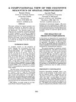
Fig. 1. Transduction of forces in ECs is a complex process involving signaling via many different molecules. This oversimplified cartoon
shows that at the luminal surface of ECs, forces due to leukocyte binding may be transmitted to the actin cytoskeleton via ICAM-1 receptors
(A), and forces due to shear stress may be transmitted via activation of stretch-activated ion channels or through displacement of the glyco-
calyx (B). Forces due to junctional cell–cell contact, whether EC–EC contact or leukocyte–EC contact during transmigration, may be transmit-
ted to the actin cytoskeleton via VE-cadherin at the cell borders (C). EC mechanosensing of the underlying substrate is probably completed
via integrin binding at FAs, leading to stretching of talin and activation of vinculin to reinforce the FA (D). The ECs respond to this interaction
by forming stress fibers that contract, allowing for measurement of the traction forces on the EC substrate. Thus, an EC contains many
mechanotransducing molecules on each of its surfaces that act to convert mechanical signals into biochemical signals within the cell. Many
of the molecules that are known to be involved in mechanotransduction are also linked to the actin cytoskeleton, which is an important
regulator of cell shape, alignment, and stiffness. Because ICAM-1 and VE-cadherin, two of the possible EC mechanotransducers, are also
involved in leukocyte transmigration, it is likely that leukocyte transmigration affects force transmission within the ECs. In (A), the force
acting on the EC (black arrow) has components both in the direction of shear stress and in the direction of pulling by leukocytes. In (B), the
force on the EC is in the direction of shear stress. In (C), the force is in the direction of tension of actin filaments, maintained with the help
of neighboring cells in contact. In (D), the force is in the direction of pulling at FAs at the substrate. See text for more details on magnitudes
of forces and the specific molecules involved. MAPK, mitogen-activated protein kinase.
K. M. Stroka and H. Aranda-Espinoza Biophysical view of transendothelial migration
FEBS Journal 277 (2010) 1145–1158 ª 2010 The Authors Journal compilation ª 2010 FEBS 1147
kinase, p160ROCK, results in decreased traction forces
and migration speed under both static and shear condi-
tions [19]. Because cell–cell contacts are important regu-
lators of cellular behavior, and these experiments were
performed on subconfluent cells, further work needs to
explore whether shear stress affects EC monolayer
migration in a similar manner. The magnitude of trac-
tion forces and stability of FAs both depend on the
flexibility of the underlying substrate [22,23], and thus,
in recent years, researchers have focused on exploring
the effects of substrate rigidity on cellular behavior.
These effects are discussed later for the case of ECs.
Another study also shows involvement of small
GTPases of the Rho family in the EC response to
shear stress [24]. RhoA, Rac and Cdc42 are rapidly
activated in response to shear stress, although the time
course and effects (rounding, spreading, elongation,
and alignment) differ. Within 5 min of application of
shear stress, RhoA is activated, leading to cell round-
ing via Rho kinase. Then, RhoA activity returns to
baseline, as Rac1 and Cdc42 reach peak activation,
leading to cell respreading, elongation, and alignment
in the direction of flow. Both Cdc42 and Rac1 are
required for cell elongation, whereas Rho and Rac1
regulate cell alignment with the direction of flow [24].
EC morphology in the vertical plane (specifically,
cell height) is carefully regulated by tension in the
cytoskeleton, as indicated by recent experiments com-
bining cytoskeletal drug treatments with atomic force
microscopy (AFM) indentation measurements [25].
Depolymerization of F-actin within subconfluent cells
results in increased cellular height. Meanwhile, disrup-
tion of microtubules lowers cell height, and stabiliza-
tion of microtubules elevates cell height [25]. Thus, the
cytoskeleton is an important structure that contributes
to determining cellular morphology, and so it makes
sense that, as shear stress affects the cytoskeletal
arrangement, cellular morphology is also affected. It is
still not clear exactly what causes the cytoskeleton
to rearrange under shear stress, but it is probably a
combination of mechanotransduction and mechano-
transmission effects.
Mechanical properties of ECs
It is believed that the mechanical state of the endothe-
lium is extremely important in maintaining vascular
homeostasis, and for this reason it is crucial to under-
stand which factors affect EC stiffness. For example,
ECs stiffen under shear stress as a function of expo-
sure time and magnitude of the shear stress [26–28].
Reducing the amount of cholesterol in untreated BAE-
Cs through methyl-b-cyclodextrin treatment increases
membrane stiffness, whereas enriching the cells with
cholesterol does not affect membrane stiffness [29].
Exposure to ox-LDLs has a similar effect in removing
cholesterol from the cell membrane, possibly through
disruption or redistribution of lipid rafts in the mem-
brane [30]. There is evidence that treatment with
ox-LDLs significantly increases the membrane stiffness
of human aortic ECs, as measured by micropipette
aspiration [30], and also the cell body stiffness of
human umbilical vein ECs (HUVECs), as measured by
AFM [31]. This increase in cell stiffness with ox-LDL
treatment is accompanied by an increase in force gen-
eration and network formation in a three-dimensional
collagen gel [30]. In addition, there is a significant
increase in the stiffness of aortic ECs isolated from
hypercholesterolemic pigs, where ox-LDL levels are
higher in the blood plasma, as compared with cells
isolated from healthy pigs [30]. These results suggest
that risk factors for atherosclerosis and stroke, such as
high cholesterol, not only lead to biological malfunction,
but are perhaps accompanied by biophysical changes
in the endothelium.
In addition to shear stress, cholesterol, and
ox-LDLs, ECs are also exposed to varying levels of
sodium in the bloodstream; this is another factor that
regulates vascular tone. ECs significantly stiffen in a
high-sodium environment in the presence of aldoste-
rone, which is a hormone that increases the reabsorp-
tion of sodium and is physiologically present in the
bloodstream. Increases in cell stiffness range from
about 10% to 50%, depending on the extracellular
sodium concentration (range of 135–160 mm) [32]. In
addition, nitric oxide production is downregulated by
aldosterone-exposed cells in a high-sodium medium
[32]. In contrast, increases in potassium soften ECs
and boost nitric oxide production, although this effect
is abrogated in the presence of high sodium levels [33].
Thus, hyperpolarization or depolarization of the cell
leads to changes in cell stiffness. Another recent study
simultaneously measured the mechanical stiffness and
electrical membrane potential of a vascular cell line
derived from BAECs, and correlated slow cell depolar-
izations with increases in cell membrane stiffness [34].
Interestingly, neutrophil adherence to ECs also
increases EC stiffness as measured by magnetic twist-
ing cytometry [35,36]. In contrast, monocyte adherence
to ECs decreases EC stiffness, as measured by AFM,
and at the same time also reduces the adhesiveness of
ECs to the substrate, as indicated by a decrease in
electric cell–substrate impedance [37]. This suggests
that leukocyte interactions with the endothelium affect
mechanotransmission events, and that these effects are
cell type-dependent.
Biophysical view of transendothelial migration K. M. Stroka and H. Aranda-Espinoza
1148 FEBS Journal 277 (2010) 1145–1158 ª 2010 The Authors Journal compilation ª 2010 FEBS
The effects that leukocytes have on the endothelium
indicate that stiffness may vary locally. Indeed, it has
been shown that ECs have a heterogeneous mechanical
surface. For example, AFM experiments have revealed
that the Young’s modulus of HUVECs ranges from
1.4 kPa near the edge of the cell to 6.8 kPa over the
nucleus of the cell [38], whereas in bovine pulmonary
aortic ECs, the Young’s modulus ranges from 0.2 to
2 kPa [39]. In contrast, Sato et al. [26] have found that
BAECs are stiffer near the edge of the cell than at the
nucleus, as measured by AFM. The discrepancies in
stiffness versus cell location in these studies may be
due to differences in the loading forces and indentation
depths used when probing with the AFM cantilever
[38], as cellular structures such as the cytoskeleton and
nucleus are positioned at different heights within the
cell. Using AFM, Engler et al. [40] probed the smooth
muscle cell-containing media layer of sectioned carotid
arteries from 6-month-old pigs, and found the Young’s
modulus to be in the range of 5–8 kPa, which is a sim-
ilar value to that for the single cultured cells discussed
above. It is obvious that the mechanical properties of
ECs are very heterogeneous and location-dependent
under normal conditions, but they are also influenced
by biophysical factors such as shear stress, cholesterol
distribution within the plasma membrane, exposure to
increased sodium, and EC–leukocyte adhesion, all of
which have been shown to be relevant in the onset and
progress of disease.
It is also possible to use AFM, in combination with
total internal reflection fluorescence microscopy, to
study the mechanotransmission of applied local forces
at the apical surface of an adherent cell to the basal
surface of the cell. Using this technique, Mathur et al.
[41] observed that exerting a local force of 0.3–0.5 nN
by an AFM probe over the nucleus of a HUVEC
results in a global rearrangement of focal contacts at
the substrate after the force is removed, including a
significant increase in FA area. Applying the same
force over the edge of the cell does not result in any
significant changes in FA cntact area after the force is
removed, suggesting that the nucleus is an important
link in force transmission between the cytoskeleton
and FAs [41]. Furthermore, application of local force
via an AFM probe also leads to mechanotransduction,
as shown by increased intracellular calcium, through
activation of stretch-activated ion channels [42].
EC–EC contacts as mechanosensors
Much biophysical characterization of cells has been
performed using single cells, for which cell–substrate
interactions are most important. However, in the case
of the endothelium, the cells are packed at high den-
sity, forming a monolayer in which cell–cell interac-
tions are as important, if not more important, than
cell–substrate interactions. As discussed above, EC
monolayers undergo global remodeling in response to
mechanical stimuli such as shear stress; recent evidence
also suggests that EC monolayers respond to local
mechanical forces [43]. When a glass needle is used to
apply local stretch to selective ECs and EC junctions,
the ECs respond by aligning and elongating parallel to
the direction of stretch, and this effect is accompanied
by a reorganization of stress fibers. At the selective
junctions where stretch is applied, Src homology-
2-containing tyrosine phosphatase-2 is recruited [43],
and this molecule is known to bind to PECAM-1 [44].
These results suggest that cell–cell junctions both sense
and transmit local forces.
Cell–cell contact has been shown to both inhibit and
stimulate cell proliferation, in different experimental
studies using different methods to regulate cell–cell con-
tact. For example, a recent study by Gray et al. [45] has
demonstrated that EC proliferation is biphasic with
regard to degree of cell–cell contact. In this study, cell–
cell contact was controlled by cell micropatterning, so
that a distinct number of cells could adhere in specific
configurations. Cells with no neighbors and cells with
more than three neighbors proliferated faster than cells
with two or three neighbors. This relationship
was mediated by RhoA, as expression of domi-
nant-negative RhoA blocked the increase in prolifera-
tion. Higher proliferation could be stimulated in single
cells with no neighbors through contact with a
VE-cadherin bead [45]. These results point to VE-cadh-
erin as an important junctional signaling molecule that
is capable of transmitting forces through cell–cell
contacts (Fig. 1).
Activation of the inflammatory
response
Both in vivo and in vitro, the immune response requires
activation of the endothelium in order to allow
leukocytes to adhere to and transmigrate through the
endothelial barrier cells. Several known cytokines are
known to induce the inflammatory response, including
tumor necrosis factor (TNF)-a and interleukin-1 (IL-1).
The pathways activated by these cytokines result in
drastic cellular behavioral changes, which create a more
permissible barrier for leukocyte transmigration.
TNF-a is produced mainly by innate immune cells,
such as macrophages, as a response to infection or
inflammation in the body. As a TNF-a molecule binds
to the TNF receptor-1 on the extracellular side of the
K. M. Stroka and H. Aranda-Espinoza Biophysical view of transendothelial migration
FEBS Journal 277 (2010) 1145–1158 ª 2010 The Authors Journal compilation ª 2010 FEBS 1149
EC, the cytosolic tails of the receptors rearrange.
A number of intracellular signaling proteins are
recruited, resulting in the possible activation of three
different pathways. These include nuclear factor-jB
(NF-jB) activation, a mitogen-activated protein kinase
cascade, and proteolysis leading to apoptosis. Activa-
tion of the NF-jB pathway leads to recruitment and
activation of IjB kinase kinase; the phosphorylation
and activation of IjB kinase by IjB kinase kinase; the
phosphorylation of IjB; and the degradation of IjB,
which releases the NF-jB. NF-jB then localizes to the
nucleus, where it initiates transcription of many genes
that contribute to the inflammatory response [46].
Following TNF-a stimulation, both intercellular
adhesion molecule-1 (ICAM-1) expression and vascular
cell adhesion molecule-1 (VCAM-1) expression are
upregulated, whereas PECAM-1 (also known as
CD31) expression is decreased, in cultured HUVECs
[47]. ICAM-1 and VCAM-1 are needed for leukocyte
firm adhesion and transmigration through the ECs. In
addition, activation of the NF-jB pathway results in a
reorganization of the EC F-actin cytoskeleton and
junctional molecules, such as VE-cadherin [48,49], as
well as changes in cell shape [50] and a decrease in cell
stiffness [51]. In particular, ECs activated by TNF-a
become more elongated and arrange into whorls [50],
and actin filaments thicken, leading to actomyosin-
mediated cell retraction and intercellular gap
formation [49]. Thus, even before leukocytes enter the
picture, the ECs have undergone significant changes in
response to activation of the inflammatory response.
Although the response is controlled by signaling path-
ways, some of the pathways are inside-out signals that
might occur through regulation of the interaction of
the cell with the extracellular matrix and through the
response to shear stress. Thus, it is important to recog-
nize the influence of these mechanical forces, not only
as possible sources of outside-in signaling, but also as
a form of feedback for the reorganization of the endo-
thelium.
Mechanical properties of the cellular
environment
In recent years, much attention has focused on the
effects of substrate stiffness on cell adhesion and
migration. Many cell types, including ECs [52–55],
smooth muscle cells [56–58], fibroblasts [23,54,59], neu-
rons [60,61], stem cells [62], neutrophils [63,64], and
macrophages [65], display behavior that changes as a
function of underlying stiffness in vitro. These in vitro
studies are quite relevant, because it is known that
pathological conditions such as cancer and atheroscle-
rosis are associated with changes in tissue and cell
stiffness [66–68]. The effects of tissue stiffness are also
important in the field of tissue engineering, where con-
structs are made to replace damaged or diseased tis-
sues in the body. Obviously, these biological
substitutes are most effective if they mimic the actual
in vivo biochemical and mechanical conditions, but
most experiments in the past have been performed on
glass, a very stiff substrate. Recently, however, poly
(dimethylsiloxane) with fibronectin micropatterning in
FA-sized circular islands has been recognized as a sub-
strate capable of achieving rapid EC confluence, cell
densities similar to those in vivo, and FA formation
[69]. Furthermore, rigidity sensing is probably accom-
plished through integrin interactions with the extracel-
lular matrix. It has been shown that substrate stiffness
directs the mechanical activation of a
5
b
1
integrin bind-
ing to fibronectin through myosin-II-generated cyto-
skeletal force, leading to internal signaling via
phosphorylation of FA kinase [70]. However, it is
unknown how the leukocyte adhesion cascade acts in
response to any engineered endothelium.
Because there is a complex interplay between the
biochemical and mechanical conditions in the body, it
is necessary first to determine how these conditions
individually affect cells, and then how they act in con-
cert. In the following section, we will review what is
known about the effects of environmental stiffness on
vascular ECs, as well as on immune cells. The sub-
strate stiffness of ECs is relevant, because changes in
the stiffness of the basement membrane or underlying
layers may affect EC structure, organization, and gene
expression. In addition, substrate stiffness may affect
EC stiffness, and because immune cells migrate on and
through ECs, it is important also to understand how
immune cells respond to changes in substrate stiffness.
Vascular ECs respond to substrate
stiffness
The effects of environmental mechanical properties on
EC behavior have been studied in both two dimen-
sions and three dimensions. Most of the previous
work on 2D substrates has focused on individual cells
or cells in networks. Single BAECs show increased
spreading areas and spreading rates on stiffer poly-
acrylamide gels in a Young’s modulus range of 6 to
165 000 Pa [54], whereas BAEC network assembly
(before monolayer formation) depends on a balance
between substrate compliance and extracellular matrix
density [52]. In general, HUVEC morphology switches
from a tube-like network to a monolayer with increas-
ing substrate stiffness, both on polyacrylamide gels
Biophysical view of transendothelial migration K. M. Stroka and H. Aranda-Espinoza
1150 FEBS Journal 277 (2010) 1145–1158 ª 2010 The Authors Journal compilation ª 2010 FEBS
and on Matrigel [71]. It is also well established that
cellular cytoskeletal organization depends on the stiff-
ness of the underlying substrate and controls the
shape of the cell. For example, severing multiple
F-actin stress fibers in bovine capillary ECs on stiff
surfaces (glass), using a laser nanoscissor, results in
very little change in cellular shape. However, severing
only one stress fiber in bovine capillary ECs on com-
pliant substrates (Young’s modulus of 3750 Pa)
results in cytoskeletal remodeling and, consequently,
dramatic changes in cellular shape [72]. Furthermore,
HUVECs on soft Matrigel surfaces contain less actin
and vinculin than the same cells on rigid Matrigel
substrates [71].
Because the F-actin network contributes to the
maintenance of prestress in the cell by regulating cellu-
lar tension, it would also be expected that the stiffness
of the ECs depends on substrate stiffness. Indeed, sin-
gle BAECs are two-fold more compliant on polyacryl-
amide gels of Young’s modulus 1700 Pa than BAECs
on 9000 Pa substrates [73]. These results are consistent
with the discovery that fibroblasts mimic the stiffness
of their substrate, up to a threshold value, and that
this response is dependent on the organization of the
F-actin cytoskeleton, whereby cells on stiff surfaces
exerting larger traction forces have a more stretched
and organized actin cytoskeleton than those on a
softer surface [74,75]. Recent work has also suggested
that BAECs can communicate with each other through
the compliance of their substrate [55]. Pairs of cells
migrate less than single cells on polyacrylamide gels
below 5500 Pa, indicating that the traction forces
exerted by one cell can be felt by another cell, resulting
in altered behavior [55]. This behavior of ECs may be
altered in a nonlinear strain-stiffening fibrin gel system,
in which recent studies have shown that fibroblasts
and human mesenchymal stem cells are influenced by
each other even when hundreds of micrometers away
from each other [76].
ECs may also be capable of sensing the mechanical
properties of their environment in 3D culture, as sug-
gested by experiments utilizing collagen gels. This
work is very promising for understanding the processes
of vasculogenesis (formation of new blood vessels) and
angiogenesis (formation of vascular trees), especially as
one of the current hurdles in the field of tissue engi-
neering is creating vascularized tissues. HUVECs
spread more, have larger lumens and exhibit less
branching when suspended in stiffer collagen gels [53].
Similarly, bovine pulmonary microvascular ECs
(BPMECs) cultured in flexible collagen gels form
dense, thin networks and have small, intracellular vac-
uoles with few actin filaments localized along the cell
membrane. In contrast, BPMECs in rigid collagen gels
form thicker and deeper networks surrounded by
intense actin filaments and with large lumens [77].
However, one must be careful in interpreting experi-
mental results involving cells on or in collagen gels, as
the strain exerted by cells on the collagen gel can mod-
ify the collagen fibers at the microscopic level [78], and
cells can enzymatically cut collagen fibers. Vinculin
expression is very low in BPMECs in soft gels, whereas
large clumps of vinculin are seen in protruding regions
at the tips of the branching networks in rigid gels [77].
Because EC morphology, stiffness, organization and
gene expression are all regulated by substrate stiffness,
manipulation of substrate mechanics is a possible
mechanism for the direction of cell migration and
wound repair.
Leukocytes respond to substrate
stiffness
Interestingly, recent studies have shown that immune
cell behavior also depends on substrate stiffness,
although the rigidity-sensing mechanism is probably
very different from that of ECs, fibroblasts, and other
tissue cells. Immune cells are highly motile cells that
must move across and through ECs at high speeds in
order to perform normal physiological functions. Both
neutrophils [63,64] and alveolar macrophages [65] dis-
play increased spreading, from rounded to flattened
morphology, with increasing substrate stiffness,
although this spreading occurs without generation of
F-actin stress fibers [65].
Recently, Stroka and Aranda-Espinoza [63] showed
that neutrophil migration speed is biphasic with regard
to substrate stiffness; that is, there exists an optimal
stiffness at which maximal migration occurs. This
optimal stiffness depends on the concentration of
extracellular matrix protein on the surface of the
substrate; at 100 lgÆmL
)1
fibronectin, the optimum
stiffness is 4 kPa, whereas with decreased fibronectin
(10 lgÆmL
)1
), the optimum stiffness increases to 7 kPa
[63]. Interestingly, smooth muscle cells also display
biphasic behavior with regard to substrate stiffness
[57]. Because neutrophils respond very differently to
substrate stiffnesses in the range 3–13 kPa, it is
expected that changes in EC structure and stiffness as
a result of varied conditions will cause significant alter-
ations in leukocyte adhesion, migration, and transmi-
gration. Consistent with this hypothesis, neutrophil
force generation during transmigration is dependent on
substrate rigidity, with larger forces being exerted on
micropillars with larger spring constants (39 ± 6 nN
versus 14 ± 4 nN) [79]. However, the use of the
K. M. Stroka and H. Aranda-Espinoza Biophysical view of transendothelial migration
FEBS Journal 277 (2010) 1145–1158 ª 2010 The Authors Journal compilation ª 2010 FEBS 1151
micropillar system for this application is questionable,
as the micropillars force ECs to adhere only in specific
locations, leading to possible differences in traction
force exertion. Finally, alveolar macrophage stiffness
is lower on softer substrates than on stiffer ones,
although cytochalasin D treatment has negligible
effects [65], suggesting that, unlike that of many tissue
cells, alveolar macrophage stiffness is not regulated
through tension of the F-actin cytoskeletal network.
Mechanotransduction during leukocyte
transmigration
Leukocytes are migrating cells in the body’s innate
immune system and constitute the first line of defense
against inflammation or infection. Infection in the
body causes activation of ECs and expression of cell
adhesion molecules [46]. Then, the leukocytes undergo
tethering to the ECs, firm adhesion, and migration,
followed by transmigration through the ECs, which
may occur in either a paracellular (through EC junc-
tions) or transcellular (through the bodies of ECs)
manner. Thorough reviews on transcellular versus
paracellular transmigration can be found elsewhere
[80,81]. Each of these steps involves interactions
between different ligand–receptor pairs [82].
Transmigration is often considered to be the least-
studied step of the leukocyte adhesion cascade. Some
work has been completed on the roles of adhesion
molecules such as ICAM-1 [83–85], VCAM-1, PE-
CAM-1 [86–88] and CD99 [89,90] in leukocyte trans-
migration. However, although some of the important
proteins have been identified, there is still a lack of
understanding of the overall process, especially its
mechanics and how forces are propagated as leuko-
cytes penetrate through the ECs. Rabodzey et al. [79]
showed that the forces that neutrophils exert on a
microfabricated pillar surface during transmigration
increase when the rigidity of the pillars is increased,
providing evidence that transmigration is a mechano-
sensitive process; furthermore, leukocytes exert three-
fold greater forces when transmigrating than adherent
leukocytes that do not transmigrate [79]. However,
because the micropillar system probably affects EC
adhesion and traction forces by constraining the ECs
to specific FA sites, much more work is needed to
determine exactly how the leukocyte transmigration
affects force propagation in ECs.
Paracellular transmigration
One method by which cells transmigrate through ECs
is in a paracellular fashion, or through the EC–EC
junctions. Several junctional adhesion receptors of ECs
are known to participate in leukocyte transmigration;
these molecules include junction adhesion molecules
(JAMs), VE-cadherin, and EC-selective adhesion mole-
cule. Nonjunctional adhesion receptors involved in
transmigration include PECAM-1, ICAM-1, intercellu-
lar adhesion molecule-2, and CD99. For a more com-
plete understanding of these molecules, see the recent
review by Vestweber [91]. VE-cadherin is largely
responsible for maintaining EC–EC contact in mono-
layers. Individual VE-cadherin to VE-cadherin bonds
have been found to have an unbinding force of
35–55 pN, as measured by single-molecule AFM [92].
VE-cadherin forms a complex with a-catenin, b-cate-
nin, c-catenin, and p120-catenin (p120). VE-cadherin is
also known to link to the actin cytoskeleton of ECs,
although the mechanism of this linkage is the subject
of much debate [93]. This controversy has been
spurred by the discovery that a-catenin cannot bind
simultaneously to b-catenin and actin [94]. A recent
study has suggested that epithelial protein lost in
neoplasm (also known as Lima-1) links actin and
a-catenin, and that a-catenin is then simultaneously
linked to b-catenin and cadherin [95]. However,
although this is true for epithelial cells, it is unknown
whether a similar protein links VE-cadherin to actin in
ECs. Somehow, however, VE-cadherin associates with
the actin cytoskeleton in ECs, maintaining tension
within the cells via cell–cell contacts.
Because of VE-cadherin’s role in cell–cell contact, it
obviously provides a physical barrier to leukocyte pen-
etration at the junction. Thus, VE-cadherin rearranges
away from the cell borders to form short-lived gaps in
the junctions during leukocyte transmigration [96].
These gaps are necessary for transmigration to occur
[97], and are induced by ICAM-1–lymphocyte func-
tion-associated antigen-1 (LFA-1) interaction [98].
Because VE-cadherin associates with the F-actin cyto-
skeleton, a rearrangement of VE-cadherin during leu-
kocyte transmigration would also be expected to affect
the F-actin arrangement within the ECs, leading to
changes in cellular prestress (Fig. 1). The expression of
VE-cadherin is mediated by p120, suggesting that p120
is an important intracellular mediator of VE-cadherin
gap formation [97].
Also maintaining EC–EC junctions are homophilic
interactions of JAM-A, and therefore these molecules
also create a physical barrier for leukocytes. Recently,
it has been shown that LFA-1 (on leukocytes) binding
to JAM-A (at EC junctions) destabilizes JAM-A
homophilic interactions [99]. AFM measurements
indicate that the interaction of JAM-A with LFA-1 is
stronger than JAM-A hemophilic interactions; the
Biophysical view of transendothelial migration K. M. Stroka and H. Aranda-Espinoza
1152 FEBS Journal 277 (2010) 1145–1158 ª 2010 The Authors Journal compilation ª 2010 FEBS
unbinding force of JAM-A–JAM-A interactions
increases from about 40 to 300 pN with increasing
loading rate, whereas the unbinding force of the
JAM-A–LFA-1 interaction increases from about 150
to 450 pN with a similar range of loading rate [99].
Dufour et al. have also recently shown that CD99 is
necessary for leukocyte transmigration in vivo [89] and
in vitro [90]. Blocking CD99 on both leukocytes and
ECs inhibits transmigration, suggesting that it is a
homophilic interaction of CD99 that mediates trans-
migration [89].
Transcellular transmigration
In addition to leukocytes crossing EC–EC junctions,
they also may take a transcellular route through the
body of the cell; see Carman and Springer [100] for a
recent review of transcellular migration of cells. Both
transmigration paths are available to leukocytes, but it
remains to be determined which is most energetically
favorable.
It is believed that leukocyte transmigration via the
transcellular route is initiated with the formation of a
cup-like ‘docking structure,’ in which the adhesion
proteins ICAM-1 and VCAM-1 localize in response
to a leukocyte present on the EC surface. This dock-
ing structure, which may be 8–12 lm wide and 1 lm
deep [101], forms as endothelial pseudopods embrace
the leukocyte, engaging ICAM-1 on the EC surface
with LFA-1 on the leukocyte surface [102], leading to
activation of RhoG downstream [103]. The inter-
action force between ICAM-1 and LFA-1 has been
measured as 100 pN, with a 50 ms contact duration
[104]. One study has shown that ICAM-1 and
VCAM-1 are recruited independently of ligand
engagement, actin cytoskeleton engagement, and hete-
rodimer formation; instead, they are included within
specialized preformed tetraspanin-enriched micro-
domains [105]. On the other hand, there is also
evidence that ICAM-1 engagement upon leukocyte
adhesion leads to EC cytoskeletal remodeling due to
tyrosine phosphorylation of cortactin, linking ICAM-
1 to the actin cytoskeleton and allowing ICAM-1 to
form clusters, facilitating transmigration [106] (Fig. 1).
Transmission electron microscopy images show that
lymphocytes concurrently send protrusive podosomes
into the ECs, and this occurs both in vivo and in vitro,
probably to probe the EC surface in order to find
regions of low resistance [107]. Thus, initiation of
leukocyte transmigration via the transcellular route
involves active involvement of both the ECs and the
leukocytes, but the molecular mechanisms are still not
well understood.
Transmigration during atherogenesis
The dynamics of leukocyte transmigration in athero-
genesis should also be considered. That is, what is the
mechanism of increased monocyte extravasation
through the endothelium, leading to formation of
raised plaques under the endothelium? Treatments of
HUVECs with ox-LDLs in vitro have recently been
shown to promote monocyte invasion of the endothe-
lium, presumably because ox-LDLs upregulate
PECAM-1, leading to enhanced homophilic interac-
tions with monocyte PECAM-1, and downregulate
VE-cadherin, leading to disrupted junctions and there-
fore increased endothelial permeability [108]. Mono-
cyte adhesion to the apical surfaces of ECs and
monocyte complete transmigration below the endothe-
lium are not affected by ox-LDL treatment [108], sug-
gesting that initiation of transmigration is the critical
step at which ox-LDL level is important.
Cytoskeletal involvement during
transmigration
Leukocyte transmigration is facilitated by increased
EC permeability. This can be accomplished through
activation of the NF-jB pathway via stimulation with
TNF-a, as discussed above. In addition, EC perme-
ability can be increased by treatment with agents such
as histamine, thrombin, vascular endothelial growth
factor-A, or hydrogen peroxide. These agents are
believed to increase tyrosine phosphorylation in the
cadherin–catenin complex [91]. Recent work suggests
that the spatial organization of the cytoskeleton, spe-
cifically F-actin, controls the permeability of ECs
in vitro [109]. For example, treating ECs with junc-
tion-disrupting agents induces stress fiber formation,
whereas treating ECs with junction-tightening agents
(such as oxidized 1-palmitoyl-2-arachidonoyl-sn-
glycero-3-phosphocholine, hepatocyte growth factor,
and iloprost) enhances the peripheral actin cytoskele-
ton [109]. These treatments will also facilitate or hin-
der leukocyte transmigration, respectively, and
therefore the spatial organization of the F-actin net-
work as a physical barrier is a crucial regulator of
leukocyte trafficking.
When AFM is used to remove neutrophils from the
endothelium during transmigration, they leave behind
footprints 8–12 lm wide and 1 lm deep [101]. The
authors claimed that these footprints are formed with-
out net depolymerization of F-actin, as ECs do not
soften at the site of adhesion [101]. However, other
work has shown that both neutrophils and ECs stiffen
during neutrophil–EC adhesion, and that this process
K. M. Stroka and H. Aranda-Espinoza Biophysical view of transendothelial migration
FEBS Journal 277 (2010) 1145–1158 ª 2010 The Authors Journal compilation ª 2010 FEBS 1153
is cytoskeleton-dependent [35,36]. Obviously, the role
of the EC cytoskeleton in leukocyte transmigration is
still not understood, and further experiments are neces-
sary to determine how it may transmit forces during
leukocyte transmigration.
Concluding remarks
The mechanical state of the endothelium is influenced
by many external factors, both chemical and mechani-
cal. Because the mechanical state of the endothelium is
probably an important regulator of vascular homeosta-
sis and leukocyte transmigration, many biophysical
tools, such as AFM, magnetic tweezers, traction force
microscopy, and immunofluorescence, are very relevant
and useful. Leukocyte transmigration through ECs is a
complex process that is involved both in the healthy
immune response and in the development of disease. It
is evident that the process involves a transmission of
physical forces as the leukocytes pass through the
endothelium. The propagation of these forces through
ECs is probably affected by interactions with neighbor-
ing ECs, interactions with the basement membrane
beneath the ECs, and shear stress. How these forces,
individually or together, translate into biochemical sig-
naling pathways is only beginning to be understood.
In the future, it will become increasingly necessary to
develop similar biophysical tools to those currently
used in vitro for more in vivo experiments, so that we
can understand how force transmission in an actual
artery differs from or is similar to that in an engi-
neered endothelium.
Acknowledgements
This work was completed under a National Science
Foundation (NSF) Graduate Research Fellowship to
K. M. Stroka and NSF award CMMI-0643783 to H.
Aranda-Espinoza. The authors thank L. Norman for
critical and thorough reading of this article.
References
1 Strell C & Entschladen F (2008) Extravasation of leu-
kocytes in comparison to tumor cells. Cell Commun
Signal 6, 10–22.
2 Lucchinetti C, Bruck W, Parisi J, Scheithauer B,
Rodriguez M & Lassmann H (2000) Heterogeneity of
multiple sclerosis lesions: implications for the patho-
genesis of demyelination. Ann Neurol 47, 707–717.
3 Noseworthy JH, Lucchinetti C, Rodriguez M & Wein-
shenker BG (2000) Multiple sclerosis. N Engl J Med
343, 938–952.
4 Bailey SL, Carpentier PA, McMahon EJ, Begolka WS
& Miller SD (2006) Innate and adaptive immune
responses of the central nervous system. Crit Rev
Immunol 26, 149–188.
5 Hickey WF (1999) Leukocyte traffic in the central
nervous system: the participants and their roles. Semin
Immunol 11, 125–137.
6 Davies PF (1995) Flow-mediated endothelial mechano-
transduction. Physiol Rev 75, 519–560.
7 Helmke BP & Davies PF (2002) The cytoskeleton
under external fluid mechanical forces: hemodynamic
forces acting on the endothelium. Ann Biomed Eng 30,
284–296.
8 Walpola PL, Gotlieb AI & Langille BL (1993) Mono-
cyte adhesion and changes in endothelial cell number,
morphology, and F-actin distribution elicited by low
shear stress in vivo. Am J Pathol 142, 1392–1400.
9 Kim DW, Gotlieb AI & Langille BL (1989) In vivo
modulation of endothelial F-actin microfilaments by
experimental alterations in shear stress. Arteriosclerosis
(Dallas, Tex) 9, 439–445.
10 Goode TB, Davies PF, Reidy MA & Bowyer DE
(1977) Aortic endothelial cell morphology observed in
situ by scanning electron microscopy during athero-
genesis in the rabbit. Atherosclerosis 27, 235–251.
11 Nerem RM, Levesque MJ & Cornhill JF (1981)
Vascular endothelial morphology as an indicator
of the pattern of blood flow. J Biomech Eng 103,
172–176.
12 Levesque MJ & Nerem RM (1985) The elongation and
orientation of cultured endothelial cells in response to
shear stress. J Biomech Eng 107, 341–347.
13 Chien S, Li S & Shyy YJ (1998) Effects of mechanical
forces on signal transduction and gene expression in
endothelial cells. Hypertension 31, 162–169.
14 Papadaki M & Eskin SG (1997) Effects of fluid shear
stress on gene regulation of vascular cells. Biotechnol
Prog 13, 209–221.
15 Braddock M, Schwachtgen JL, Houston P, Dickson
MC, Lee MJ & Campbell CJ (1998) Fluid shear stress
modulation of gene expression in endothelial cells.
News Physiol Sci 13, 241–246.
16 Jiang G, Giannone G, Critchley DR, Fukumoto E &
Sheetz MP (2003) Two-piconewton slip bond between
fibronectin and the cytoskeleton depends on talin.
Nature 424, 334–337.
17 del Rio A, Perez-Jimenez R, Liu R, Roca-Cusachs P,
Fernandez JM & Sheetz MP (2009) Stretching single
talin rod molecules activates vinculin binding. Science
(NY) 323, 638–641.
18 Davies PF, Robotewskyj A & Griem ML (1994)
Quantitative studies of endothelial cell adhesion.
Directional remodeling of focal adhesion sites in
response to flow forces. J Clin Invest 93, 2031–2038.
Biophysical view of transendothelial migration K. M. Stroka and H. Aranda-Espinoza
1154 FEBS Journal 277 (2010) 1145–1158 ª 2010 The Authors Journal compilation ª 2010 FEBS
19 Shiu YT, Li S, Marganski WA, Usami S, Schwartz MA,
Wang YL, Dembo M & Chien S (2004) Rho mediates
the shear-enhancement of endothelial cell migration and
traction force generation. Biophys J 86, 2558–2565.
20 Helmke BP, Goldman RD & Davies PF (2000) Rapid
displacement of vimentin intermediate filaments in
living endothelial cells exposed to flow. Circ Res 86,
745–752.
21 Helmke BP, Rosen AB & Davies PF (2003) Mapping
mechanical strain of an endogenous cytoskeletal
network in living endothelial cells. Biophys J 84,
2691–2699.
22 Wang N, Tolic-Norrelykke IM, Chen J, Mijailovich
SM, Butler JP, Fredberg JJ & Stamenovic D (2002)
Cell prestress. I. Stiffness and prestress are closely
associated in adherent contractile cells. Am J Physiol
282, C606–C616.
23 Pelham RJ Jr & Wang Y (1997) Cell locomotion and
focal adhesions are regulated by substrate flexibility.
Proc Natl Acad Sci USA 94, 13661–13665.
24 Wojciak-Stothard B & Ridley AJ (2003) Shear stress-
induced endothelial cell polarization is mediated by
Rho and Rac but not Cdc42 or PI 3-kinases. J Cell
Biol 161, 429–439.
25 Deguchi S, Fukamachi H, Hashimoto K, Iio K &
Tsujioka K (2009) Measurement and finite element
modeling of the force balance in the vertical section
of adhering vascular endothelial cells. J Mech Behav
Biomed Mater 2, 173–185.
26 Sato M, Nagayama K, Kataoka N, Sasaki M & Hane
K (2000) Local mechanical properties measured by
atomic force microscopy for cultured bovine endo-
thelial cells exposed to shear stress. J Biomech 33,
127–135.
27 Theret DP, Levesque MJ, Sato M, Nerem RM &
Wheeler LT (1988) The application of a homogeneous
half-space model in the analysis of endothelial cell
micropipette measurements. J Biomech Eng 110,
190–199.
28 Sato M, Levesque MJ & Nerem RM (1987) Micro-
pipette aspiration of cultured bovine aortic endothelial
cells exposed to shear stress. Arteriosclerosis (Dallas,
Tex) 7, 276–286.
29 Byfield FJ, Aranda-Espinoza H, Romanenko VG,
Rothblat GH & Levitan I (2004) Cholesterol depletion
increases membrane stiffness of aortic endothelial cells.
Biophys J 87, 3336–3343.
30 Byfield FJ, Tikku S, Rothblat GH, Gooch KJ &
Levitan I (2006) OxLDL increases endothelial stiffness,
force generation, and network formation. J Lipid Res
47, 715–723.
31 Chouinard JA, Grenier G, Khalil A & Vermette P
(2008) Oxidized-LDL induce morphological changes
and increase stiffness of endothelial cells. Exp Cell Res
314, 3007–3016.
32 Oberleithner H, Riethmuller C, Schillers H, MacGre-
gor GA, de Wardener HE & Hausberg M (2007)
Plasma sodium stiffens vascular endothelium and
reduces nitric oxide release. Proc Natl Acad Sci USA
104, 16281–16286.
33 Oberleithner H, Callies C, Kusche-Vihrog K, Schillers
H, Shahin V, Riethmuller C, MacGregor GA & de
Wardener HE (2009) Potassium softens vascular endo-
thelium and increases nitric oxide release. Proc
Natl Acad Sci USA 106, 2829–2834.
34 Callies C, Schon P, Liashkovich I, Stock C, Kusche-
Vihrog K, Fels J, Strater AS & Oberleithner H (2009)
Simultaneous mechanical stiffness and electrical poten-
tial measurements of living vascular endothelial cells
using combined atomic force and epifluorescence
microscopy. Nanotechnology 20, 175104–175111.
35 Wang Q & Doerschuk CM (2000) Neutrophil-induced
changes in the biomechanical properties of endothelial
cells: roles of ICAM-1 and reactive oxygen species.
J Immunol 164, 6487–6494.
36 Wang Q, Chiang ET, Lim M, Lai J, Rogers R, Janmey
PA, Shepro D & Doerschuk CM (2001) Changes in the
biomechanical properties of neutrophils and endothelial
cells during adhesion. Blood 97, 660–668.
37 Kataoka N, Iwaki K, Hashimoto K, Mochizuki S,
Ogasawara Y, Sato M, Tsujioka K & Kajiya F (2002)
Measurements of endothelial cell-to-cell and cell-
to-substrate gaps and micromechanical properties of
endothelial cells during monocyte adhesion. Proc Natl
Acad Sci USA
99, 15638–15643.
38 Mathur AB, Collinsworth AM, Reichert WM, Kraus
WE & Truskey GA (2001) Endothelial, cardiac muscle
and skeletal muscle exhibit different viscous and elastic
properties as determined by atomic force microscopy.
J Biomech 34, 1545–1553.
39 Pesen D & Hoh JH (2005) Micromechanical architec-
ture of the endothelial cell cortex. Biophys J 88,
670–679.
40 Engler AJ, Richert L, Wong JY, Picart C & Discher D
(2004) Surface probe measurements of the elasticity of
sectioned tissue, thin gels and polyelectrolyte multi-
layer films: correlations between substrate stiffness and
cell adhesion. Surface Sci 570, 142–154.
41 Mathur AB, Truskey GA & Reichert WM (2000)
Atomic force and total internal reflection fluorescence
microscopy for the study of force transmission in
endothelial cells. Biophys J 78, 1725–1735.
42 Charras GT & Horton MA (2002) Single cell
mechanotransduction and its modulation analyzed by
atomic force microscope indentation. Biophys J 82,
2970–2981.
43 Ueki Y, Sakamoto N, Ohashi T & Sato M (2009)
Morphological responses of vascular endothelial cells
induced by local stretch transmitted through intercellu-
lar junctions. Exp Mech 49, 125–134.
K. M. Stroka and H. Aranda-Espinoza Biophysical view of transendothelial migration
FEBS Journal 277 (2010) 1145–1158 ª 2010 The Authors Journal compilation ª 2010 FEBS 1155
44 Osawa M, Masuda M, Kusano K & Fujiwara K
(2002) Evidence for a role of platelet endothelial cell
adhesion molecule-1 in endothelial cell mechanosignal
transduction: is it a mechanoresponsive molecule?
J Cell Biol 158, 773–785.
45 Gray DS, Liu WF, Shen CJ, Bhadriraju K, Nelson
CM & Chen CS (2008) Engineering amount of cell–cell
contact demonstrates biphasic proliferative regulation
through RhoA and the actin cytoskeleton. Exp Cell
Res 314, 2846–2854.
46 Alberts B, Johnson A, Lewis J, Raff M, Roberts K &
Walter P (2002) Molecular Biology of the Cell, 4th edn.
Garland Science, Taylor & Francis Group, New York,
NY.
47 Sawa Y, Sugimoto Y, Ueki T, Ishikawa H, Sato A,
Nagato T & Yoshida S (2007) Effects of TNF-alpha
on leukocyte adhesion molecule expressions in cultured
human lymphatic endothelium. J Histochem Cytochem
55, 721–733.
48 Blum MS, Toninelli E, Anderson JM, Balda MS,
Zhou J, O’Donnell L, Pardi R & Bender JR (1997)
Cytoskeletal rearrangement mediates human micro-
vascular endothelial tight junction modulation by
cytokines. Am J Physiol 273, H286–H294.
49 Wojciak-Stothard B, Entwistle A, Garg R & Ridley
AJ (1998) Regulation of TNF-alpha-induced reorgani-
zation of the actin cytoskeleton and cell–cell junctions
by Rho, Rac, and Cdc42 in human endothelial cells.
J Cell Physiol 176, 150–165.
50 Stolpen AH, Guinan EC, Fiers W & Pober JS (1986)
Recombinant tumor necrosis factor and immune inter-
feron act singly and in combination to reorganize
human vascular endothelial cell monolayers. Am J
Pathol 123, 16–24.
51 Kang I, Panneerselvam D, Panoskaltsis VP, Eppell
SJ, Marchant RE & Doerschuk CM (2008) Changes
in the hyperelastic properties of endothelial cells
induced by tumor necrosis factor-alpha. Biophys J 94 ,
3273–3285.
52 Califano JP & Reinhart-King CA (2008) A balance of
substrate mechanics and matrix chemistry regulates
endothelial cell network assembly. Cell Mol Bioeng 1,
122–132.
53 Sieminski AL, Hebbel RP & Gooch KJ (2004) The
relative magnitudes of endothelial force generation and
matrix stiffness modulate capillary morphogenesis
in vitro. Exp Cell Res 297, 574–584.
54 Yeung T, Georges PC, Flanagan LA, Marg B, Ortiz
M, Funaki M, Zahir N, Ming W, Weaver V & Janmey
PA (2005) Effects of substrate stiffness on cell
morphology, cytoskeletal structure, and adhesion. Cell
Motil Cytoskeleton 60, 24–34.
55 Reinhart-King CA, Dembo M & Hammer DA (2008)
Cell–cell mechanical communication through compli-
ant substrates. Biophys J 95, 6044–6051.
56 Harley BA, Kim HD, Zaman MH, Yannas IV, Lauf-
fenburger DA & Gibson LJ (2008) Microarchitecture
of three-dimensional scaffolds influences cell migration
behavior via junction interactions. Biophys J 95, 4013–
4024.
57 Peyton SR & Putnam AJ (2005) Extracellular matrix
rigidity governs smooth muscle cell motility in a
biphasic fashion. J Cell Physiol 204, 198–209.
58 Engler A, Bacakova L, Newman C, Hategan A,
Griffin M & Discher D (2004) Substrate compliance
versus ligand density in cell on gel responses. Biophys
J 86, 617–628.
59 Lo C-M, Wang H-B, Dembo M & Wang Y-L (2000)
Cell movement is guided by the rigidity of the
substrate. Biophys J 79, 144–152.
60 Flanagan LA, Ju YE, Marg B, Osterfield M & Janmey
PA (2002) Neurite branching on deformable
substrates. Neuroreport 13, 2411–2415.
61 Gunn JW, Turner SD & Mann BK (2005) Adhesive
and mechanical properties of hydrogels influence
neurite extension. J Biomed Mater Res A 72, 91–97.
62 Engler AJ, Sen S, Sweeney HL & Discher DE (2006)
Matrix elasticity directs stem cell lineage specification.
Cell 126, 677–689.
63 Stroka KM & Aranda-Espinoza H (2009) Neutrophils
display biphasic relationship between migration and
substrate stiffness. Cell Motil Cytoskeleton 66,
328–341.
64 Oakes PW, Patel DC, Morin NA, Zitterbart DP,
Fabry B, Reichner JS & Tang JX (2009) Neutrophil
morphology and migration are affected by substrate
elasticity. Blood 7, 1387–1395.
65 Fereol S, Fodil R, Labat B, Galiacy S, Laurent VM,
Louis B, Isabey D & Planus E (2006) Sensitivity of alve-
olar macrophages to substrate mechanical and adhesive
properties. Cell Motil Cytoskeleton 63, 321–340.
66 Erler JT & Weaver VM (2009) Three-dimensional
context regulation of metastasis. Clin Exp Metastasis
26, 35–49.
67 Paszek MJ, Zahir N, Johnson KR, Lakins JN, Rozen-
berg GI, Gefen A, Reinhart-King CA, Margulies SS,
Dembo M, Boettiger D et al. (2005) Tensional homeo-
stasis and the malignant phenotype. Cancer Cell 8,
241–254.
68 Majno G & Joris I (1996) Cells, Tissues, and Disease:
Principles of General Pathology. Blackwell Science,
Worcester, MA.
69 Feinberg AW, Schumacher JF & Brennan AB (2009)
Engineering high-density endothelial cell monolayers
on soft substrates. Acta Biomater 5, 2013–2024.
70 Friedland JC, Lee MH & Boettiger D (2009) Mechani-
cally activated integrin switch controls alpha5beta1
function. Science (NY) 323, 642–644.
71 Deroanne CF, Lapiere CM & Nusgens BV (2001)
In vitro tubulogenesis of endothelial cells by relaxation
Biophysical view of transendothelial migration K. M. Stroka and H. Aranda-Espinoza
1156 FEBS Journal 277 (2010) 1145–1158 ª 2010 The Authors Journal compilation ª 2010 FEBS
of the coupling extracellular matrix–cytoskeleton.
Cardiovasc Res 49, 647–658.
72 Kumar S, Maxwell IZ, Heisterkamp A, Polte TR, Lele
TP, Salanga M, Mazur E & Ingber DE (2006) Viscoelas-
tic retraction of single living stress fibers and its impact
on cell shape, cytoskeletal organization, and extracellu-
lar matrix mechanics. Biophys J 90, 3762–3773.
73 Byfield FJ, Reen RK, Shentu TP, Levitan I & Gooch
KJ (2009) Endothelial actin and cell stiffness is modu-
lated by substrate stiffness in 2D and 3D. J Biomech
42, 1114–1119.
74 Ghosh K, Pan Z, Guan E, Ge S, Liu Y, Nakamura T,
Ren XD, Rafailovich M & Clark RA (2007) Cell
adaptation to a physiologically relevant ECM mimic
with different viscoelastic properties. Biomaterials 28,
671–679.
75 Solon J, Levental I, Sengupta K, Georges PC &
Janmey PA (2007) Fibroblast adaptation and stiffness
matching to soft elastic substrates. Biophys J 93,
4453–4461.
76 Winer JP, Oake S & Janmey PA (2009) Non-linear
elasticity of extracellular matrices enables contractile
cells to communicate local position and orientation.
PLoS ONE 4, e6382, doi:10.1371/journal.pone.
0006382.
77 Yamamura N, Sudo R, Ikeda M & Tanishita K (2007)
Effects of the mechanical properties of collagen gel on
the in vitro formation of microvessel networks by
endothelial cells. Tissue Eng 13, 1443–1453.
78 Vader D, Kabla A, Weitz D & Mahadevan L (2009)
Strain-induced alignment in collagen gels. PLoS ONE
4, e5902, doi:10.1371/journal.pone.0005902.
79 Rabodzey A, Alcaide P, Luscinskas FW & Ladoux B
(2008) Mechanical forces induced by the transendothe-
lial migration of human neutrophils. Biophys J 95,
1428–1438.
80 Hordijk PL (2006) Endothelial signalling events during
leukocyte transmigration. FEBS J 273, 4408–4415.
81 Petri B & Bixel MG (2006) Molecular events during
leukocyte diapedesis. FEBS J 273, 4399–4407.
82 Ley K, Laudanna C, Cybulsky MI & Nourshargh S
(2007) Getting to the site of inflammation: the leuko-
cyte adhesion cascade updated. Nat Rev 7, 678–689.
83 Yang L, Froio RM, Sciuto TE, Dvorak AM, Alon R
& Luscinskas FW (2005) ICAM-1 regulates neutrophil
adhesion and transcellular migration of TNF-alpha-
activated vascular endothelium under flow. Blood 106,
584–592.
84 Dustin ML & Springer TA (1988) Lymphocyte func-
tion associated antigen-1 (Lfa-1) interaction with inter-
cellular-adhesion molecule-1 (Icam-1) is one of at least
3 mechanisms for lymphocyte adhesion to cultured
endothelial-cells. J Cell Biol 107, 321–331.
85 Lawson C & Wolf S (2009) ICAM-1 signaling in
endothelial cells. Pharmacol Rep 61, 22–32.
86 Muller WA (1995) The role of PECAM-1 (CD31) in
leukocyte emigration: studies in vitro and in vivo.
J Leukoc Biol 57, 523–528.
87 O’Brien CD, Lim P, Sun J & Albelda SM (2003)
PECAM-1-dependent neutrophil transmigration is
independent of monolayer PECAM-1 signaling or
localization. Blood 101, 2816–2825.
88 Feng D, Nagy JA, Pyne K, Dvorak HF & Dvorak AM
(2004) Ultrastructural localization of platelet endothe-
lial cell adhesion molecule (PECAM-1, CD31) in vascu-
lar endothelium. J Histochem Cytochem 52, 87–101.
89 Dufour EM, Deroche A, Bae Y & Muller WA (2008)
CD99 is essential for leukocyte diapedesis in vivo.
Cell
Commun Adhes 15, 351–363.
90 Lou O, Alcaide P, Luscinskas FW & Muller WA
(2007) CD99 is a key mediator of the transendothelial
migration of neutrophils. J Immunol 178, 1136–1143.
91 Vestweber D (2007) Adhesion and signaling molecules
controlling the transmigration of leukocytes through
endothelium. Immunol Rev 218 , 178–196.
92 Baumgartner W, Hinterdorfer P, Ness W, Raab A,
Vestweber D, Schindler H & Drenckhahn D (2000)
Cadherin interaction probed by atomic force micro-
scopy. Proc Natl Acad Sci USA 97, 4005–4010.
93 Vestweber D, Winderlich M, Cagna G & Nottebaum
AF (2009) Cell adhesion dynamics at endothelial junc-
tions: VE-cadherin as a major player. Trends Cell Biol
19, 8–15.
94 Yamada S, Pokutta S, Drees F, Weis WI & Nelson
WJ (2005) Deconstructing the cadherin–catenin–actin
complex. Cell 123, 889–901.
95 Abe K & Takeichi M (2008) EPLIN mediates linkage
of the cadherin catenin complex to F-actin and stabi-
lizes the circumferential actin belt. Proc Natl Acad Sci
USA 105, 13–19.
96 Shaw SK, Bamba PS, Perkins BN & Luscinskas FW
(2001) Real-time imaging of vascular endothelial-
cadherin during leukocyte transmigration across
endothelium. J Immunol 167 , 2323–2330.
97 Alcaide P, Newton G, Auerbach S, Sehrawat S,
Mayadas TN, Golan DE, Yacono P, Vincent P,
Kowalczyk A & Luscinskas FW (2008) p120-Catenin
regulates leukocyte transmigration through an effect
on VE-cadherin phosphorylation. Blood 112,
2770–2779.
98 Wee H, Oh HM, Jo JH & Jun CD (2009) ICAM-
1 ⁄ LFA-1 interaction contributes to the induction of
endothelial cell–cell separation: implication for
enhanced leukocyte diapedesis. Exp Mol Med 41,
341–348.
99 Wojcikiewicz EP, Koenen RR, Fraemohs L,
Minkiewicz J, Azad H, Weber C & Moy VT (2008)
LFA-1 binding destabilizes the JAM-A homophilic
interaction during leukocyte transmigration. Biophys J
96, 285–293.
K. M. Stroka and H. Aranda-Espinoza Biophysical view of transendothelial migration
FEBS Journal 277 (2010) 1145–1158 ª 2010 The Authors Journal compilation ª 2010 FEBS 1157
100 Carman CV & Springer TA (2008) Trans-cellular
migration: cell–cell contacts get intimate. Curr Opin
Cell Biol 20, 533–540.
101 Riethmuller C, Nasdala I & Vestweber D (2008)
Nano-surgery at the leukocyte–endothelial docking
site. Pflugers Arch 456, 71–81.
102 Carman CV, Jun CD, Salas A & Springer TA (2003)
Endothelial cells proactively form microvilli-like mem-
brane projections upon intercellular adhesion molecule
1 engagement of leukocyte LFA-1. J Immunol 171,
6135–6144.
103 van Buul JD, Allingham MJ, Samson T, Meller J,
Boulter E, Garcia-Mata R & Burridge K (2007) RhoG
regulates endothelial apical cup assembly downstream
from ICAM1 engagement and is involved in leukocyte
trans-endothelial migration. J Cell Biol 178, 1279–
1293.
104 Wojcikiewicz EP, Abdulreda MH, Zhang X & Moy
VT (2006) Force spectroscopy of LFA-1 and its
ligands, ICAM-1 and ICAM-2. Biomacromolecules 7,
3188–3195.
105 Barreiro O, Zamai M, Yanez-Mo M, Tejera E, Lopez-
Romero P, Monk PN, Gratton E, Caiolfa VR &
Sanchez-Madrid F (2008) Endothelial adhesion recep-
tors are recruited to adherent leukocytes by inclusion
in preformed tetraspanin nanoplatforms. J Cell Biol
183, 527–542.
106 Yang L, Kowalski JR, Yacono P, Bajmoczi M, Shaw
SK, Froio RM, Golan DE, Thomas SM & Luscinskas
FW (2006) Endothelial cell cortactin coordinates inter-
cellular adhesion molecule-1 clustering and actin
cytoskeleton remodeling during polymorphonuclear
leukocyte adhesion and transmigration. J Immunol
177, 6440–6449.
107 Carman CV, Sage PT, Sciuto TE, de la Fuente MA,
Geha RS, Ochs HD, Dvorak HF, Dvorak AM &
Springer TA (2007) Transcellular diapedesis is initiated
by invasive podosomes. Immunity 26, 784–797.
108 Hashimoto K, Kataoka N, Nakamura E, Tsujioka K
& Kajiya F (2007) Oxidized LDL specifically promotes
the initiation of monocyte invasion during transendo-
thelial migration with upregulated PECAM-1 and
downregulated VE-cadherin on endothelial junctions.
Atherosclerosis 194, e9–e17.
109 Birukova AA, Arce FT, Moldobaeva N, Dudek SM,
Garcia JG, Lal R & Birukov KG (2009) Endothelial
permeability is controlled by spatially defined cyto-
skeletal mechanics: atomic force microscopy force
mapping of pulmonary endothelial monolayer.
Nanomedicine 5, 30–41.
Biophysical view of transendothelial migration K. M. Stroka and H. Aranda-Espinoza
1158 FEBS Journal 277 (2010) 1145–1158 ª 2010 The Authors Journal compilation ª 2010 FEBS