RHEOLOGY - NEW CONCEPTS, APPLICATIONS AND METHODS potx
Bạn đang xem bản rút gọn của tài liệu. Xem và tải ngay bản đầy đủ của tài liệu tại đây (6.98 MB, 114 trang )
RHEOLOGY - NEW
CONCEPTS,
APPLICATIONS AND
METHODS
Edited by Rajkumar Durairaj
Rheology - New Concepts, Applications and Methods
/>Edited by Rajkumar Durairaj
Contributors
Rajkumar Durairaj, Abu-El Hassan, Martin Williams, Talero, Trofimov, Jeshwanth K. Rameshwaram
Published by InTech
Janeza Trdine 9, 51000 Rijeka, Croatia
Copyright © 2013 InTech
All chapters are Open Access distributed under the Creative Commons Attribution 3.0 license, which allows users to
download, copy and build upon published articles even for commercial purposes, as long as the author and publisher
are properly credited, which ensures maximum dissemination and a wider impact of our publications. After this work
has been published by InTech, authors have the right to republish it, in whole or part, in any publication of which they
are the author, and to make other personal use of the work. Any republication, referencing or personal use of the
work must explicitly identify the original source.
Notice
Statements and opinions expressed in the chapters are these of the individual contributors and not necessarily those
of the editors or publisher. No responsibility is accepted for the accuracy of information contained in the published
chapters. The publisher assumes no responsibility for any damage or injury to persons or property arising out of the
use of any materials, instructions, methods or ideas contained in the book.
Publishing Process Manager Ana Pantar
Technical Editor InTech DTP team
Cover InTech Design team
First published January, 2013
Printed in Croatia
A free online edition of this book is available at www.intechopen.com
Additional hard copies can be obtained from
Rheology - New Concepts, Applications and Methods, Edited by Rajkumar Durairaj
p. cm.
ISBN 978-953-51-0953-2
free online editions of InTech
Books and Journals can be found at
www.intechopen.com
Contents
Preface VII
Chapter 1 A Practical Review of Microrheological Techniques 1
Bradley W. Mansel, Stephen Keen, Philipus J. Patty, Yacine Hemar
and Martin A.K. Williams
Chapter 2 Rheological Characterisation of Diglycidylether of Bisphenol-A
(DGEBA) and Polyurethane (PU) Based Isotropic Conductive
Adhesives 23
R. Durairaj, Lam Wai Man, Kau Chee Leong, Liew Jian Ping, N. N.
Ekere and Lim Seow Pheng
Chapter 3 Heliogeophysical Aspects of Rheology: New Technologies and
Horizons of Preventive Medicine 39
Trofimov Alexander and Sevostyanova Evgeniya
Chapter 4 Performance of Fresh Portland Cement Pastes – Determination
of Some Specific Rheological Parameters 57
R. Talero, C. Pedrajas and V. Rahhal
Chapter 5 Rheology - New Concepts, Applications and Methods 81
Jeshwanth K. Rameshwaram and Tien T. Dao
Chapter 6 Unsteady Axial Viscoelastic Pipe Flows of an
Oldroyd B Fluid 91
A. Abu-El Hassan and E. M. El-Maghawry
Preface
Rheology is the study of the flow and deformation of matter. Rheology is also used to de‐
scribe the flow and deformation of complex materials such as rubber, molten plastics, poly‐
mer solutions, slurries and pastes, electro-rheological fluids, blood, muscle, composites,
soils, and paints. The study of the rheology of materials is very important for two main rea‐
sons. Firstly, rheology could be used to determine the process window in which operations
such as mixing, transportation, dispensing and storage in the production process could be
carried out. Secondly, rheology can be used as a quality control tool in the processing and
production stages for identifying batch-to-batch variation. As a quality control tool, the sen‐
sitivity of rheological measurements to minor structural differences in materials can provide
a useful aid for quality control engineers when deciding whether to accept or reject an in‐
coming material.
In this InTech book, 6 chapters on various rheology related aspects are written by experts
from the industry and academia. The first chapter, by Mansel et al. reviews the set-up and
calibration procedures of four different modern microrheological techniques, namely: dy‐
namic light scattering (DLS), diffusing wave spectroscopy (DWS), multiple particle tracking
(MPT) and probe laser tracking using a quadrant photodiode (QPD) in combination with
optical trapping. Chapter 2 by Durairaj et al., focuses on the oscillatory rheometric character‐
isation of isotropic conductive adhesives. In Chapter 3, Trofimov and Sevostyanova discuss
heliogeophysical aspects of rheology. Chapter 4 by Talero el at., studies the physical-chemi‐
cal interaction of Portland cement paste. Chapter 5, by Rameshwaram and Dao, investigates
capillary rheological measurement at high shear rates and used Time-temperature Superpo‐
sition (TTS) to predict the real viscosities of materials at extremely high shear rates. In Chap‐
ter 6, Hassan and Maghawry investigate analytically the flow of an Oldroyd-B fluid in an
infinite pipe of circular cross-section.
Rajkumar Durairaj
Department of Mechanical and Material Engineering
Faculty of Engineering and Science
Universiti Tunku Abdul Rahman (UTAR)
Kuala Lumpur
Chapter 1
A Practical Review of Microrheological Techniques
Bradley W. Mansel, Stephen Keen, Philipus J. Patty,
Yacine Hemar and Martin A.K. Williams
Additional information is available at the end of the chapter
/>1. Introduction
Microrheology is a method for the study of the viscoelastic properties of materials [1, 2]. It
has many potential benefits including requiring only microlitres of sample and applying on‐
ly microscopic strains, making it ideal for costly, rare or fragile samples. Ever since the earli‐
est papers began emerging in the biophysical arena some ten to fifteen years ago [3,4], to
more current publications [5-8] fascinating insights into the material properties of the cell
and its constituent biopolymers have been revealed by microrheological studies. It can ex‐
tract information about the underlying heterogeneities in soft materials of interest, and can
measure viscoelastic properties to high frequencies compared to traditional rheological
measurements [9]. This paper reviews the limits of speed and accuracy achievable with cur‐
rent advances in instrumentation, such as state-of-the-art correlators and cameras, by direct‐
ly comparing different methodologies and equipment.
2. Basic principles
2.1. Extracting traditional rheological parameters
To use microrheology to obtain the traditional storage and loss moduli, (G’, G’’), of complex
soft materials of interest, the mean square displacement (MSD) of microscopic tracer parti‐
cles must be measured, defined in three dimensions as:
( ) ( ) ( ) ( ) ( ) ( ) ( )
2 2 2
2
r x t x t y t y t z t z t
t t t t
é ù é ù é ù
D = + - + + - + + -
ë û ë û ë û
(1)
© 2013 Mansel et al.; licensee InTech. This is an open access article distributed under the terms of the Creative
Commons Attribution License ( which permits unrestricted use,
distribution, and reproduction in any medium, provided the original work is properly cited.
where, τ is the lag time, t is the time and x, y and z represent position data [10]. There are a
number of experimental techniques to measure the MSD, each with its own advantages and
disadvantages that will be described in due course.
If a material is purely viscous, the MSD of an ensemble of thermally-driven tracer particles
will increase linearly with time, yielding a logarithmic plot having a slope of one. In con‐
trast, tracers embedded in a purely elastic material will show no increase in the MSD with
time and the particle’s location will simply fluctuate around some equilibrium position.
While these two limiting cases are intuitive many materials of interest, particularly in the bi‐
ophysical arena, are viscoelastic, both storing and dissipating energy as they are deformed.
This is signaled by a slope between the extreme cases of zero and one on a logarithmic plot
of MSD versus time. Additionally materials often display differing viscoelastic properties on
different time-scales so that the slope of such a plot can change throughout the experimen‐
tally observed range. Indeed, the range of lag times over which the MSD is measured is
equivalent to probing the viscoelastic properties as a function of frequency. Whilst the basic
idea of using the dynamic behavior of such internal colloidal probes as an indication of the
viscoelasticity of the surrounding medium has a long history, it took the relatively recent
availability of robust numerical methods to transform the raw MSD versus time data into
traditional viscoelastic spectra to drive the field forwards [10].
Tracer particles embedded in a purely viscous medium have an MSD defined by:
( )
2
2r dD
t t
D =
(2)
where
τ
is the lag time,
d
is the dimensionality and
D
is the diffusion coefficient, which is
defined by the ratio of thermal energy to the friction coefficient, as embodied by the famous
Einstein-equation:
B
k T
D
f
=
(3)
where, T, is the temperature and f is the friction coefficient. For added spherical tracers in
low Reynolds number fluids, f can be calculated by the Stokes drag equation for a sphere:
6
f R
ph
=
(4)
where
η
is the viscosity of the surrounding material and
R
, the radius of the tracer.
Tracer particles embedded in a viscoelastic medium do not have such a simple relation be‐
tween the MSD and diffusion coefficient. However, a Generalized Stokes-Einstein Relation
(GSER) can be used, that accommodates the viscoelasticity of a complex fluid as a frequency
dependent viscosity, yielding [1, 10, 11]:
Rheology - New Concepts, Applications and Methods
2
( )
( )
2
B
k T
G s
as r s
p
=
%
%
(5)
where r
˜
2
(
s
)
is the Laplace transform of the MSD and, G
˜
(
s
)
is the viscoelastic spectrum as a
function of Laplace frequency, s [1]. This relationship provides a method to quantify the
rheological properties of a viscoelastic medium and calculate the storage and loss modulus
from the MSD measurement. Many methods are available to implement this scheme, al‐
though the numerical method of Mason and Weitz is possibly the most popular method,
due to its simplicity and ability to handle noise [10]. Briefly, the MSD plot is fitted to a local
power law and the logarithmic differential is then calculated:
( )
( )
( )
2
ln
ln
d r
d
t
a t
t
D
=
(6)
which is used with, Γ, the gamma function in an algebraic form of the GSER:
( ) ( )
*
2
1 1 1
B
k T
G
a r
p t w a t w
»
é ù
D = G + =
ë û
(7)
Finally, defining δ
(
ω
)
as:
( )
( )
*
ln
2 ln
d G
d
w
p
d w
w
=
(8)
then the storage and loss moduli with respect to frequency can be obtained:
( ) ( ) ( )
( )
*
cosG G
w w d w
¢
=
(9)
( ) ( ) ( )
( )
*
sinG G
w w d w
¢¢
=
(10)
Thus, with the framework of microrheology clear and modern methods in place to obtain
traditional viscoelastic spectra from the movement of internalized tracer particles, the dis‐
cussion switches to reviewing experimental methods for the extraction of their mean
squared displacement.
A Practical Review of Microrheological Techniques
/>3
2.2. Measuring the MSD
In order to facilitate the review of the available techniques four different modern techniques
have been used to measure the positions of micron sized particles embedded in soft materi‐
als, namely: Dynamic Light Scattering (DLS), Diffusing Wave Spectroscopy (DWS), Multiple
Particle Tracking (MPT), and probe laser tracking with a Quadrant Photo Diode (QPD) and
the use of Optical Traps (OT).
2.3. Light scattering techniques
Dynamic light scattering (DLS) techniques for microrheology use a coherent monochromat‐
ic light source and detection optics to measure the intensity fluctuations in light scattered
from tracer particles of a known size, which are embedded in a material of unknown viscoe‐
lastic properties. Light passing through the sample produces a speckle pattern that fluctu‐
ates as the scattering probe moves. Thus, by measuring the intensity fluctuations of the
dynamic speckle, at a single spatial position, information about the diffusion of particles in
the sample can be gathered [12]. A correlation function is defined by:
( )
( ) ( )
( )
(2)
2
I t I t
g
I t
t
t
+
=
(11)
With τ, the lag time, t the time and the angular bracket denoting a time average. For ergodic
samples the auto-correlation function can be simply converted to the so-called field auto-
correlation function,
g
(1)
, using the Siegert relation:
( ) ( )
2
(2) (1)
1g g
t b t
= +
(12)
The coherence factor,
β
, in this relationship, is related to the experimental setup, and for a
properly aligned system should be close to unity. DLS uses a sample containing a low num‐
ber of probe scatterers to ensure that each photon exiting the sample has been scattered only
a single time. Using recently developed techniques such as multiple scattering suppression
[13] one can still extract some information if multiple scattering cannot be avoided, but these
are not commonly used as sample optimization can often provide a simpler solution. Cen‐
tral to DLS experiments is the scattering vector defined by:
4
sin
2
n
q
p q
l
æ ö
=
ç ÷
è ø
(13)
where λ represents the wavelength of the incident laser light, n, the refractive index of the
medium surrounding the scatterer and θ the angle the incident beam makes with the detec‐
Rheology - New Concepts, Applications and Methods
4
tor. Ultimately, for traditional DLS experiments, the q vector must be known to extract infor‐
mation about the displacements made by the particles. For more information see Dasgupta
[14] or Pecora [12].
Practically, light emitted from a continuous wave, vertically-polarized laser is directed
through the sample held in a goniometer. Using a polarized laser combined with a crossed
polarizer on the detection optics helps to reduce the chance of light that has not been scat‐
tered entering the detection optics, which helps improve the signal. As well as providing an‐
gular control the goniometer typically has a bath surrounding the sample that is filled with
a fluid of a similar refractive index to the cuvette in which the sample is housed, to help
eliminate light reflections from the surface. In the case of the DLS setup used in our studies,
detection optics in the form of a gradient index (GRIN) lens directs photons scattered at a
particular angle into a single-mode optical fiber that incorporates a beam splitter. The two
beams thus produced are taken to two different photo multiplier tubes (PMTs) that produce
electronic signals. These are interrogated by a correlator interfaced to a computer that con‐
verts fluctuations in the scattered light falling onto the PMTs into a correlation function.
When two photomultiplier tubes are used the cross-correlation function can be formed, as
opposed to an auto-correlation function that can be measured with a single PMT. Cross cor‐
relation help circumvent dead time in the electronics as well as helping eliminate after-puls‐
ing effects. A schematic of a typical experimental setup is shown in figure 1(a).
Laser
Correlator
F. O.B. S
G.R.I.N Lens
P.M.Ts
Water bath
Polarizer
θ
G.R.I.N Lens
Polarizer
Beam expander
Sample in curvette
(b)
(a)
G
G
oniome
oniomet
er
er
Figure 1. Schematic of light scattering apparatus used, showing a) goniometer for DLS and b) the DWS setup.
A Practical Review of Microrheological Techniques
/>5
In DLS, where single scattering events dominate, the decay of the field correlation function,
g
(1)
, is related to the diffusion of the particles in the sample by:
( )
(1) 2
, exp( )g q Dq
t t
= -
(14)
where τ represents the lag time, q, the scattering vector and D the diffusion coefficient
which, by equation (2) can be written as [12]:
( )
( )
2 2
(1)
, exp
6
q r
g q
t
t
æ ö
- D
ç ÷
=
ç ÷
ç ÷
è ø
(15)
By inverting this equation one obtains the MSD versus lag time directly from the field corre‐
lation function.
Diffusing Wave Spectroscopy: At high frequencies DLS is limited by the sensitivity of the
correlator. This limitation can be overcome by adding many scatterers to the sample. The
light now diffuses through the sample taking a random walk with mean-free path, l [15].
The diffusion of light through the sample means that even if each individual scatterer was
only to move a very small amount, the overall path that the light travels is changed very
dramatically, resulting in a much higher sensitivity than DLS. However, when making
measurements in materials with a very large number of scatterers a statistical approach
must be used to derive the form of the correlation function. To ensure the accuracy of the
statistical approach the number of scatterers must be large enough so that the photon paths
can be themselves described by a random walk. Light scattering in this high scattering limit
is known as Diffusing Wave Spectroscopy (DWS) [15].
The equipment used for DWS is very similar to that used for DLS. The main difference is
that no goniometer is required as, provided that all the photons studied have traversed the
cell, there is no angular dependence of the intensity of scattered light. Additionally the inci‐
dent beam is first expanded to distribute the intensity of the light across the width of the
sample cuvette, (in the case described here to around 8 millimetres). Otherwise, as in DLS, a
continuous wave, vertically polarized, laser is used as the light source; the scattered light is
coupled to a single mode optical fibre using a GRIN lens; split by a single mode fiber-optic
beam-splitter (FOBS) and sent to two PMTs. The correlation function is then calculated us‐
ing a cross-correlation method in software on a standard personal computer. A schematic of
the experimental setup used here can be seen in figure 1 (b).
The measurement of the length a photon must travel before its direction is completely
randomized,
l
*
, is fundamental to DWS. Firstly, comparing it to the pathlength of the cell
reveals if the number of scatterers present in a sample is large enough to validate the diffu‐
sive criterion, and secondly, it is needed in order to extract the MSD from the correlation
function.
l
*
, being at least four times smaller than the thickness of the sample ensures that
Rheology - New Concepts, Applications and Methods
6
the light is strongly scattered, producing statistically viable results. To calculate the MSD in
transmission geometry, with uniform illumination over the face of the sample, an inversion
is performed on the following equation [15]:
( )
*
2 2 2 2 2 2
0 0
0 0 0
* * *
0
(1)
2 2 2 2 2 2
0 0 0
* *
4 / 3
2
sinh cosh
3
2 / 3
8 4
1 sinh cosh
3 3
z z
L l
k r k r k r
z l l l
g
t L L
k r k r k r
l l
t
t
æ ö
æ ö æ ö
+
+
ç ÷
ç ÷ ç ÷
ç ÷
+
è ø è ø
è ø
=
æ ö æ ö æ ö
+ +
ç ÷ ç ÷ ç ÷
è ø è ø è ø
(16)
Here L represents the thickness of the sample and l
*
the transport mean free path of the
medium. It is assumed that the source of diffusing intensity is a distance, z
0
, inside the sam‐
ple, which is routinely assumed to be equal to l
*
. k
0
is the wave vector of the incident light,
equal to 2πλ. For a detailed description on the theory of DWS and the mathematics behind
equation (16) see chapter 16 of Dynamic light scattering: The Method and Some Applications
edited by Wyn Brown, which covers this extensively [15].
Summary: Generally light scattering techniques have the advantage that they have a low
setup cost, are well known and produce reliable results. DLS, one of the more common light
scattering techniques, cannot measure to the high frequencies of DWS and also has a slightly
higher setup cost as a goniometer is required for angular control. However, if many meas‐
urements can be taken and averaged, DLS can produce very consistent results, and if the
scattering angle is reduced it is possible to obtain particle dynamics out to tens of seconds.
DWS is also a robust, well proven technique that can achieve higher frequency measure‐
ments than any other method, due to a small displacement of the bead causing an additive
effect in each successive scatter through the sample. Traditional light scattering experiments
do not however have the ability to extract any information about the homogeneity of the
sample, although this can be accomplished to some extent using modified techniques such
as multispeckle DWS, where the PMT is replaced by a camera [13, 16].
2.4. Real space tracking techniques
Multiple particle tracking typically consists of visually tracking tens to hundreds of probe
particles embedded in the material to be studied [9]. Commonly, an epifluorescence micro‐
scope with a CCD or CMOS camera is used to record a series of images of fluorescent tracer
particles as they undertake random walks due to Brownian motion. Fluorescence microsco‐
py has many advantages over simple bright field microscopy; it produces images with the
particles represented as bright spots on a dark background, facilitating the use of many dif‐
ferent tracking algorithms, and allows the position of particles smaller than the wavelength
of light to be obtained. Image series taken from the chosen microscopy technique are subse‐
quently processed using tracking software, turning the images into a time-course of x-y co‐
ordinate data for each particle. From this data the MSD can be calculated and hence the
rheological information extracted. MPT is mainly limited by the temporal resolution of the
A Practical Review of Microrheological Techniques
/>7
camera (typically 45 Hz), meaning that lower frequency rheological information is accessible
when compared with other light-scattering based microrheology techniques. It has advan‐
tages however of measuring information about the spatial homogeneity of the sample, and
is one of the few techniques capable of studying the viscoelastic properties of living samples
where, for example, naturally occurring particles (liposomes and organelles) might be
tracked.
Information about spatial homogeneity:
A plot of the probability of displacements of a certain value observed at each time lag gives
an indication of homogeneity. If the sample is homogenous then one would expect this to
result in a Gaussian function centred on the origin at each time lag, with the variances of the
distributions being the MSDs. The plotting of the frequency with which a measured dis‐
placement falls in a particular displacement-range is known as a Van Hove plot [17], and is
shown for particles diffusing in water in figure 2.
Figure 2. A Van Hove plot measured for 505nm polystyrene fluorescence particles diffusing in a glycerol water mix‐
ture, a homogenous medium, as can be seen by a good agreement to a Gaussian fit (solid line). Data obtained using a
CMOS camera at 45Hz.
If significant heterogeneities exist then the Van Hove function will be non-Gaussian, indicat‐
ing that the differences in the distances travelled by different beads in the same time does
not simply represent the sampling of a stochastic process, but that differences in the local
viscoelastic properties exist. The Van Hove plots of such heterogeneous systems can be
quantified by a so-called non-Gaussian parameter that reports how much the ratio of second
to fourth moments of the distribution differs from the Gaussian expectation [18, 19]. Infor‐
Rheology - New Concepts, Applications and Methods
8
mation about the underlying structure of the sample can also be extracted by observing the
behavior of the MSD when probe particles of different sizes are used [20].
The so-called one-point microrheology (OPM) described thus far simply extracts the dis‐
placements of each probe particle by comparing their co-ordinates in time-stamped frames
recorded by the camera using a tracking algorithm. This is the simplest form of analysis and
is often sufficient. However, the results can be highly dependent on the nature of interac‐
tions existing between the tracer particles and the medium, and effects of any specific bind‐
ing, or depletion interactions can produce spurious measurements of the viscoelastic
properties of the medium [21]. That is, OPM can be thought of as a superposition of the bulk
rheology and the rheology of the material at the particle boundary [22]. With video-micro‐
scopy, where multiple probe-particles are tracked simultaneously, a method to overcome
these difficulties has been developed, known as two-point microrheology (TPM). TPM only
differs in the way the data is analysed, in that, rather than just looking at one particle TPM
measures the cross-correlation of the movement of pairs of particles [22]. In some cases TPM
has been shown to measure viscoelastic properties in better agreement with those measured
using a bulk rheometer, due to the elimination of dependence on particle size, particle
shape, and coupling between the particle and the medium [23]. TPM is a fairly intuitive
technique if the two limiting cases are considered; the probe particles in an elastic solid will
exhibit completely correlated motion throughout the sample, while in a simple fluid they
would exhibit very little correlated motion. In between these extremes the viscoelasticity can
be quantified by knowledge of the distance between particles, the thermal energy and the
cross-correlation function [24]. While it does have potential advantages, two-point micro‐
rheology is very susceptible to any drift or mechanical vibration; which appears as com‐
pletely correlated motion [24]. If the material of interest is homogenous, incompressible,
isotropic on length-scales significantly smaller than the probe particle, and connected to the
tracers by uniform no-slip boundary conditions over the whole surface, then the one- and
two- point MSDs should be equal [24].
To perform two-point microrheology first the ensemble average tensor product is calculated:
( ) ( ) ( )
(
)
,
( , ) , ,
ij
i i
i j t
D r r t r t r R t
ab a b
t t t d
¹
= D D -
(17)
where i and j label different particles, and label different coordinates, and
R
ij
is the distance
between particle i and j. The distinct MSD can be defined by rescaling the two-point correla‐
tion tensor by a geometric factor [22-24]:
( ) ( )
2
2
,
rr
D
r
r D r
a
t t
D =
(18)
where a is the diameter of the probe particles. Further information on the mathematics be‐
hind the method can be obtained from Crocker (2007) [24] and Levine (2002) [23].
A Practical Review of Microrheological Techniques
/>9
Tracking software: A plethora of different programs and algorithms exist to track objects in
successive images. Both commercial and freeware programs exist. Commercial software
such as Image Pro Plus, can track images straight out of the box with little fuss, although it
is reasonably costly. One can also write their own program to cater to their own needs, and
kindly many research groups have made free software available that generally works as
well as many commercial packages. There are four main tracking algorithms, namely: centre
of mass, correlation, Gaussian fit and polynomial fit with Gaussian weight. Ready to use
programs are available on the following web pages:
This web page is a great resource with links to
many different programs written in many different programming languages.
This code uses the centroid algorithm for sub-pixel
tracking, it is the code used for the majority of the particle tracking in this work. Some
knowledge of programming in MATLAB is needed to implement the code.
This resource has code available for
calculating the MSD, two-point microrheology, and many other useful programs imple‐
mented in MATLAB.
This page has links to a 2D and 3D
particle tracking algorithm, as published in [25]. The code is implemented using ImageJ a
popular Java-based open source image processing and analysis program.
Polyparticle tracker
uses a polynomial fit with Gaussian weight. This powerful tracking algorithm has a good
graphical user interface and is easy to implement. Details of the algorithm can be viewed in
the following publication [26].
Theoretically it can be seen that the selection of tracking algorithm could play a large role in
multiple particle tracking experiments. In reality the differences in performance between
different tracking algorithms can largely be overcome by optimizing the experimental setup.
Indeed Cheezum (2001) [27] have shown that at a high signal-to-noise ratio the different
tracking algorithms produce very similar bias and standard deviations. The lower limit
where differences in the algorithms do become important is a signal-to-noise of around 4,
which roughly corresponds to imaging single fluorescent molecules. The fluorescent micro‐
spheres imaged in multiple particle tracking experiments are many tens of times brighter
than the background fluorescence, generally providing a high signal-to-noise. Additionally,
modern cameras have photo-detector arrays consisting of many megapixels, resulting in a
particle diameter in the order of tens of pixels, so that effects from noise on the edge of a
particle often have little effect. Oscillation and drift in an experimental setup can however
create large sources of error and often are the hardest errors to remove. For a more in depth
description see references Rogers (2007) [26] and Cheezum (2001) [27]. Errors in particle
tracking can be placed in 4 different categories: Random error, systematic error, dynamic er‐
ror and sample drift. A thorough discussion is given in Crocker (2007) [24] and further pre‐
cise methods with which to estimate the static and dynamic errors present in particle
tracking are given by Savin (2005, 2007) [28, 29]. Practically multiple experimental techni‐
Rheology - New Concepts, Applications and Methods
10
ques are often used and the comparison of results quickly reveals if significant errors in the
MPT are present.
Optimizing experimental set-up for microscope based experiments: The camera used for
MPT is the central apparatus limiting the temporal and spatial resolution. Current CMOS
technology allows the fastest frame rate of any off-the-shelf camera designed for microscopy
[30, 31]. The main problem with this technology is the sensitivity, although these issues are
beginning to be addressed [32]. Cameras with a high sensitivity, large detector size, higher
speed and small pixel size can obtain a larger amount of information from the sample. To
supply the tracking algorithm with enough information to calculate the position of a probe
particle to sub-pixel accuracy, the particle must be represented by a sufficient number of
pixels. The size representation of fluorescent particles is dependent on the size of the parti‐
cles, the intensity of the excitation fluorescent lamp, the size of each pixel on the sensor, and
the magnification of the objective lens used. The strength of the fluorescent lamp that can be
used is ultimately limited by the speed at which it photo-bleaches the fluorophore. One pos‐
sible method to overcome the photo-bleaching difficulties is to use quantum dots.
Magnification: For high signal-to-noise applications, it is advantageous to have the highest
possible magnification, resulting in the particles being represented by the maximum num‐
ber of pixels, and so enhancing the accuracy of the tracking algorithm subsequently applied
[33]. However, as the magnification is increased the illumination of each pixel decreases as
the square of the magnification. This results in a decrease in the signal-to-noise proportional
to the magnification, if the illumination is not increased [33] and therefore for low signal-to-
noise applications, the highest possible magnification will not always result in the best im‐
age sequence for tracking. As a result care must be taken in selecting the correct
magnification objective lens. A simple method to check that the selected objective is of the
correct magnification before recording an image sequence is to record a single image, then
using an image analysis program such as ImageJ ( to find the
brightness of an individual pixel on a particle. This can then be compared to the background
brightness of the image. By comparing the two intensity values one can roughly estimate the
signal-to-noise. If the signal to noise is too low (< ~10) then a lower power objective lens can
be chosen. This basic method will suffice to quickly give an indication of what objective is
appropriate for the sample. A lower magnification objective will also result in a larger field
of view in the sample, thus, the positions of more individual particles can be measured, and
better statistics of the ensemble averaged MSD will result.
Numerical aperture: A high numerical aperture (NA) objective lens creates a higher resolu‐
tion image than the equivalent lower NA objective lens. This would suggest that a high NA
objective lens would create a superior image for tracking, although, a high NA objective also
results in a small point-spread function, meaning a smaller image. In reality these two com‐
peting effects relating to the NA lens used usually cancel out. A simple calculation shows
that if the signal-to-noise is high (around 30) then there is no effect of the NA used. No rela‐
tion between the NA and accuracy of tracking was found in an experiment performed using
different tracking algorithms and comparing data for a 0.6 NA and 1.3 NA lens [33].
A Practical Review of Microrheological Techniques
/>11
Allan variance: If no drift is present in an experimental setup, a very unlikely situation, then
the longer the experiment is run the better the accuracy of the measurement. However, if
drift is present, as in nearly every experimental setup, running the experiment for the lon‐
gest duration will not result in the highest accuracy measurement, it will actually result in a
worse measurement than if the measurement was taken for a shorter duration. One can
check the optimum length of time for which to record an experiment by using the Allan Var‐
iance. Defined as:
( ) ( )
2
2
1
1
2
x i i
x x
t
s t
+
= -
(19)
where τ is the time lag, x
i
is the mean over the time interval defined as
(
τ
)
= f
acq
m, where m
is the number of elements in that interval acquired at
f
acq
, and the angle brackets denote
arithmetic mean [34]. Most commonly the Allan variance is used in optical tweezers experi‐
ments, and the tracking performed using a Quadrant Photodiode (QPD) discussed in the fol‐
lowing section, although with modern CMOS cameras approaching the kilohertz regime one
can optimize particle-tracking experiments in this way. The Allan variance can be seen in
figure 3 to decrease as the number of measurements (number of lag times evaluated) in‐
creases, until such long lag times are used that drift becomes a significant effect on the meas‐
urement.
Figure 3. Schematic showing the relation between a particles mean position and the Allan Variance. It can be seen
that in a perfect experiment, with no drift, the Allan variance decreases for the duration of the experiment, but where
drift is present the Allan variance has a minimum corresponding to when the effects of sampling statistics and drift are
balancing out.
Rheology - New Concepts, Applications and Methods
12
2.5. QPD measurements using optical traps
The movement of individual probe particles can also be tracked using a probe laser and a
quadrant photodiode (QPD) (a photodiode that is divided into four quadrants). A probe la‐
ser is used to scatter light from the selected particle and this produces an interference pat‐
tern that is arranged to fall on the QPD. Two output voltages are produced from the
difference- signals generated by light falling on different quadrants and therefore any move‐
ment of the interference pattern on the QPD is detected by a change in output voltages.
Thus, if the probe particle is located between the laser and QPD, any motion of the particle
will be detected. Once calibrated these recorded voltages correspond directly to a measure‐
ment of the x and y co-ordinates of the probe particle - so that ultimately the output is equiv‐
alent to that which would be obtained by a video-microscopy tracking experiment. While
the calibration requires an extra step in the measurements, a QPD has the advantage that
measurements are not limited by a camera frame rate and for commercial QPDs can be tak‐
en on the order of tens of microseconds, subsequently giving access to rheological informa‐
tion up to the 100 kHz regime, albeit one probe particle at a time.
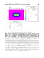
Figure 4. Schematic of the microscope and optical tweezers apparatus.
There is, however, an additional complication in making such measurements. Clearly a fixed
QPD is limited in the maximum particle displacement it can measure and as probe particles are
diffusing in 3 dimensions it is essential to provide a mechanism that ensures the particle being
tracked stays within the range of the QPD. This can be carried out with an optical tweezers ar‐
A Practical Review of Microrheological Techniques
/>13
rangement that uses a tightly focused higher-power laser to hold and manipulate micron-
sized particles [35-37]. Figure 4 shows a typical holographic optical tweezer setup (HOT) that
employs a spatial light modulator (SLM), which provides the ability to make multiple steera‐
ble traps and move objects in three dimensions using a single laser, in real time [38, 39].
Optical traps [40] formed by such an arrangement can be utilized to restrict larger-scale
movements of probe particles so they stay in the detection region of the QPD / laser appara‐
tus - essentially fencing them in, while leaving the smaller scale Brownian-motion unpertur‐
bed. At longer time lags the effect of the trap can be seen in the MSD plot, as a plateau
indicating the effect of the trap, as shown in figure 5.
Calibration of the raw photodiode voltages in order to obtain actual bead displacements are
routinely carried out by moving a probe particle a set distance across the QPD detection
area. This can be carried out either by locating a particle that is stuck to the coverslip of the
sample cell and translating the chamber a known amount using a piezo-electric stage; or by
moving a particle using a pre-calibrated optical trap. An average piezoelectric stage current‐
ly available for microscopy is able to provide nanometer resolution to displacements up to
300 microns.
Figure 5. MSD plot of a particle undertaking Brownian motion within optical traps formed with three different laser
intensities. The insert shows a particle optically trapped.
2.6. Standard experimental studies
Having described the setup and calibration of four microrheological techniques, results ob‐
tained from 3 different fluids are described and compared. Water, a glycerol-water mixture,
and several polyethylene oxide (PEO) solutions were utilized to provide three different en‐
Rheology - New Concepts, Applications and Methods
14
vironments, namely; low viscosity, high viscosity and viscoelastic fluids, to test and com‐
pare the different methods. Such samples are standards that can be quickly used to ensure
the proper functioning of the equipment and analysis before more complex biological sys‐
tems are investigated.
Samples:Water has a lower viscosity than most biological materials of interest; and thus pro‐
vides a good test of how the methodologies cope with fast particle dynamics. Glycerol is a
homogenous, purely viscous fluid and was used in combination with water (results shown
here for 62 wt%) to generate a highly viscous solution. Solutions were made by mixing glyc‐
erol (99.9% from Ajax Laboratory Chemicals) and MilliQ water, using a magnetic flea, for
approximately 2 hours. PEO, an electrically-neutral water-soluble polymer available in a
range of molecular weights was used to generate a viscoelastic polymer solution. PEO starts
to exhibit viscoelasticity at concentrations higher than the overlap concentration (approxi‐
mately 0.16 wt% for the 900 kDa PEO samples used in the following experiments). Solutions
were made by adding dry PEO powder (Acros Organics) in MilliQ water, and then slowly
mixing over approximately a 7 day period to help homogenize the solution. Solutions were
prepared at 2.2 wt% and 4 wt%, around 14 and 25 times the overlap concentration, to ensure
significant viscoelasticity [14]. The mesh size of PEO solutions at these concentrations have
been calculated to be the order of a few nanometres. As there is little evidence of surface ef‐
fects between the particles and these solutions, and the solution is then homogenous on the
length scale smaller than the particle size, it was expected that the one-and two-point micro‐
rheology should produce very similar results, and as such the system forms an ideal test of
those two methodologies.
Probe Particles: DWS measurements require a high bead concentration producing a turbid
solution and ensuring strong multiple scattering. On the other hand, optical tweezers ex‐
periments and DLS measurements require that the concentration of particles in the solution
is very low. For optical tweezers one must ensure that that there is only one particle present
in the imaging plane, any more and there is a chance that a second additional particle might
get sucked into the trap, and as discussed DLS requires that photons only be scattered a sin‐
gle time. For DLS and DWS polystyrene particles are chosen due to their low density and
good scattering properties. Silica particles are used for optical tweezers due to the high re‐
fractive index of silica, which ensures a strong trapping force. DLS and DWS experiments
were carried out for all samples with solid polystyrene probe particles (Polysciences) at con‐
centrations of 0.01% and 1%, respectively. Solutions for optical tweezers and MPT experi‐
ments were made to concentrations of 10
-6
% solid silica (Bangs Laboratories) and 10
-3
%
solid fluorescent polystyrene particles (Polysciences).
DLS experiments were performed using a set-up as shown in figure 1, specifically using a
35 milli-watt Helium Neon laser (Melles Griot) and a goniometer (Precision Devices) set
nominally to measure a 90 degree scattering angle. Measurements were taken for approxi‐
mately 40 minutes.
DWS experiments were performed using a set-up based on work originally published in
[41] and as shown in figure 1. Initially experiments were conducted using a flex99 correlator
from correlators.com and a 35 milli-watt Helium Neon laser (Melles Griot). In the quest for
A Practical Review of Microrheological Techniques
/>15
shorter lag times and higher accuracy a flex02 correlator (correlator.com) was purchased.
Experiments were first run using water to obtain l
*
of the standard solution, and then repeat‐
ed on the sample solution containing the same phase volume of scatterers. DWS experi‐
ments were typically run for approximately 40 minutes to one hour.
1
2
(a)
Figure 6. (a) Plot showing the agreement of measurements between multiple techniques in water (circles) and 62%
glycerol water mixture (squares). (b) MSD plot for 4 wt% PEO showing an agreement between data obtained using
DWS MPT and 2 point analysis (Inset: Extracted rheological properties).
MPT experiments were carried out with an inverted microscope (Nikon Eclipse TE2000-U)
on an air damped table (Photon Control) equipped with a mercury fluorescent lamp (X-cite
Series 120PC EXFO), and a 60x 1.2 NA (Nikon, Plan Apo VC 60x WI) water immersion ob‐
jective lens was used for MPT experiments. A range of different cameras were trialled: Focu‐
lus FO124SC (CCD), prototype DSI-640-mt smartcam (high speed CMOS), Hamamatsu Orca
Flash 2.8 (CMOS large detector size and pixel number). Image series were taken for approxi‐
Rheology - New Concepts, Applications and Methods
16
mately ten seconds; and x-y coordinate data extracted using a homebuilt program written
using algorithms obtained from: In-house programs
to calculate the MSD and Van Hove correlation function were used in combination with a
program to extract the rheological information obtained from: />~kilfoil/downloads.html.
QPD experiments were also carried out. The microscope used for MPT was additionally uti‐
lized to tightly focus a 2 watt 1064 nm Nd:YAG laser (spectra physics) to produce optical
traps. Particle displacements were recorded using a 2.5 mW probe laser (Thorlabs S1-
FC-675) and a QPD (80 kHz) for approximately 10 seconds. Calibration was aided using pie‐
zoelectric multi-axis stage (PI P-517.3CD).
Figure 6(a) shows a log-log plot of the three dimensional mean-square displacement of
probe-particles as a function of time; for 500 nm polystyrene particles and 1.86 micron silica
beads (optical tweezers data, normalized to 500nm) in either water or a 62 wt% glycerol/
water mixture. The mean-square displacement data shown shows an excellent agreement
between different methods and also with the expected result of a slope of one (for diffusion
in a viscous medium). Figure 6 (b) shows a similar log-log plot of the mean-square displace‐
ment versus time for 4 wt%, PEO solutions, together with a fit to a sum of power laws with
exponents of ~0.4 and ~0.9, in good agreement with previous work. The inset shows the ex‐
tracted frequency dependent viscoelastic properties that appear in good agreement with
previously published work [14].
2.7. Comparison of with bulk rheometry
The efficiency of these microrheological methods can be assessed against conventional rhe‐
ometry. Figure 7 reports the elastic modulus G’ and the loss modulus G” for a 30 wt% aque‐
ous dextran solution. G’ and G” were obtained using DWS or by the use of a commercial
rheometer (TA 2000 rheometer, fitted with a cone-and-plate geometry).
The experimental data were fitted using the Maxwell model:
2 2
0 0
2 2 2 2
' ; '
1 1
G G
G G
w t w t
w t w t
= =
+ +
(20)
where G
0
is the plateau elastic modulus, τ is the relaxation time, and ω (ω=2 f, f the frequen‐
cy) the angular frequency. The fit using the Maxwell model allows showing the continuation
in the experimental data using the two methods. Further the combination of the two techni‐
ques allows the determination of the rheological behaviour over more than 7 decades in fre‐
quency. However, at low frequencies, some discrepencies between G’ obtained by rheology
and the Maxwell model can be observed. This is likely due to the geometry inertia affecting
rheological measurements.
A Practical Review of Microrheological Techniques
/>17