Báo cáo khoa học: Molecular identification of adrenal inner zone antigen as a heme-binding protein potx
Bạn đang xem bản rút gọn của tài liệu. Xem và tải ngay bản đầy đủ của tài liệu tại đây (536.93 KB, 12 trang )
Molecular identification of adrenal inner zone antigen
as a heme-binding protein
Li Min
1
*, Natallia V. Strushkevich
1,2
*, Ivan N. Harnastai
2
, Hiroko Iwamoto
3
, Andrei A. Gilep
1,2
,
Hiroshi Takemori
1
, Sergey A. Usanov
2
, Yasuki Nonaka
3
, Hiroshi Hori
4
, Gavin P. Vinson
5
,
Mitsuhiro Okamoto
1,6
1 Department of Molecular Physiological Chemistry, Graduate School of Medicine, Osaka University, Japan
2 Institute of Bioorganic Chemistry, National Academy of Sciences of Belarus, Minsk, Belarus
3 College of Nutrition, Koshien University, Hyogo, Japan
4 Graduate School of Engineering Science, Osaka University, Japan
5 School of Biological Sciences, Queen Mary University of London, UK
6 Laboratories for Biomolecular Networks, Graduate School of Frontier Biosciences, Osaka University, Japan
Distinguished histologically, the three zones in the
mammalian adrenal cortex have distinct functions. In
man, the outermost zona glomerulosa secretes aldo-
sterone, the intermediate zona fasciculata, cortisol,
and the innermost zona reticularis is the main site
for dehydroepiandrosterone formation, whereas in
the rat, corticosterone is the main product of the
fasciculata and reticularis, with little if any dehydro-
epiandrosterone. The molecular mechanisms under-
lying the functional differentiation of the three zones
have been a focus of numerous investigations [1–3].
To facilitate the study of zonal function, Laird et al.
[4] produced a monoclonal antibody that recognizes
an antigen, named inner zone antigen (IZA), which
is present in the zonae fasciculata ⁄ reticularis in the
rat, but not in the zona glomerulosa. Here we call
this antigen, which was originally identified in rat
tissue, ‘rIZA1’. The monoclonal antibody was
capable of inhibiting dose-dependently adrenal 21-
hydroxylation of progesterone and 18-hydroxylation
Keywords
adrenal inner zone antigen; heme-binding
protein; membrane-associated progesterone
receptor; steroidogenesis; zonae fasciculata
and reticularis
Correspondence
M. Okamoto, Department of Biochemistry
and Molecular Biology, Graduate School of
Medicine (H-1), Osaka University,
2-2 Yamadaoka, Suita, Osaka 565-0871,
Japan
Fax: +81 6 6879 3289
Tel: +81 6 6879 3280
E-mail:
*Note
These two authors contributed equally to
this paper
(Received 21 July 2005, revised 12 September
2005, accepted 16 September 2005)
doi:10.1111/j.1742-4658.2005.04977.x
The adrenal inner zone antigen (IZA), which reacts specifically with a
monoclonal antibody raised against the fasciculata and reticularis zones of
the rat adrenal, was previously found to be identical with a protein vari-
ously named 25-Dx and membrane-associated progesterone receptor. IZA
was purified as a glutathione S-transferase-fused or His
6
-fused protein, and
its molecular properties were studied. The UV-visible absorption and EPR
spectra of the purified protein showed that IZA bound a heme chromo-
phore in high-spin type. Analysis of the heme indicated that it is of the
b type. Site-directed mutagenesis studies were performed to identify the
amino-acid residues that bind the heme to the protein. The results suggest
that two Tyr residues, Tyr107 and Tyr113, and a peptide stretch, D99–
K102, were important for anchoring the heme into a hydrophobic pocket.
The effect of IZA on the steroid 21-hydroxylation reaction was investigated
in COS-7 cell expression systems. The results suggest that the coexistence
of IZA with CYP21 enhances 21-hydroxylase activity.
Abbreviations
IZA, inner zone antigen; GST, glutathione S-transferase; MPR, membrane-associated progesterone receptor.
5832 FEBS Journal 272 (2005) 5832–5843 ª 2005 FEBS
of 11-deoxycorticosterone. When rat adrenal homo-
genates were subjected to SDS ⁄ PAGE followed by
immunoblot analysis, two proteins of molecular mass
27–28 kDa and 55–60 kDa reacted with the mono-
clonal antibody [5]. The larger protein was thought
to be a dimer of the smaller protein. rIZA1
appeared to be distributed not only in the adrenal
cortex but also in other tissues [6,7].
Using the monoclonal antibody immobilized to Seph-
arose beads, Raza et al. [8] successfully purified rIZA1
and determined its N-terminal amino-acid sequence.
The sequence was found to be consistent with that of a
protein reported previously as ‘25-Dx’ (GenBank acces-
sion number U63315) [9] or ‘membrane-associated prog-
esterone receptor (MPR)’ (GenBank accession number
AJ005837) [10]. In the human genome sequence, two
genes encode IZA; one, Hpr6.6 (accession number
NM_006667), encodes a protein corresponding to
rIZA1, which we name here hIZA1, and the other, Dg6
(accession number NM_006320), encodes a protein sim-
ilar to, although distinctly different from, the protein
named hIZA2 here [11]. Complementary DNA encoding
25-Dx was isolated as one of the dioxin-inducible genes
in rat liver [9], whereas MPR had been purified from
porcine liver [12] and its cDNA from porcine vascular
smooth muscles [10]. The cloned sequence of rIZA1 was
identical with that of MPR, although somewhat differ-
ent from that of 25-Dx at the 3¢-terminal. It is possible
that a splicing error occurred during the preparation of
25-Dx cDNA. rIZA1 has also been reported as ‘ventral
midline antigen’, a protein expressed in the rat central
nervous system [13]. A yeast ortholog of IZA was
recently reported as ‘Damage response protein related
to membrane-associated progesterone receptors I pro-
tein’ (Dap1p) [14]. As this brief review indicates, IZA
has been studied by many investigators from a variety of
viewpoints and a variety of biological functions have
been attributed to it. However, its precise physiological
role is still unclear. To investigate this point further,
IZA was purified to homogeneity to examine its mole-
cular nature. Our preliminary results suggest that IZA
contains a heme chromophore [15]. Mallory et al. [16]
also reported recently that Dap1p, the yeast homolog of
IZA, is a heme-binding protein. Here we report our
further characterization of human and rat IZAs.
Results and Discussion
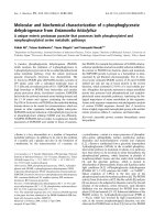
The domain structure of IZA was explored by inputting
its amino-acid sequence into a protein domain structure
prediction program in the website, ger.
ac.uk/cgi-bin/Pfam/nph-search.cgi. The results illustra-
ted in Fig. 1A suggested that IZA contains a heme ⁄
steroid-binding domain similar to a heme-binding
domain of cytochrome b
5
. The 134-amino-acid protein
human cytochrome b
5
has a heme-binding domain of
80 residues near its N-terminus in which His44 and
His68 act as the sixth-axial and fifth-axial ligands for
the heme iron, respectively. The transmembrane region
of 20 amino acids is located near the C-terminus. Con-
versely, hIZA1, a protein of 195 amino acids, has a
transmembrane region at its N-terminal side, and the
predicted heme ⁄ steroid-binding domain is located in the
central portion. The aligned amino-acid sequences of
hIZA1, rIZA1, and hIZA2 are shown in Fig. 1B, in
which the transmembrane regions and the heme ⁄ steroid-
binding domains are highlighted in yellow and red,
respectively. Amino acids in the heme ⁄ steroid-binding
domain are well conserved among the three proteins
(shown in bold letters). This strongly suggests that this
domain plays an important role in the physiological
function of IZA. The amino-acid sequence of the heme-
binding domain of cytochrome b
5
(shown in blue) was
aligned with those of the heme ⁄ steroid-binding domain
of IZA. Surprisingly, the similarity between IZA and
cytochrome b
5
was rather weak, and only 15 out of 82
residues are identical (highlighted in green; the residues
covered with dark green shade are identical residues,
whereas those with light green are similar). It should be
noted that hIZA1 contains only three His residues, all
located outside the heme ⁄ steroid-binding domain: one,
His23, near the N-terminus and the others, His165 and
His166, near the C-terminus (the numbering is that of
hIZA1). hIZA2 has only one His near the C-terminus.
These findings raised the question whether the
heme ⁄ steroid-binding domain of IZA actually functions
as a specific heme-binding site. We therefore purified
IZA and examined its molecular properties.
IZA was expressed as either a His
6
-fused protein or
a glutathione S-transferase (GST)-fused protein in
Escherichia coli and purified to homogeneity. The puri-
fied protein was tinged with brown, a color clearly
distinct from the bright red color of the similarly
expressed and purified cytochrome b
5
(not shown). The
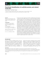
UV and visible light absorption spectra of His
6
-rIZA1
are shown in Fig. 2A, revealing the oxidized form of
the heme chromophore, with a sharp c-absorption
peak at 402 nm and broad absorptions between
497 nm and 616 nm (shown in green). When the sam-
ple was treated with sodium dithionite, the spectra
were converted into those of the reduced heme chro-
mophore with distinct a and c peaks at 559 nm, and
426 nm, respectively (shown in red). The addition of
CO to the reduced sample changed the spectra into a
CO-binding form with a, b and c peaks at 567 nm,
538 nm and 420 nm respectively (shown in blue). The
L. Min et al. Molecular properties of adrenal inner zone antigen
FEBS Journal 272 (2005) 5832–5843 ª 2005 FEBS 5833
incubation of the oxidized form with either NADH
and NADH-cytochrome b
5
reductase or NADPH and
NADPH-cytochrome P450 reductase did not influence
the absorption spectra. As shown in supplementary
material Tables S1 and S2, hIZA1 and rIZA1 had
essentially similar spectral properties, no matter whe-
ther they were expressed as His
6
-tagged proteins or
GST-tagged proteins.
The nature of the heme bound to IZA was further
studied by measuring EPR spectra (shown in Fig. 2B).
The spectra of rat GST-rIZA1 at either 5 K or 15 K
showed high-spin type signals with g values near 6.0
and 2.0. Unlike those of oxidized myoglobin, the EPR
signals showed strong anisotropy; the signals near g ¼
6.0 appeared to be a mixture of two components. The
major component had larger anisotropy (g
1
¼ 6.44
and g
2
¼ 5.57) and the minor, smaller anisotropy
(g
1
¼ 6.10 and g
2
¼ 5.90). When
14
NO was added to
the reduced form (Fig. 2C), the EPR spectra revealed
a
14
NO-bound penta-co-ordinated heme, indicating
that the co-ordination between heme iron and an
amino acid was disrupted upon binding of NO. hIZA1
and hIZA2 yielded spectra essentially similar to those
of rIZA1 whether purified as (His)
6
-fused proteins or
GST-fused proteins. Taken together these EPR proper-
ties suggest that IZA, like myoglobin, contains a high-
spin type heme. However, unlike myoglobin which has
His as the fifth ligand for heme iron, the ligand of
IZA may not be a single amino acid. Rather, it is poss-
ible that two amino acids each partially contribute to
binding the heme, producing the mixture of two aniso-
tropic EPR signals. The fact that His
6
-IZA showed
essentially the same EPR spectra as GST-IZA excludes
the possibility that the heme is nonspecifically bound
to an imidazole group contained in the His tag.
Acid ⁄ acetone treatment of rIZA1 released heme
from the protein. Aliquots of hemin were added to the
apoprotein thus prepared, and A
402
was monitored
A
B
C
Fig. 1. Primary structure of IZA reveals a
heme ⁄ steroid-binding domain. (A) Outline of
primary structures of IZA and cytochrome b
5
.
The predicted heme ⁄ steroid-binding domain
and transmembrane domain are illustrated.
(B) Alignment of entire sequences of hIZA1,
hIZA2 and rIZA1. A sequence of the heme-
binding region of cytochrome b
5
was also
aligned (blue). Asterisks indicate amino-acid
residues that were mutated in this study. (C)
Prediction of the 3D structure of hIZA1. The
structure of bovine cytochrome b
5
(1CYO) is
shown in the left panel [34,35]. The numbers
of the two axial ligand His residues indicate
those predicted from cDNA (M63326). The
model of hIZA1 was illustrated by using
the
LOOPP program (.
cornell.edu/loopp.aspx). As a template for the
modeling, the most similar protein, a cyto-
chrome b
5
homolog of Ectothiorhodospira
vacuolata (1CXY), was used [36]. The model
of hIZA1 is shown in the right panel in an
orientation similar to that of bovine cyto-
chrome b
5
. The secondary structures of the
two molecules are colored similarly, except
the red stretch of b-sheet in hIZA1 highligh-
ted to indicate the region important for heme
binding. Two Tyr residues that may play a
role in heme binding are shown in yellow.
Molecular properties of adrenal inner zone antigen L. Min et al.
5834 FEBS Journal 272 (2005) 5832–5843 ª 2005 FEBS
(Fig. 2D). This titration revealed a reflection point
where 4 lm hemin was added to 5 lm apoprotein,
apparently indicating that one molecule of rIZA1
maximally bound 0.8 molecule of heme. However, the
absorption coefficient of the rIZA1-bound heme at
402 nm may be different from that of free heme.
Therefore, we examined this point further.
The A
280
of a protein molecule can be calculated
based on the content of aromatic amino acids, and our
calculation suggested that 10 lm GST-hIZA1 would
have an A
280
of 0.357 absorbance unit. On the other
hand, when we added small amounts of hemin drop-
wise to a 20 lm apo-GST-hIZA1 solution and recorded
A
402
, the difference in absorbance between the sample
added with 1 lm hemin and that added with 5 lm was
0.263, suggesting that 1 lm bound heme would have
an A
402
of 0.0658 absorbance unit. Thus, if heme
bound to 10 lm GST-hIZA1 stoichiometrically, the
A
402
of the holoprotein would be 0.658 absorbance
unit, and we can determine the value A
402
⁄ A
280
of the
A
BD
C
E
Fig. 2. IZA1 contains a protoheme. (A) UV and visible light absorption spectra of His
6
-rIZA1. The oxidized form absorption spectrum (green),
the reduced spectrum taken after the addition of sodium dithionite (red) and the CO-bound spectrum (blue) are shown. (B) EPR spectra of
the oxidized forms of GST-rIZA1, horse heart myoglobin and human cytochrome b
5
are shown. (C) EPR spectrum of the
14
NO-bound form
of GST-rIZA1 at 35 K. (D) Titration of apo-rIZA1 with hemin. (E) GST-rIZA1 was treated under various conditions, subjected to SDS ⁄ PAGE,
and stained by the peroxidase reaction (left panel). The right panel shows Coomassie blue staining of the same gel.
L. Min et al. Molecular properties of adrenal inner zone antigen
FEBS Journal 272 (2005) 5832–5843 ª 2005 FEBS 5835
holoprotein as 0.658 ⁄ 0.357 ¼ 1.84. In the meantime
the maximal value of A
402
⁄ A
280
that we obtained for
several purified samples was 1.07. This suggested that
one molecule of the purified GST-hIZA1 contained
about 0.6 molecule of heme at most. This value was
reasonably consistent with the approximate value
obtained from the result of Fig. 2D.
Several heme-binding proteins are known to bind
heme tightly, so that the proteins can be detected as
peroxidase reaction-stained bands even when subjected
to electrophoresis in SDS-containing gels. To charac-
terize the heme-binding nature of IZA1, purified GST-
rIZA1 was subjected to SDS ⁄ PAGE, and then the gel
was stained by the peroxidase reaction (Fig. 2E left
panel). As shown in the first lane from the left, three
bands appeared, with molecular masses of 50 kDa,
85 kDa and 130 kDa, suggesting that heme was still
bound to the monomeric, dimeric, and trimeric forms
of GST-rIZA1. (The theoretical molecular mass of
GST-rIZA1 is 60.2 Da.) Similar bands appeared in the
lane loaded with heat-denatured GST-rIZA1 (the sec-
ond from the left). Thus, heme bound to GST-rIZA1
seemed not to be released from the protein even when
treated in boiling water. In a lane loaded with the
sample pretreated with heat in the presence of dithio-
threitol, the relevant peroxidase-reaction-stained bands
disappeared, suggesting that heme was released from
the protein after these treatments, although another
interpretation may be that dithiothreitol treatment
reduced the heme iron, making it negative to peroxi-
dase activity. In any case, these results indicate that
IZA binds heme relatively tightly.
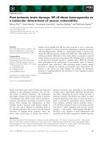
When treated with pyridine under alkaline condi-
tions, the heme molecule produces a pherochrome
complex with characteristic absorption spectra. Fig-
ure 3A illustrates the redox difference absorption spec-
tra of pyridine pherochromes prepared from rIZA1,
myoglobin and cytochrome c oxidase. The spectra of
rIZA1-derived pherochrome, like those derived from
myoglobin, but unlike those derived from cyto-
chrome c oxidase, had peaks at 419 nm, 525 nm and
556 nm, suggesting that heme bound to rIZA1 is of
type b, not of type a. To confirm this point, heme
extracted from rIZA1 was subjected to HPLC analysis
(Fig. 3B). The results show that heme derived from
rIZA1 had the same retention time as that from myo-
globin. These results again suggest that rIZA1 contains
type b heme, not type a heme.
Fig. 3. IZA1 contains a type b heme. (A) The redox-difference adsorption spectra of pyridine-hemochromogens prepared from His
6
-rIZA1,
GST-rIZA1, myoglobin and cytochrome c oxidase. The absorption spectra of hemochromogens derived from myoglobin and rIZA1s had peaks
at 556 nm, 524 nm and 419 nm, whereas that derived from cytochrome c oxidase, at 588 nm, 536 nm and 431 nm. (B) The hemes released
from GST-rIZA1, myoglobin and cytochrome c oxidase, and free hemin were subjected to HPLC analysis. The retention times of the hemes
extracted from rIZA1 and myoglobin were 28.9 min and 29.2 min, that of hemin 28.4 min, and that of cytochrome c oxidase 38.4 min.
Molecular properties of adrenal inner zone antigen L. Min et al.
5836 FEBS Journal 272 (2005) 5832–5843 ª 2005 FEBS
To determine which amino-acid residue interacts with
heme in IZA1, a variety of mutant IZA1s were produced
in which amino acids thought to bind the heme ligand
had been disrupted. The purified mutants were then
evaluated for their heme absorption. Because an imida-
zole group often plays a role in binding heme in many
heme proteins, we first introduced mutations into
His165 and His166 in hIZA1, even though they are
located outside the predicted heme ⁄ steroid-binding
domain. H165N-hIZA1 and H166N-hIZA1, however,
were found capable of binding heme as strongly as the
wild-type (not shown). Amino-acid side-chain groups
other than imidazole that could interact with heme
molecule are thiol and phenol. Noting that Tyr107,
Tyr113, Tyr139, and Cys129 are present in the
heme ⁄ steroid-binding region, and moreover are con-
served in hIZA1, hIZA2, and rIZA1, mutants Y107F,
Y113F, Y139F, and C129A were produced. The heme
absorptions of these mutants, however, again seemed
not significantly diminished compared with that of the
wild-type. We tested further mutants, such as Y43F,
Y164F, Y180F and P109A, but none of these single-
amino-acid mutants seemed to lose heme-binding capa-
bility completely. When two phenol groups, Tyr107 and
Tyr113, were disrupted, the mutant appeared substan-
tially to lose its capacity to bind heme (Fig. 4A). In con-
trast, another double mutant, Y164F ⁄ H166N-hIZA1,
retained heme-binding capacity (not shown). When
mutations were introduced into a four consecutive
amino-acid stretch from Asp99 to Lys102, the mutant
bound heme at a level of 10% of the wild-type (Fig. 4A).
It should be noted that three amino-acid residues in this
tetrapeptide, Asp99, Thr101 and Lys102, are conserved
in IZA and cytochrome b
5
.
The 3D structure of the heme-binding pocket of
bovine cytochrome b
5
was adopted from the previously
published crystallographic study (left panel in Fig. 1C).
Next the 3D structure of the heme ⁄ steroid-binding
domain of IZA was modeled based on that of the
Ectothiorhodospira vacuolata cytochrome b
5
homolog
(accession number 1CXY) and shown in the same ori-
entation as that of bovine cytochrome b
5
(right panel
in Fig. 1C). The simulated structure revealed a heme-
binding pocket surprisingly similar to that of cyto-
chrome b
5
, with a space large enough to accommodate
a heme molecule. Interestingly, if a heme were inserted
into this pocket, those residues mentioned above for
their importance in the interaction with heme, i.e.
Tyr107, Tyr113 and the tetrapeptide, D99–K102, seem
to be located at one side of the heme molecule (the
upper side in this orientation), constituting the ceiling
of the heme-binding pocket.
Determination of the intracellular localization of
IZA would provide insights into its physiological
function. IZA1 was first reported as MPR and puri-
fied from the membrane fractions of rat liver homo-
genates. Immunohistochemical observations of other
investigators revealed that this protein forms vesicle-
like structures in cells. Moreover, the predicted
domain structure of IZA indicated that it contains a
transmembrane region (Fig. 1A). All these previous
reports indicate that IZA1 is a membrane-associated
protein. However, to which intracellular membrane
compartment IZA1 is associated is not clear. To
determine the intracellular localization of IZA1 more
precisely, we expressed rIZA1, cytochrome b
5
,an
endoplasmic reticulum-associated protein, and
CYP11B1, a mitochondrial inner membrane-associated
protein, in HeLa cells. The cells were stained with the
specific antibodies directed against the respective pro-
teins. As shown in Fig. 5, rIZA1 was distributed dif-
fusely in the cell, forming vesicular structures,
suggesting its association with the membrane compart-
ments [8]. In addition, rIZA1 appeared to be some-
what concentrated at a perinuclear region. The
intracellular location of rIZA1 was completely consis-
tent with that of coexpressed cytochrome b
5
, but not
with that of coexpressed CYP11B1. These results
suggest that IZA1 is associated with the endoplasmic
reticulum membrane.
A
B
Fig. 4. (A) The heme-binding capacities of the wild-type hIZA1 and
mutants. The capacities were estimated by measuring the
A
402
⁄ A
280
ratios (mean and SD of triplicate expression). The purifi-
cation of the hIZA1s was given in Experimental procedures. (B) The
amounts of protein used for the heme absorbance measurement
were shown by immunoblot analysis.
L. Min et al. Molecular properties of adrenal inner zone antigen
FEBS Journal 272 (2005) 5832–5843 ª 2005 FEBS 5837
Given that IZA1 is abundantly present in the endo-
plasmic reticulum of zona fasciculata cells, it would be
reasonable to speculate that it is involved in the physi-
ology of the adrenal cortex. We indeed reported previ-
ously that the steroid 21-hydroxylation reaction, which
is essential for biosynthesis of corticosteroids, was
enhanced in the presence of rIZA1 [10]. We re-exam-
ined this by expressing CYP21 together with hIZA1 or
its mutants in COS-7 cells (Fig. 6A). Secretion of
11-deoxycorticosterone, the CYP21 reaction product
from progesterone, was increased about twofold by
coexpressing the wild-type hIZA1, whereas it was
depressed by 75% by coexpressing the D99–K102-
mutated hIZA1, and increased by 60% by coexpress-
ing the Y107F ⁄ Y113F-hIZA1 (Fig. 6A). When the
levels of expressed hIZA1s in cell homogenates
were examined, the D99–K102-mutated hIZA1 was
expressed at a higher level than the wild-type hIZA1
(Fig. 6A, lower panel), suggesting that this mutant was
fairly stable in the cells, although in this experiment
we could not confirm the mutant’s intracellular local-
ization as the endoplasmic reticulum. On the other
hand, the level of expressed CYP21 in these cells
seemed to be slightly lower than in the wild-type
hIZA1-expressing cells.
To exclude the possibility that D99–K102-mutated
hIZA1 repressed the expression of CYP21 protein by
inhibiting the promoter used for CYP21 expression,
firefly luciferase cDNA was introduced into the vector
instead of CYP21 cDNA, and the promoter activities
were measured in the wild-type hIZA1-expressing cells
and the mutant hIZA1-expressing cells. Overexpression
of hIZA1, whether wild-type or mutant, did not influ-
ence the promoter activity of the expression vector
(Fig. 6B), suggesting that the slightly lower concentra-
tion of CYP21 protein in the D99–K102-mutated
hIZA1-expressing cells may be due to a post-trans-
lational event; possibly, CYP21 protein instability is
induced by the coexistence of the D99–K102-mutated
hIZA1.
Next, we tested the possibility that IZA directly regu-
lates the CYP21-dependent steroid hydroxylation reac-
tion by using the microsomal P450 electron-transport
reconstitution system. As shown in Fig. 6C, the addi-
tion of rIZA1 failed to stimulate the CYP21-catalyzed
21-hydroxylation of progesterone in the reconstituted
system. Rather, the hydroxylation activity seemed to be
inhibited in the presence of a large amount of rIZA1.
Although we cannot explain this phenomenon beyond
doubt, the involvement of a hydrophobic protein such
as rIZA1 in the reconstitution system may disturb the
smooth conduct of the electron transport to the CYP21
molecule. The effect of rIZA1 on the CYP17-dependent
hydroxylation reactions was also tested in comparison
with the effect of cytochrome b
5
, because the latter is
well known to regulate the CYP17-mediated 17a-hy-
droxylation reaction and the consecutively occurring
17,20-lyase reaction [17]. As reported previously, the
presence of cytochrome b
5
in the CYP17-reconstitution
system seemed not to influence the 17-hydroxylation of
progesterone, but it indeed activated the lyase reaction
of 17a -hydroxyprogesterone (Fig. 6D). In contrast, the
presence of rIZA1 seemed to influence neither one of
the reactions. We surmised therefore that IZA1 could
activate the CYP21-dependent reaction in the trans-
formed cells, but this activation may not be caused by
the direct interaction of IZA1 with the microsomal
P450 electron-transport components, as seems to be the
case for cytochrome b
5
.
Taken together, the results presented here show that
IZA1 is a heme-binding protein present in the endoplas-
mic reticulum membrane. The primary structure of its
heme-binding region looks slightly similar to that of
cytochrome b
5
, presumably forming a hydrophobic
pocket. The heme in IZA1 is type b, and binds to the
protein in high-spin type. To identify the amino-acid
residues involved in binding to the heme, extensive site-
directed mutation studies were conducted. However, the
results remain somewhat ambiguous. Nevertheless, it is
possible to conclude that the heme ⁄ steroid-binding
region in IZA1 constitutes a hydrophobic pocket that
could accommodate a heme molecule, and, in this
pocket, two Tyr residues, Tyr107 and Tyr113, and a
peptide stretch D99–K102 play important roles in
attaching the heme iron to one side of the protoporphy-
rin ring. Mallory et al. [16] recently reported the nature
of Dap1p, the yeast homolog of IZA1, which also seems
to bind heme.
Fig. 5. Intracellular localization of rIZA1, rat CYP11B1 and human
cytochrome b
5.
HeLa cells were cotransformed with rIZA1 and
CYP11B1 or cytochrome b
5.
After 24 h, cells were fixed and sub-
jected to immunocytochemistry by using anti-rIZA1 monoclonal
antibody [4], anti-CYP21 polyclonal antibody [32], or anti-(cyto-
chrome b
5
) Ig [33].
Molecular properties of adrenal inner zone antigen L. Min et al.
5838 FEBS Journal 272 (2005) 5832–5843 ª 2005 FEBS
As IZA1 cDNA was first isolated as MPR, we tried
assaying its progesterone-binding activity under var-
ious conditions. For instance, [
3
H]progesterone was
incubated with GST-rIZA1 and the extent of isotope
binding to the protein was estimated by GST pull-
down assays. The results failed to show specific bind-
ing of the isotope to rIZA1 (supplementary Table S3).
The addition of an IZA1 antibody to the incubation
mixture of the radioactive progesterone and rIZA1
also failed to show specific isotope binding. Therefore
our investigation has so far failed to establish that
rIZA1 specifically binds progesterone.
Although the results presented here cannot conclu-
sively establish the precise physiological role(s) played
A
C
D
B
Fig. 6. Effects of IZA1 on the CYP21 reactions. (A) Wild-type hIZA1 and mutant hIZA1s were coexpressed with CYP21 in COS-7 cells. The
conversion of progesterone into 11-deoxycorticosterone was measured as given in Experimental procedures. Lower panels show the levels
of expressed CYP21 and hIZA1s by immunoblot analysis. (B) To test the effect of hIZA1 on the promoter activity of CYP21 plasmid, pSVL-
fLuc plasmid was cotransformed with hIZA1s expression vectors and Renilla luciferase vector as an internal standard. The promoter activity
of pSVL was normalized to Renilla luciferase activity. (C) Effect of IZA on the CYP21 reaction in the reconstitution system. The reaction mix-
ture contained, in a final volume of 1 mL, various amounts of (His)
6
-rIZA1, 25 pM CYP21, 50 pM NADPH-P450 reductase, 50 lM progester-
one, 0.5 m
M NADPH, 8 mM isocitrate, 0.1 U isocitrate dehydrogenase, 50 mM Tris ⁄ HCl, pH 7.4, and 10 mM MgCl
2
. The reactions were
carried out at 37 °C for 2 min. (D) Effect of rIZA1 or cytochrome b
5
on guinea pig CYP17 reactions in the reconstitution system. The reaction
mixture contained, in a final volume of 1 mL, 0.25 l
M CYP17, 0.5 lM NADPH-P450 reductase, 50 lM progesterone or 17a-hydroxyprogester-
one, 0.5 m
M NADPH, 8 mM isocitrate, 0.1 U isocitrate dehydrogenase, 50 mM Tris ⁄ HCl, pH 7.4, and 10 mM MgCl
2
. The reactions were
carried out with or without 0.25 l
M His
6
-rIZA1, or 0.25 lM cytochrome b
5
,at37°C for 2 min. After the reactions, steroids were extracted
and analyzed as described previously [36].
L. Min et al. Molecular properties of adrenal inner zone antigen
FEBS Journal 272 (2005) 5832–5843 ª 2005 FEBS 5839
by IZA in the adrenal cortex, we surmise that this
heme-containing microsomal protein may have a role
in supplying heme molecules to cytochrome P450-
involved reactions and eventually influence adrenal
steroidogenesis. A similar role of Dap1p in the
CYP51-catalyzed reaction in yeast has been suggested
by Mallory et al. [16].
Experimental procedures
Materials
A plasmid containing hIZA1 cDNA (IMAGE clone, No.
5300612) and a transfection reagent, lipofectamine 2000
tm
,
were purchased from Invitrogen (Carlsbad, CA, USA).
pGEX-6p-3 vector was from Amersham Bioscience (Piscat-
away, NJ, USA). pTargeT vector and pGL3 luciferase
reporter assay system were obtained from Promega (Madi-
son, WI, USA). Restriction endonuclease and E. coli
strains, JM109 and Bl21, were purchased from Takara
(Kyoto, Japan) and Toyobo (Osaka, Japan), respectively.
QuikChange XL Site-Directed Mutagenesis Kit was from
Stratagene (La Jolla, CA, USA).
Construction of plasmids
A BamHI site (GAATTC) was created at a point before the
starting Met codon of hIZA1 cDNA by site-directed muta-
genesis. The cDNA containing both coding and 3¢ noncoding
regions was prepared by BamHI and NotI digestion, and sub-
cloned into the BamHI ⁄ NotI site of pGEX-6P-3 or pTargeT.
The resultant plasmids were named pGEX-hIZA1 and pTar-
geT-hIZA1, respectively. Point mutations of hIZA1 were
produced by site-directed mutagenesis using pGEX-hIZA1
as a template. To obtain N-terminal His
6
rat IZA, cDNA
was PCR amplified with 5¢ primer containing the NcoI site
(TACCATGGCTGCCGAGGATG) and 3¢ primer contain-
ing the HindIII site (CAAGCTTCAGTCACTCTTCC
GAGC). The PCR product was digested with NcoI and Hin-
dIII and subcloned into the NcoI ⁄ HindIII site of pRSET-B.
The resulting construct containing N-terminal His
6
was
recloned into pCW vector using NdeI and HindIII.
Expression and purification of IZA1
IZA1 was expressed in E. coli JM109 as GST-fused or
(His)
6
-fused protein as reported previously [18,19] with
some modifications. For the purification of the GST-fused
protein, E. coli JM109 transformed with pGEX-hIZA1 was
grown in 1 L 2YT (yeast ⁄ tryptone) medium containing
0.2 mm d-aminolevulinic acid hydrochloride at 30 ° C. When
culture growth reached D
600
0.7, 0.1 mm isopropyl thio-
b-d-galactopyranoside was added to the medium, and cul-
ture was continued for 16 h at 25 °C. E. coli was harvested
from the culture solution, and GST-hIZA1 expressed was
purified as described previously [19]. (His)
6
-rIZA1 was
coexpressed with glutamyl-tRNA reductase (hemA ⁄ gtrA)
[20]. E. coli JM109 cotransformed with the pCW-rIZA1
and pHg2 (hemA) was grown in 3000 mL TB (terrific
broth) medium containing 100 lgÆmL
)1
ampicillin,
25 lgÆmL
)1
chloramphenicol and microelements. The cul-
ture was performed at 37 °C until the cell density reached
D
600
¼ 0.6–0.8. Then 0.5 mm isopropyl thio-b-d-galacto-
side, 100 lgÆmL
)1
ampicillin and 25 lgÆmL
)1
chlorampheni-
col were added to induce protein expression. The cells were
grown for a further 24 h at 29 °C, harvested, and frozen at
)70 °C for later use. The frozen cells were thawed in
100 mL 50 mm Tris ⁄ HCl buffer, pH 7.5, containing 20%
(v ⁄ v) glycerol and 0.3 m NaCl, and sonicated on ice using
a Tomy Ultrasonic disruptor UD-200. Proteins associated
with the membrane fraction of sonicates were solubilized
by the dropwise addition of 10% (w ⁄ v) sodium cholate to
the final concentration of 1%. To the solution containing
the solubilized proteins, imidazole was added to a final
concentration of 5 mm. The solution was loaded on to a
Ni ⁄ nitrilotriacetate ⁄ agarose column to absorb the His
6
-
fused proteins. The proteins were eluted with the 50 mm
Tris ⁄ HCl buffer, pH 7.5, containing 20% (v ⁄ v) glycerol,
0.2% (w ⁄ v) sodium cholate and 50 mm histidine, and His
6
-
rIZA1 eluted in colored fractions was further purified by
hydroxyapatite column chromatography.
Preparation of mutated hIZA1s used for
measuring heme-binding capacity
E. coli BL21 was used for expressing GST-hIZA1 and its
mutants. The transformed E. coli was cultured in 2YT med-
ium without d-aminolevulinic acid, and induction of protein
expression was initiated as described above. The cells har-
vested were suspended in buffer A (50 mm Tris ⁄ HCl, 1 mm
EDTA, 300 mm NaCl, pH 8.0), sonicated on ice, and lysed
with 2% (v ⁄ v) Triton X-100. The lysate was then centri-
fuged at 8000 g for 30 min, and the supernatant recovered
was applied to a glutathione–Sepharose column (Amersham
Bioscience) pre-equilibrated with buffer B [buffer A con-
taining 0.1% (v ⁄ v) Triton X-100 and 5% (v ⁄ v) glycerol].
The column was washed with 2 column vol. buffer B. GST-
hIZA1 protein was retained on the column at this step,
although it was uncolored because it had been expressed
without d-aminolevulinic acid. To obtain the protein in a
heme-bound form, 0.05 mm hemin chloride dissolved in
buffer B containing 1% dimethyl sulfoxide was loaded on
to the column. The column was then washed with 5 times
the column volume of buffer B and 5 times the column vol-
ume of buffer C [50 mm Tris ⁄ HCl, 1 mm EDTA, 100 mm
NaCl, 5% (v ⁄ v) glycerol, and 0.5% (w ⁄ v) sodium cholate].
GST-hIZA1 was finally eluted from the column with buffer
C containing 10 mm glutathione. The purified protein was
Molecular properties of adrenal inner zone antigen L. Min et al.
5840 FEBS Journal 272 (2005) 5832–5843 ª 2005 FEBS
dialyzed against buffer C, and its absorption spectrum
recorded to evaluate its heme-binding capacity.
Spectrophotometric analysis
UV-visible absorption spectra were measured using a JASCO
V-550 UV ⁄ VIS spectrophotometer system. Photometric
determination of heme type was performed by a pyridine
hemochrome method [21]. The heme type was also confirmed
by HPLC analysis. Heme bound to IZA1 was extracted by
acetone ⁄ HCl followed by ethyl acetate treatment, and sub-
jected to HPLC using the Shimadzu CL-10A HPLC system
equipped with a reverse-phase column (YMC-Pack, ODS-
A303, S-5, 250 · 4.6 mm) as described by Fromwald et al.
[22]. Heme a, which was used as the standard, was extracted
from bovine heart cytochrome c oxidase purified by the
method of Yonetani [23]. Horse skeletal muscle myoglobin
and hemin were obtained from Wako Pure Chemical indus-
tries, Ltd (Osaka, Japan) and ICN Pharmaceuticals, Inc.
(Irvine, CA, USA), respectively.
Preparation of apo-rIZA1
Cold acid ⁄ acetone solution [0.2% (v ⁄ v) HCl; )20 °C;
10 mL] was added dropwise to 20 nmol rIZA1 dissolved in
0.5 mL potassium phosphate buffer (10 mm, pH 7.4). The
mixture was stirred for 1 h at 4 °C, and then centrifuged at
5000 g for 10 min at 4 °C. The precipitate recovered was
dried under a stream of N
2
and solubilized in 0.5 mL
potassium phosphate buffer (100 mm, pH 7.4) containing
0.5% 3-[(3-cholamidopropyl)dimethylammonio]propanesulf-
onic acid (CHAPS), 1 mm EDTA and 1 mm dithiothreitol.
The solution was then dialyzed against three changes of
1 L potassium phosphate buffer (100 mm, pH 7.4), contain-
ing 0.1% (v ⁄ v) CHAPS, 1 mm EDTA, and 1 mm dithio-
threitol. The apo-rIZA1 obtained mobilized as a single
band in SDS ⁄ PAGE and did not contain any heme absorb-
ance in the Soret region. The binding of heme to apo-
rIZA1 was monitored by adding dropwise 1-lL aliquots of
hemin (1 mm, dissolved in dimethyl sulfoxide) into the sam-
ple cuvette, which contained 5 lm protein in potassium
phosphate buffer (50 mm, pH 7.4) and 50 mm NaCl. The
reference cuvette contained the same solution without the
protein. A
402
was monitored after each addition of the ali-
quot, and plotted against the amounts of hemin added [24].
EPR measurements
EPR measurements were carried out at X-band (9.23 GHz)
microwave frequency with a Varian E-12 spectrometer with
100-kHz field modulation. An Oxford flow cryostat (ESR-
900) was used for measurements at cryogenic temperatures.
The microwave frequency was calibrated with a microwave
frequency counter (model TR5212; Takeda Riken Co. Ltd,
Osaka, Japan). The strength of the magnetic field was
determined with an NMR field meter (model EFM
2000AX; ECHO Electronics Co. Ltd, Hong Kong). Sam-
ples were loaded into EPR tubes at 4 °C and frozen imme-
diately in liquid nitrogen. Other conditions were as
described previously [25,26].
Cells culture, immunofluorescence microscopy,
and steroid secretion
COS-7 and HeLa cells were grown in Dulbecco’s modified
Eagle’s medium (Sigma, St Louis, MO, USA) supplemented
with 10% (v ⁄ v) fetal bovine serum and antibiotics at 37 °C
under an atmosphere of 5% CO
2
⁄ 95% air (v ⁄ v). To deter-
mine the subcellular localization of IZA1, it was coex-
pressed with either human cytochrome b
5
or rat CYP11B1
in HeLa cells, IZA-nonexpressing cells. The proteins
expressed were visualized using fluorophore-labeled anti-
bodies as described previously [27,28]. For measurement of
steroid production, COS-7 cells (2 · 10
5
) plated on a 10-cm
dish were transfected with 2.0 lg pTargetT-hIZA1 plasmid
or its mutants and 1.0 lg pSVL-CYP21 plasmid using lipo-
fection transfection. The cells were incubated in Dulbecco’s
modified Eagle’s medium for 24 h, and then the medium
was replaced with fresh medium containing 100 l m pro-
gesterone. The incubation was continued for 24 h, and the
medium was harvested. Steroid products were extracted
from the medium into dichloromethane and analyzed using
HPLC with 60% (v ⁄ v) ethanol as described previously [18].
Reporter assays
COS-7 cells were transfected with a reporter plasmid pSVL-
luc, pTargetT-hIZA1 and its mutants, pRL-TK (Promega)
using Escort V (Sigma) reagent. The cells were incubated
for 16 h and harvested. The preparation of cell lysates and
the assay for luciferase activity using the Dual-Luciferase
Reporter Assay System were performed according to the
manufacturer’s instructions (Promega).
In vitro reconstitution assay
In vitro reconstitution assays for CYP21 and CYP17 activit-
ies were as described in [17,28–31].
Acknowledgements
We thank Dr Shiro Kominami and Dr Takeshi
Yamazaki (Hiroshima University, Higashi-Hiroshima,
Japan) for providing us with antibodies against cyto-
chrome b
5
and CYP21 and IgG against CYP17, and
cytochrome b
5
[32,33]. We also acknowledge that the
preliminary work on the progesterone-binding assays
L. Min et al. Molecular properties of adrenal inner zone antigen
FEBS Journal 272 (2005) 5832–5843 ª 2005 FEBS 5841
of rIZA1 was performed by Dr Nanao Horike at
our laboratory. A part of this work was supported
by Grants-in-Aid for Scientific Research from the
Ministry of Education, Science, Sports and Culture,
Japan and Technology and Ministry of Health,
Labor and Welfare Japan.
References
1 Vinson GP (2003) Adrenocortical zonation and ACTH.
Microsc Res Tech 61, 227–239.
2 Rainey WE, Rehman KS & Carr BR (2004) The human
fetal adrenal: making adrenal androgens for placental
estrogens. Semin Reprod Med 22, 327–336.
3 Conley AJ, Pattison JC & Bird IM (2004) Variations in
adrenal androgen production among (nonhuman) pri-
mates. Semin Reprod Med 22, 311–326.
4 Laird SM, Vinson GP & Whitehouse BJ (1988)
Monoclonal antibodies against rat adrenocortical
cell antigens. Acta Endocrinol (Copenhagen) 119,
420–426.
5 Barker S, Laird SM, Ho MM, Vinson GP & Hinson JP
(1992) Characterization of a rat adrenocortical inner
zone-specific antigen and identification of its putative
precursor. J Mol Endocrinol 9, 95–102.
6 Ho MM & Vinson GP (1993) 11 beta-Hydroxylase gene
expression in the rat adrenal cortex. J Endocrinol 139,
301–306.
7 Ho MM, Barker S & Vinson GP (1994) Distribution of
the adrenocortical inner zone antigen. J Endocrinol 141,
459–466.
8 Raza FS, Takemori H, Tojo H, Okamoto M & Vinson
GP (2001) Identification of the rat adrenal zona
fasciculata ⁄ reticularis specific protein, inner zone anti-
gen (IZAg), as the putative membrane progesterone
receptor. Eur J Biochem 268, 2141–2147.
9 Selmin O, Lucier GW, Clark GC, Tritscher AM,
Vanden Heuvel JP, Gastel JA, Walker NJ, Sutter TR
& Bell DA (1996) Isolation and characterization
of a novel gene induced by 2,3,7,8-tetrachloro-
dibenzo-p-dioxin in rat liver. Carcinogenesis 17, 2609–
2615.
10 Falkenstein E, Meyer C, Eisen C, Scriba PC &
Wehling M (1996) Full-length cDNA sequence of a
progesterone membrane-binding protein from porcine
vascular smooth muscle cells. Biochem Biophys Res
Commun 229, 86–89.
11 Gerdes D, Wehling M, Leube B & Falkenstein E (1998)
Cloning and tissue expression of two putative steroid
membrane receptors. Biol Chem 379, 907–911.
12 Meyer C, Schmid R, Scriba PC & Wehling M (1996)
Purification and partial sequencing of high-affinity pro-
gesterone-binding site (s) from porcine liver membranes.
Eur J Biochem 239, 726–731.
13 Runko E, Wideman C & Kaprielian Z (1999) Cloning
and expression of VEMA: a novel ventral midline anti-
gen in the rat CNS Mol Cell Neurosci 14, 428–443.
14 Hand RA, Jia N, Bard M & Craven RJ (2003)
Saccharomyces cerevisiae Dap1p, a novel DNA damage
response protein related to the mammalian membrane-
associated progesterone receptor. Eukaryot Cell 2,
306–317.
15 Min L, Takemori H, Nonaka Y, Katoh Y, Doi J,
Horike N, Osamu H, Raza FS, Vinson GP & Okamoto
M (2004) Characterization of the adrenal-specific anti-
gen IZA (inner zone antigen) and its role in the steroi-
dogenesis. Mol Cell Endocrinol 215, 143–148.
16 Mallory JC, Crudden G, Johnson BL, Mo C, Pierson
CA, Bard M & Craven RJ (2005) Dap1p, a heme-bind-
ing protein that regulates the cytochrome P450 protein
Erg11p ⁄ Cyp51p in Saccharomyces cerevisiae. Mol Cell
Biol 25, 1669–1679.
17 Gilep AA, Estabrook RW & Usanov SA (2003) Mole-
cular cloning and heterologous expression in E. coli of
cytochrome P45017a. Comparison of structural and
functional properties of substrate-specific cytochromes
p450 from different species. Biochemistry (Mosc) 68,
86–98.
18 Nonaka Y, Fujii T, Kagawa N, Waterman MR,
Takemori H & Okamoto M (1998) Structure ⁄ function
relationship of CYP11B1 associated with Dahl’s salt-
resistant rats: expression of rat CYP11B1 and CYP11B2
in Escherichia coli. Eur J Biochem 258, 869–878.
19 Lin X, Takemori H, Katoh Y, Doi J, Horike N,
Makino A, Nonaka Y & Okamoto M (2001) Salt-indu-
cible kinase is involved in the ACTH ⁄ cAMP-dependent
protein kinase signaling in Y1 mouse adrenocortical
tumor cells. Mol Endocrinol 15, 1264–1276.
20 Harnastai I, Gilep AA & Usanov SA (2004) Engineer-
ing of self-sufficient for heme biosynthesis E. coli strain
suitable for efficient cytochrome P450 heterologuos
expression. 15th International Symposium on Micro-
somes and Drug Oxidations p. 116.
21 Berry EA & Trumpower BL (1987) Simultaneous deter-
mination of hemes a, b, and c from pyridine hemo-
chrome spectra. Anal Biochem 161, 1–15.
22 Fromwald S, Zoder R, Wastyn M, Lubben M &
Peschek GA (1999) Extended heme promiscuity in the
cyanobacterial cytochrome c oxidase: characterization
of native complexes containing hemes A, O, and D,
respectively. Arch Biochem Biophys 367, 122–128.
23 Yonetani T (1960) Studies on cytochrome oxidase. I.
Absolute and difference absorption spectra. J Biol
Chem. 235, 845–852.
24 Guryev OL, Gilep AA, Usanov SA & Estabrook RW
(2001) Interaction of apo-cytochrome b5 with cyto-
chromes P4503A4 and P45017A: relevance of heme
transfer reactions. Biochemistry 40, 5018–5031.
Molecular properties of adrenal inner zone antigen L. Min et al.
5842 FEBS Journal 272 (2005) 5832–5843 ª 2005 FEBS
25 Takeuchi K, Tsubaki M, Futagawa J, Masuya F &
Hori H (2001) Adrenodoxin–cytochrome P450scc inter-
action as revealed by EPR spectroscopy: comparison
with the putidaredoxin-cytochrome P450cam system.
J Biochem (Tokyo) 130, 789–797.
26 Makino R, Obayashi E, Homma N, Shiro Y & Hori H
(2003) YC-1 facilitates release of the proximal His resi-
due in the NO and CO complexes of soluble guanylate
cyclase. J Biol Chem 278, 11130–11137.
27 Takemori H, Katoh Y, Horike N, Doi J & Okamoto M
(2002) ACTH-induced nucleocytoplasmic translocation
of salt-inducible kinase. Implication in the protein
kinase A-activated gene transcription in mouse adreno-
cortical tumor cells. J Biol Chem 277, 42334–42343.
28 Katoh Y, Takemori H, Min L, Muraoka M, Doi J,
Horike N & Okamoto M (2004) Salt-inducible kinase-1
represses cAMP response element-binding protein activ-
ity both in the nucleus and in the cytoplasm. Eur J
Biochem 271, 4307–4319.
29 Shen AL, Porter TD, Wilson TE & Kasper CB (1989)
Structural analysis of the FMN binding domain of
NADPH-cytochrome P-450 oxidoreductase by site-
directed mutagenesis. J Biol Chem 264, 7584–7589.
30 Guzov VM, Zel’ko IN, Chudaev MV, Guzova I, Bon-
Chu C & Usanov SA (1996) Expression of functionally
active hyman cytochrome p-450c21 (cypxxia2) in
Escherichia coli and single-stage purification of it using
metal-affinity chromatography. Biokhimiia 61, 1758–
1771.
31 Chudaev MV & Usanov SA (1997) Expression of func-
tionally active cytochrome b
5
in Escherichia coli:
isolation, purification, and use of the immobilized
recombinant heme protein for affinity chromatography
of electron-transfer proteins. Biochemistry (Mosc) 62,
401–411.
32 Kominami S, Tagashira H, Ohta Y, Yamada M,
Kawato S & Takemori S (1993) Membrane topology of
bovine adrenocortical cytochrome P-450C21: structural
studies by trypsin digestion in vesicle membranes.
Biochemistry 32, 12935–12940.
33 Kominami S, Ogawa N, Morimune R, De-Ying H &
Takemori S (1992) The role of cytochrome b5 in adre-
nal microsomal steroidogenesis. J Steroid Biochem Mol
Biol. 42, 57–64.
34 Mathews FS, Argos P & Levine M (1972) The structure
of cytochrome b5 at 2.0 Angstrom resolution. Cold
Spring Harbor Symp Quant Biol 36, 387–395.
35 Abe K, Kimura S, Kizawa R, Anan FK & Sugita Y
(1985) Amino acid sequences of cytochrome b5 from
human, porcine, and bovine erythrocytes and compari-
son with liver microsomal cytochrome b5. J Biochem
(Tokyo) 97, 1659–1668.
36 Kostanjevecki V, Leys D, Van Driessche G, Meyer TE,
Cusanovich MA, Fischer U, Guisez Y & Van Beeumen J
(1999) Structure and characterization of Ectothiorhodos-
pira vacuolata cytochrome b (558), a prokaryotic homo-
logue of cytochrome b5. J Biol Chem 274, 35614–35620.
Supplementary material
The following material is available online for this
article:
Table S1. Peaks (nm) of heme absorbance of the oxi-
dized forms of GST-fused IZAs.
Table S2. Peaks (nm) of heme absorbance of the
reduced forms of GST-fused IZAs.
Table S3. Results of GST pull-down assay of the
incubation mixture of GST-rIZA1 and [
3
H]progester-
one.
L. Min et al. Molecular properties of adrenal inner zone antigen
FEBS Journal 272 (2005) 5832–5843 ª 2005 FEBS 5843