Báo cáo khoa học: Antisense glutaminase inhibition decreases glutathione antioxidant capacity and increases apoptosis in Ehrlich ascitic tumour cells docx
Bạn đang xem bản rút gọn của tài liệu. Xem và tải ngay bản đầy đủ của tài liệu tại đây (257.42 KB, 9 trang )
Antisense glutaminase inhibition decreases glutathione antioxidant
capacity and increases apoptosis in Ehrlich ascitic tumour cells
Jorge Lora, Francisco J. Alonso, Juan A. Segura, Carolina Lobo, Javier Ma
´
rquez and Jose
´
M. Mate
´
s
Departamento de Biologı
´
a Molecular y Bioquı
´
mica, Laboratorio de Quı
´
mica de Proteı
´
nas, Facultad de Ciencias, Universidad de
Ma
´
laga, Spain
Glutamine is an essential amino acid in cancer cells and is
required for the growth o f many o ther cell t ypes. Glutami-
nase activity is positively correlated with malignancy in
tumours and with growth rate in normal cells. In the present
work, Ehrlich ascites tumour cells, and their derivative,
0.28AS-2 cells, expressing antisense glutaminase mRNA,
were assayed for apoptosis induced by methotrexate and
hydrogen peroxide. I t is s hown t hat E hrlich a scites tu mour
cells, expressing antisense mRNA for glutaminase, contain
lower levels of glutathio ne than normal ascites cells. In
addition, 0.28AS-2 cells contain a higher number of apop-
totic cells and are more sensitive to both methotrexate
and hydrogen p eroxide toxicity than normal cells. T aken
together, these results provide insights into the role of
glutaminase i n a poptosis by demonstrating that t he expres-
sion of antisense mRNA for glutaminase alters apoptosis
and g lutathione antioxida nt c apacity.
Keywords: antisense; apoptosis; glutaminase; glutamine;
glutathione.
Phosphate-activated glutaminase (GA, EC 3.5.1.2) has a
critical role in tumours and rapidly dividing cells, and its
activity is correlated with malignancy [1]. Ehrlic h ascites
tumour cells (EATC), transfected with the pcDNA3 vector
containing an antisense s egment (0.28 kb) of rat k idney GA,
showed impairment in the rate of growth and a reduction in
the GA protein level, when compared with the parental
cells. Cells were selected after culture for 2–3 weeks.
Following G418 selection, 12 drug-resistant (neo
+
) clones
were picked randomly from different plates and studied
after expansion [2]. The transfected cells, named 0.28AS-2,
displayed remarkable changes in their morphology and
interestingly had lost their t umorigenic capacity in vivo [2].
Glutamine is one of the precursor amino acids i n the
biosynthesis of glutathione (GSH) [3]. In a ddition, gluta-
mine is a source of glutamate in many locations, and has
been shown to p reserve total GSH levels after oxidative
damage [4]. GSH (c-glutamyl-cysteinyl-glycine) is the
most abundant low-molecular-mass thiol, and GSH/
oxidized glutathione (GSSG) is the major redox couple
that determines the antioxidative capacity of cells [5 ]. GSH
plays important roles in antioxidant defense, and in the
regulation of cellular events such as cell proliferation and
apoptosis [6]. On the other hand, its deficiency contributes
to oxidative s tress [ 7], w hich plays a key role i n t he
pathogenesis of many diseases, including cancer [8].
Glutamine is p articularly associated with increased
proliferation and decreased apoptosis in intestinal epith elial
cells [9,10] and w hite blood cells [11]. Augmentation of cell
apoptosis and i nhibition of tumour growth by glutamine
depletion seems to be associated with decreased antioxida-
tive GSH-dependent activity and its requirement during cell
proliferation [12]. Recent findings support the fact that that
the extracellular glutamine level affects the susceptibility
of cells to different apoptosis triggers. In fact, glutamine-
starving cells contain a reduced level of the antioxidant
GSH [13].
0.28AS-2 cells have been used in this study as a model
with reduced GA ac tivity in comparison to the parental
EATC. We have characterized the effect of GA inhibition
on both GSH-dependent antioxidant capacity and apopt-
osis. The cellular redox potential and the intrac ellular
GSH : GSSG ratio was measured in this study and the
possible i mplications of their d ifferent le vels in cells
expressing high or low levels of GA will be discussed.
Different inducers of apoptosis [methotrexate (MTX) and
H
2
O
2
) have been used to facilitate a better understanding of
the molecular basis of the ir toxicity in r elation to GS H levels
and GA inhibition. In fact, MTX, a structural analogue of
folic acid, i s widely used in antimetabolite c ancer therapy,
demonstrating consistent activity against several malignant
tumours [14]. Another a spect of this work is the discovery of
new insights into the role of MTX in apoptosis. To illustrate
this point, annexin V–fluorescein isothiocyanate (FITC)
assays, caspase-3 activity and DNA ladder experiments
were carried out with and without MTX. These results,
discussed in more detail below, suggest that the use of
Correspondence to J. M. Mate
´
s, Department Biologı
´
aMoleculary
Bioquı
´
mica, Facultad de Ciencias, Campus de Teatinos, Universidad
de Ma
´
laga, 29071 Ma
´
laga, Spain. Fax: +34 952 132000,
Tel.: +34 952 133430, E-mail:
Abbreviations: EATC, Ehrlich ascitic tumour cells; DCF, 2¢,7¢-
dichlorofluorescein; DCFH-DA, 2¢,7¢-dichlorodihydrofluorescein
diacetate; FITC, fluorescein isothiocyanate; FSC, forward scatter;
GA, phosphate-activated glutaminase; GR, glutathione reductase;
GSH, glutathione; GSSG, oxidized glutathione; MTT, 3-[4,5-di-
methylthiazol-2-yl]-2,5-diphenyltetrazolium bromide; MTX,
methotrexate; PI, propidium iodide.
Enzymes: phosphate-activated glutaminase (EC 3.5.1.2); glutathione
reductase (EC 1.6.4.2).
(Received 11 June 2004, revised 13 S eptember 2004,
accepted 20 September 2004)
Eur. J. Biochem. 271, 4298–4306 (2004) Ó FEBS 2004 doi:10.1111/j.1432-1033.2004.04370.x
chemotherapeutic agents, in combination with GA
inhibitors, should be taken into account when design ing
treatment strategies for cancer.
Materials and methods
Cell lines
EATC (ATCC, Manassas, VA, USA) and i ts derivative,
0.28AS-2, were grown in RPMI medium (Sigma) sup-
plemented w ith 10% F BS, 100 units ÆmL
)1
penicillin,
100 mgÆmL
)1
streptomycin and 1.25 mgÆmL
)1
amphoteri-
cin (BioWhittaker, W alkersville, MD, USA). Cultures were
incubated in a humidified atmosphere, in 5% CO
2
/95% air,
at 37 °C. 0.28AS-2 cells were obtained b y EATC lipo-
fection, using the lipid Dosper (Boehringer Mannheim,
Mannheim, Germany), with the plasmid pcDNA3 contain-
ing a n antisense 3¢ cDNA segment (0.28 kb) of rat kidney
GA [2].
Assessment of cell growth and viability
Cells were enumerated by using a haemocytometer and
a ZM Coulter Counter (Coulter, Luton, UK). Prior to
apoptosis evaluations, i nhibitory dose 50% (IC
50
)values
were determined in clonogenic survival assays of EATC and
0.28AS-2 cells at various concentrations of MTX and H
2
O
2
.
In order to characterize the time-course action of concen-
tration o f t hese chemicals on cell proliferation, cell viability
was examined by using the 3-[4,5-dimethylthiazol-2-yl]-2,5-
diphenyltetrazolium bromide (MTT)/cytotoxicity t est assay.
Cells were seeded at a concentration of 2 · 10
4
cellsÆmL
)1
in 96-well culture plates. After 24 h of incubation, cells
were rinse d with NaCl/P
i
(PBS) a nd further incubated with
fresh medium containing MTX or H
2
O
2
. Finally, c ells were
treatedwithMTXorH
2
O
2
at 24, 48, 72 or 96 h, and cell
viability was assayed b y using the MTT method. Briefly,
MTT (Sigma) was added to the cells at a concentration of
0.5 mgÆmL
)1
and the cells were then incubated at 37 °Cina
CO
2
incubator f or 3 h. V iable c ells gener ate insoluble
crystal, but cells float and are loosely attached to the surface
of culture plates. Therefore, to avoid the potential loss of
cells, and to dissolve the insoluble crystal generated by the
cells, 100 lLof0.04
M
HCl in 2-propanol was a dded
directly to each well. After 30 min, the sample absorbance
was measured at 570 nm by the use of an ELISA microplate
reader, and the results were analysed by using
SOFTMAX PRO
software (Molecular Devices, Sunnyvale, CA, USA).
GSH antioxidant system
Reduced glutathione (GSH) plus oxidized glutathione
(GSSG) levels were determined b y using a m ethod described
previously [15]. Approximately 5 · 10
6
cells were disrupted
in ice-cold 1
M
perchloric acid containing 2 m
M
EDTA. The
homogenate was then centrifuged at 15 000 g for 5 min at
4 °C to obtain the supernatant. An aliquot (0.5 mL) of the
acidic supernatant was neutralized with 0.5 mL of 2
M
KOH containing 0.3
M
Mops. The sample was assayed
for GSH and GSSG immediately after neutralization [16].
The reaction mixture comprised 0.26 m
M
NADPH, 76 l
M
5,5¢-dithiobis(2-nitrobenzoic acid) (DTNB) and t he sample,
in 0.2 mL. The final volume was adjusted to 0.3 mL with
NaCl/P
i
(0.1
M
containing 1 m
M
EDTA, pH 7 .0) and the
absorbance was monitored at 412 nm for 120 s after the
addition of glutathione reductase (GR) (0.06 unitsÆmL
)1
).
To determine the amount of GSSG in the sample, an
identical 0.3 mL neutralized sample was i ncubated f or
60minwith1lL of 4-vinilpyridine, and assayed as
described above [17].
GR activity was evaluated by using a method based on
that previously described by Carlberg & Mannervik, with
minor modifications [18]. We used 3 · 10
6
cells that were
washed with ice-cold 200 m
M
NaCl/P
i
(pH 7 .0) and frozen
at )80 °C. Cells were thawed in 0.5 mL of buffer containing
1m
M
EDTA, 50 l
M
phenylmethanesulfonyl fluoride,
0.1 m
M
dithioerythritol an d 200 m
M
NaCl/P
i
(pH 7 .0).
The homogenate was refrozen until required for use. The
first reaction mixture c omprised 0.1 m
M
NADPH, 1 m
M
EDTA, 100 m
M
NaCl/P
i
(pH 7.0) and 100 lLofsample
in 1 mL of final v olume. In this assay, the oxidation of
NAPH, independently of GR, was determined as the
decrease of absorbance at 340 nm for 60 s. Finally, we
added 50 lLof20m
M
GSSG, and the decrease in
absorbance at 340 nm was monitored for an additional
120 s . One unit of GR activity was defined as 1 lmol of
NADPH oxidized per minute.
Quantification of reactive oxygen species (ROS)
EATC and 0.28AS-2 cells (5 · 10
5
perwell),tobeusedfor
ROS quantification, were exposed to 10 l
M
2¢,7¢-dichloro-
dihydrofluorescein diacetate (DCFH-DA) at 37 °Cfor
10 min . They were then washed twice with NaCl/P
i
and
lysed with 10 m
M
Tris/HCl buffer, pH 7.4, containing 0.5%
Tween-20. The homogenates were centrifuged at 10 000 g
for 10 min to remove cell debris. This method is based on
the oxidation of DCFH-DA by ROS, resulting in the
formation of t he fluorescent c ompound, 2¢,7¢-dichlorofluo-
rescein (DCF). D CF fluorescence in the supernatant w as
measured by using a spectrofluorometer with excitation of
500 n m and emission by scanning from 500 to 550 nm.
GA, glutamine and glutamate assays
GA activities were determined by using a previously
described method [ 2]. The contents of glutamine and
glutamate w ere determined according to the method
described by Baverel & Lund, with slight modifications
[19]. Blanks with samples o mitted were also run in parallel.
Intracellular concentrations of glutamine and glutamate
were calculated as previously described [ 20].
DNA ladder assay
For DNA ladder assays, 1 · 10
6
cells were harvested and
washed with NaCl/P
i
. Alternatively, cells were collected
after i ncubation with 100 n
M
MTX f or 48 h, or with
100 l
M
H
2
O
2
for 24 h , and washed with NaCl/P
i
.Cell
pellets were r esuspended in l ysis buffer [100 n
M
NaCl,
10 m
M
Tris/HCl, 24 m
M
EDTA, and 0.5% (v/v) SDS]
containing 0.1 mgÆmL
)1
proteinase K, and then incubated
at 55 °C overnight [21]. DNA was extracted by using an
equal volume of phenol/chloroform (v/v) and p recipitated
Ó FEBS 2004 Effect of glutaminase on glutathione and apoptosis ( Eur. J. Biochem. 271) 4299
by adding absolute ethanol and 0.3
M
ammonium acetate at
)20 °C overnight. The DNA was resuspended in sterile
water, treated with RNase at 3 7 °C for 1 h, and then
analysed by gel electrophoresis on a 1% (w/v) agarose gel
containing 0.5 lgÆmL
)1
ethidium bromide i n both gel and
running buffer.
Phosphatidylserine membrane asymmetry assay
For the detection of annexin V, 5 · 10
5
cells were incubated
with the respective chemicals, harvested, washed with NaCl/
P
i
and then suspended in binding buffer. Cells were stained
with FITC-labelled annexin V, a s described by the manu-
facturer (annexin V-FITC assay kit purchased from MBL
Co. Ltd, Nagoya, Japan), and 10 lL of a stock s olution o f
propidium iodide (PI, 20 lgÆmL
)1
). PI is excluded from
viable cells and commonly used f or identifying dead cells as
a counterstain in multicolor fluorescent techniques [22].
After staining, cells were maintained on ice until analysis.
Flow cytometric determinations were performed by using a
FACSort analyzer (Becton-Dickinson, Franklin Lakes, NJ,
USA)andanair-cooled,15mWargon-ionlasertunedat
448 n m. For each cell suspension, 10
4
events we re collected
and the following parameters were recorded simultaneously
for each cell: forward scatter (FSC ), as an estimation of cell
size and refractive index, right-angle light scatter (NaCl/
Cit), as an estimation of cell complexity, green fluorescence
(FL1, 525 nm) to determine annexin V -FITC binding, and
orange (FL2, 5 75 nm) and red ( FL3, 6 60–675 nm) fluores-
cence to quantify the incorporation of P I [23]. FSC a nd
NaCl/Cit signals were amplified lineally, and FL1, FL2, and
FL3 signals were amplifie d logarithmically. All measure-
ments were recorded and stored as listmode files. Numerical
analysis was performed off-line by using
CELLQUEST
software (Becton-Dickinson), as described previously [24].
FSC vs. NaCl/Cit dot-plots were used to exclude debris
and aggregates from the analysis. Annexin V is detected
on the cell membrane i n early p hase apoptotic cells,
which is followed by the formation of both annexin
V-positive and PI-positive late apoptotic cells. FACS
analysis, using anti-annexin V immunoglobulin and P I,
revealed the formation of both annexin V-positive and
PI-negative early apoptotic cells and annexin V-positive and
PI-positive late apoptotic cells [25]. This discrimination is
currently in use in flow cytometric analysis, and so early and
late apoptotic cells were included in our analysis.
Measurement of caspase-3 activity
The enzymatic activity of caspase-3 was determined by
using the chromogenic s ubstrate DEVD-pNA, containing a
specific cleavage site (DEVD) linked to p-nitroanilide
(Caspase-3/CPP32 colorimetric assay kit; M BL Co. L td).
EATC and 0.28AS-2 untreated cells, or cells treated f or 48 h
with 100 n
M
MTX, were washed twice w ith NaCl/P
i
,
suspended in lysis buffer and then incubated on ice for
10 min after which cell debris was removed by centrifuga-
tion and s upernatants were used to determine enzyme
activity and p rotein content [26]. Sample protein c oncen-
trations were measured by using the Bio-Rad Protein Assay
System (Hercules, CA, USA). Samples were normalized
for protein concentration, added to a reaction buffer
containing 400 l
M
DEVD-pNA and incubated for 60 min
at 37 °C. The p-nitroanilide was quantified by using a
spectrophotometer (Shimadzu, Kyoto, Japan) to determine
absorbance (A) values a t 405 nm. Calculations were
performed to determine the caspase activity (min
)1
Ælg
)1
of
protein) of each sample.
Statistical analysis
In the MTT assay, and i n experiments carried out to
determine GR a ctivity, GSH and GSSG contents and
caspase-3 activity, the result of each experiment is expressed
as the m ean and SD from at least three independent values.
Other data are representative of three individual experi-
ments, where s imilar r esults were obtain ed o n e ach occasion.
The statistical significance of e xperimental data was eval-
uated b y using the Mann–Whitney U-test. A P value of
< 0.05 was considered as statistically signific ant.
Results
Effect of the antisense targeting GA expression on the
GSH-dependent antioxidant system and on apoptosis
GSH-dependent antioxidant levels, in response t o the
inhibition of GA expression, are presented in Table 1. The
inhibition of GA expression decreases the level of GSH,
resulting in a 40% re duction of the GSH/GSSG r atio. This
effect was accompanied by a lower activity of GR (60%)
compared w ith the control values of GR activity in E ATC.
In addition, cells transfected with random antisense mRNA,
which did not affect GA activity, did not show lower GSH
levels (results not shown). Therefore, specific inhibition of
GA expression using a ntisense technology may result in a
reduced or dysfunctional GSH-dependent antioxidant sys-
tem. As 0.28AS-2 cells have a decreased GSH antioxidant
capacity, they have an increase i n ROS, as shown i n
Table 1 . Additionally, a fourfold increase in the c oncentra-
tion of glutamine is found in 0.28AS-2 cells compared with
EATC (Table 2). Surprisingly, no differences w ere d etected
in the intracellular concentrations of glutamate, despite the
higher glutamine level detected in 0.28AS-2 cells (Table 2).
To evaluate apoptosis, flow cytometric quantification w ith
annexin V-FITC w as employed. A smaller p roportion of
EATC than of 0.28AS-2 cells were apoptotic. In fact, twice as
many apoptotic cells were found in EATC than in 0.28AS-2
Table 1. Effect of antisense mRNA glutaminase expression on the
glutathione ( GSH) antioxidant capacity and on the reactive oxygen
species (ROS) levels. Results r epresent the mean values ± SD for three
different determinations. Assays were performed on individual cell
culture plates, and the mean o f duplicate analysis w as used for s tatis-
tical calculations. GSSG, o xidized glutathione.
Cells
GSH
(nmolÆg
)1
)
GSSG
(nmolÆg
)1
) GSH/GSSG
ROS
(relative units)
EATC 4320 ± 1390 33 ± 0.6 131 ± 27 1.00 ± 0.08
0.28AS-2 2670 ± 210* 34 ± 4 78 ± 5* 1.35 ± 0.07*
*P< 0.01 comparing control Ehrlich ascitic tumour cell (EATC)
values with those of 0.28AS-2 cells.
4300 J. Lora et al. (Eur. J. Biochem. 271) Ó FEBS 2004
cells (Table 3). Consequently, the effect of constitutive
apoptosis represents a higher degree o f apoptosis for
0.28AS-2 cells, a s twice as many apoptotic cells are present
despite their reduced proliferation rate [2]. On t he other
hand, EATC transfected with random antisense m RNA,
which did not affect GA activity, did not show a greater
tendency for apoptosis than control cells (results not shown).
Hence, our model strongly suggests that the inhibition of
antisense GA is linked to the activation o f apoptosis.
To further confirm the observation that antisense GA
expression induces apoptosis, we tested caspase-3 activity in
0.28AS-2 c ells and EATC. This enzyme plays a critical role
in the execution of apoptosis and it is r esponsible for many
of the biochemical and morphological changes associated
with it; consequently, caspase-3 activity has been widely
used to diagnose cells undergoing apoptosis [27]. In
agreement with this finding, caspase-3 activity was 65%
higher in 0.28AS-2 cells compared to the EATC control cell
line (Table 3).
GR activity in response to MTX action
The inhibitory actio n of MTX o n several NAD
+
(P)-
dependent dehydrogenases gave rise to the hypothesis t hat
this drug could promote alterations in the redox state [28].
GR is a protein found in both cytosol and mitochondria,
whose activity is NADPH dependent [29]. Being also a
dehydrogenase, in spite of the decreased l evels of NADPH
caused by MTX in p reviously published r eports [30], the
activity of GR could b e directly affected by the d rug. In fact,
the specific activity of GR present in cells treated with
100 n
M
MTX was significantly diminished (P <0.01for
EATC, and P < 0 .05 f or 0.28AS-2 cells) compared to
untreated cells (Table 3). In our study, GR activity of
EATC was strongly decreased to l ess than h alf of that f ound
when the drug was present. Interestingly, GR activity was
only s lightly reduced in 0.28AS-2 cells when 100 n
M
of GR
was present for 48 h (Table 3). As the remaining activity
was minimal in both c ases, it is presumed that both c ytosolic
and mitochondrial forms of GR are i nhibited by 100 n
M
MTX. This result reinforces the h ypothesis that MTX
interferes with the maintenance of intracellular levels of
reduced GSH, suggesting that the cells could be more
sensitive to ROS [31].
Dose rate effect of MTX and H
2
O
2
on cell viability
In order to a nalyse the biological effect associated with the
down-regulation of GA expression, the growth of EATC
and 0.28AS-2 cells during treatment with MTX and H
2
O
2
was investigated by using the MTT assay. First, we analysed
the growth curve of EATC and 0 .28AS-2 cells at baseline,
and at 24, 48, 72 and 96 h, to illustrate the differences in
their growth after seeding with similar number of cells.
Clones transfected with the 0.28 kb antisense GA cDNA
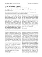
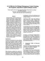
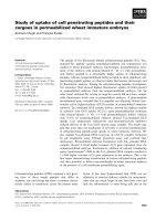
segment showed a decrease in g rowth rate (Fig. 1). Chem-
icals were administered to the cells 24 h after plating, and
the effects o f MTX and H
2
O
2
were examined after 24, 48, 72
and 96 h of exposure, respectively. As shown by the MTT
assay, the l ong-term action of both MTX and H
2
O
2
(up to
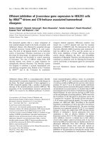
96 h) resulted in a dose-dependent loss of viability ( Figs 2
and 3). The concentrations employed were 16, 32, 64, 128,
256, 512, 1024 and 2048 n
M
. MTX, at c oncentrations higher
than 64 n
M
, s ignificantly i nhibited the proliferation of both
EATC and 0.28AS-2 cells after 48 h of exposure (results not
shown). This effect was also observed at a lower concen-
trations of the drug (16 n
M
,32n
M
and 64 n
M
)onthe
0.28AS-2 cells. When cells were treated for 96 h, at
concentrations lower than 128 n
M
, there was a larger
decrease in the proliferation of 0.28AS-2 cells in comparison
to EATC (Fig. 2).
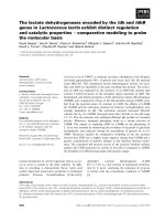
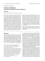
The H
2
O
2
concentrations used in this experiment were
0.78, 1.56, 3.13, 6 .25, 12.5, 25, 50 and 100 l
M
.After96hof
incubation with H
2
O
2
, the differences between 0.28AS-2
cells and EATC were significant (0.78, 1.56, 12.5 and 25 l
M
:
P < 0 .01; and 3.13 and 6.25 l
M
: P < 0.05) (Fig. 3).
Treatment of E ATC with > 50 l
M
H
2
O
2
resulted in the
destruction of the cell population by 24 h, with no change at
25 l
M
, similarly to baseline (results not shown). 0.28AS-2
cells are less resistant than EATC to H
2
O
2
, as it was found
Table 2. Effect of antisense mRNA glutaminase expression on gluta-
minase activity and on the intracellular contents of glutamine and glu-
tamate. Results represent the mean value ± S D for three different
determinations. Assays were perf or med on individual cell culture
plates, and the mean of duplicate analysis was used for statistical
calculations.
Cells
Glutaminase
(mU per 10
6
cells)
Glutamine
(m
M
)
Glutamate
(m
M
)
EATC 23.0 ± 2.8 1.3 ± 0.5 6.7 ± 1.0
0.28AS-2 5.0 ± 0.9* 5.2 ± 1.7* 6.9 ± 1.1
*P< 0.01 comparing control Ehrlich ascitic tumour cell (EATC)
values with those of 0.28AS-2 cells.
Table 3. Effect of antisense mRNA glutaminase e xpression on the apoptosis lev el determined by fluorescence-activated cell sorter ( FACS) anal ysis, a nd
caspase-3 activity, and on glutathione reductase (GR) activity. Results represent the mean values ± SD for three different determinations. Ehrlich
ascitic tumour cells (EATC) and 0.28AS-2 cells, untreated or treated for 48 h with 100 n
M
methotrexate ( MTX) were used for experiments.
Experimental conditions are as described in the Materials and methods.
Cells
Annexin V/FITC
+
cells (%) Caspase-3 (lmolÆmg
)1
protein) GR (unitsÆmg
)1
protein)
–MTX +MTX –MTX +MTX –MTX +MTX
EATC 4.1 ± 0.5 4.2 ± 0.4 139 ± 12 329 ± 31 0.34 ± 0.05 0.16 ± 0.03
0.28AS-2 8.0 ± 0.7* 14.0 ± 0.9* 214 ± 27* 397 ± 18* 0.20 ± 0.02* 0.14 ± 0.02
*P< 0.01 comparing the values of control EATC with those of 0.28AS-2 cells.
Ó FEBS 2004 Effect of glutaminase on glutathione and apoptosis ( Eur. J. Biochem. 271) 4301
that incubation of 0.28AS-2 cells for 24 h with 25 l
M
H
2
O
2
resulted in a similar effect to that seen in EATC incubated
with 50 l
M
H
2
O
2
during the same time-period (results not
shown). T hese findings strongly agree with the sensitization
to drugs of the cell line expressing antisense GA.
After 72 h of treat ment with H
2
O
2
, the growth of EATC
and 0.28AS-2 cells was reduced in a time-dependent
manner, y ielding I C
50
values of 2 3 and 22 l
M
, respectively.
Although 0.28AS-2 cells showed a greater sensitivity to
H
2
O
2
than EATC (Fig. 3), the MTX effect was even greater
(Fig. 2 ). In fact, 72 h after the addition of MTX, EATC and
0.28AS-2cellsshowedareducedcellgrowthalsoinatime-
dependent manner, with IC
50
values of 52 and 12 n
M
,
respectively. It must also be emphasized that the basal levels
of absorban ce at 570 n m (time zero in the absence of a ny
drug treatment) for EATC were twice as high as the values
for 0.28AS-2 cells (Fig. 1 ). After t reatment of both c ell lines
with 100 n
M
MTX or 100 l
M
H
2
O
2
, neither were able to
grow after the removal of MTX or H
2
O
2
.
Apoptosis in response to MTX and H
2
O
2
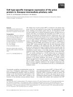
MTX-induced apoptosis was first demonstr ated in the
nucleus by DNA fragmentation, which is a biochemical
hallmark o f a poptosis. F igure 4 shows typical DNA
fragmentation in cells undergoing apoptosis induced by
100 n
M
MTX, indicating that this antineoplastic molecule
also causes cell death by apoptosis. Similar DNA frag-
mentation w as observed i n cells treated with H
2
O
2
,a
chemical that at the assayed concentration has been
reported to trigger both morphological change and intra-
nucleosomal DNA fragmentation, indicative of apoptosis
in many cell types [32]. Not surprisingly, c onsidering the
higher toxicity of MTX, this compound induced DNA
fragmentation at lower concentrations than seen for H
2
O
2
.
In any c ase, DNA fragmentation took pla ce, giving
remarkable DNA degradation after exposure of EATC
control cells and 0.28AS-2 cells to both 100 n
M
MTX and
100 l
M
H
2
O
2
(Fig. 4 ).
Fig. 1. Growth curve of Ehrlich ascitic tumour cells (EATC) and
0.28AS-2 cells. Cells were see ded at a c oncentration of 2 · 10
4
mL
)1
in
a 96-well culture plate. After 24, 48, 72 and 96 h , 3-[4,5-dimethyl-
thiazol-2-yl]-2,5-diphenyltetrazoliumbromide(MTT)wasaddedata
concentration of 0.5 mg ÆmL
)1
and inc ubated f or 3 h, as detailed i n t he
Materials and met hods. After 3 0 min, the absorbance of t he sampl e
was measured at 570 nm, and the results are shown in the figure. The
results represent the mean values f or at least three different wells and
are representative of at least three individual experiments, with an SD
value of < 10%.
Fig. 2. Effect of methotrexate (MTX) on the viability of Ehrlich ascitic
tumour cells (EATC) and 0 .28AS-2 c ells. Cells were seeded at a con-
centration of 2 · 10
4
mL
)1
in a 96-well culture plate, and incubated for
96 h. Then, 3-[4,5-dimethylthiazol-2-yl]-2,5-diphenyltetrazolium bro-
mide(MTT)wasaddedataconcentrationof0.5mgÆmL
)1
and incu-
bated for 3 h, as detailed in the Materials and methods. After 30 m in,
the absorbance o f t he samp le was measured a t 5 70 nm, and the results
are depicted in the figure . Data were normalized to 100% of the
untreated control. The results represent the mean values for at least
three different wells an d are rep resent ative of at least three in dividual
experiments, with an SD of < 10%.
Fig. 3. Effect of H
2
O
2
on the viability of Ehrlich ascitic tumour cells
(EATC) and 0.28AS-2 cells. Cells were seeded at a co ncentration of
2 · 10
4
mL
)1
in a 96-well culture plate, and incubated for 96 h. Then,
3-[4,5-dimethylthiazol-2-yl]-2,5-diphenyltetrazolium bromide (MTT)
was added at a conc entratio n of 0 .5 mgÆmL
)1
and incubated fo r 3 h, as
detailed in the M aterials and methods section. After 30 min, the
absorbance of the sample was measured at 57 0 nm, and the results a re
depicted in the fi gure. Data are nor malized to 100% o f the untreated
control. Re sults represent the mean values for at least three d ifferent
wells and are representative of at least three individual experiments,
with an SD of < 10%.
4302 J. Lora et al. (Eur. J. Biochem. 271) Ó FEBS 2004
To support th e DNA fragmentation assays, we used
annexin V-binding assays to detect the l oss of phospholipid
plasma membrane asymmetry and exposure of phosphat-
idylserine at the cell s urface, which is an early eve nt in the
sequence of e vents that leads to apoptotic cell death. This
experiment shows that 48 h of treatment with 100 n
M
MTX
does not affect the r elative number of apo ptotic cells in
EATC, but enh ances the ratio of apoptosis (P < 0.01) in
0.28AS-2 cells (Table 3). When comparing EATC and
0.28AS-2 cells in the absence of MTX, twice as many
apoptotic cells were found. This effect was found to be
augmented at 100 n
M
MTX, when fourfold more apoptotic
0.28AS-2 c ells were found than control EATC cells
(Table 3).
Additionally, basal levels (at time zero and in the absence
of any added chemical) of MTT staining in 0.28AS-2 cells
were found to be lower than those of control EATC (results
not shown ). A s observed, EATC cells are more resistant to
MTX-induced apoptosis than 0.28AS-2 cells (Fig. 2). On
the other h and, antisense GA expression induces apoptosis
and sensitizes Ehrlich t umour cells to MTX action.
In this study we have confirmed (by assaying the
activation of caspase-3, one of the caspase effector s) that
MTX induces apoptosis [33]. As shown in Table 3, caspase-
3 activity in both cell lines – control EATC a nd 0.28AS-2 –
treatedfor48hwith100n
M
MTX is highly increased.
Caspase-3 activity is augmented, in the presence of MTX,
by 2.4-fold and 1.9-fold in EATC and 0 .28AS-2 cells,
respectively (Table 3). Of interest, these findings are in
agreement with results from the DNA fragmentation and
annexin V-FITC a ssays.
Discussion
The results presented a bove expand on previous work
exploring the redox imbalance, consequent to GSH deple-
tion, and t he possibility of a relationship b etween apoptosis
and tumour proliferation [34]. This work is consistent with
others providing evidence of a ntisense technology i nducing
apoptosis [35], involving GSH depletion [36], and sensitizing
cancer cells to chemotherapy [37]. I n several cases, the
findings i n this w ork set the stage for detailed future
investigation into various aspects of the role of glutamine
and GSH in apoptosis and its implication in selective cancer
therapy.
The reduced GSH l evels found in 0.28AS-2 cells could be
caused by energy depletion and/or a reduced availability of
its precursor metabolites as glutamate. Inhibition of GA
activity should not result in a straightforward energy
depletion, because g lucose is present in the m edium and
this metabolite is preferred as an energy substrate by EATC
[38]. Consequently, energy status alone might not explain
the difference i n GSH le vels. In s pite of simil ar intracellular
glutamate concentrations, 0 .28AS-2 cells have significantly
higher glutamine levels, reflecting the strong inhibition of
GA expression (Table 2). The diminished mitochondrial
glutamine catabolism in 0.28AS-2 cells is consistent with the
lower GSH levels found in these cells: a positive c orrelation
has been found between glutamine supplementation and
enhanced production of both intracellular ROS and GSH
levels [11]. Moreover, b locking glutamine metabolism
by inh ibiting GA causes a significant decrease in ROS
production [39]. We reported previously that there exists a
mutual dependence b etween glutamine metabolism a nd
oxidative stress [4]. c-Glutamylc ysteine is the limiting
substrate in the synthesis of GSH. Additionally, c-glut-
amylcysteine synthethase, the enzyme that catalyzes the
formation of the substrate c-glutamylcysteine, is the rate-
limiting enzyme fo r GSH synthesis and i s f eedback inhibited
by the production of GSH itself [40]. We reasoned that if an
attenuated oxidative metabolism of glutamine is active in
these cells, there would b e a lower requirement for GSH as
an oxidative scavenger in 0.28AS-2 cells, and a putatively
lower c-glutamylcysteine content w ould account for their
reduced levels of GSH.
In previou s work we demonstrated that epithelial mucin-1
(MUC1) was markedly d iminished in 0.28AS-2 c ells.
MUC1 shedding has a protective function agains t the
humoral immune response developed against the tumour, so
0.28AS-2 cells were more susceptible to t he immune system
response than EATC [41]. It has been reported that MUC1
expression is up-regulated by oxidative stress, demonstra-
ting that MUC1 expression is associated with the attenu-
ation of ROS levels. It has also been shown that the
apoptotic response to o xidative stress is attenuated by a
MUC1-dependent mechanism. These r esults suggest a
model in which activation of MUC1 by oxidative stress
provides a protective function against increased intracellular
oxidant levels and ROS-induced apoptosis [42]. Therefore,
depleted MUC1 expression of 0.28AS-2 cells agrees with our
model, which proposes an inhibition of GSH-dependent
antioxidant defence as well as an activation of apoptosis in
cells with decreased GA expression.
Very re cently, it has b een proposed that glutamine may
be protective to cells during periods of oxidative stress,
increasing survival in some cell lines through an
up-regulation of GSH levels [12]. In addition, the thiol/
disulfide redox state in the intestinal epithelium is an
Fig. 4. Agarose gels s howing DNA fragmentation. Cells undergo
apoptosis following exposure to 100 l
M
H
2
O
2
and 100 n
M
metho-
trexate (MTX). Results are representative of three in dividual e xp eri-
ments. Control lanes (Ctl) r epresent DNA extracted from untreated
Ehrlich ascitic t umour cells (EATC) a nd 0.28AS-2 cells, respectively.
H
2
O
2
lanes represent DNA extracted from H
2
O
2
-treated (24 h)
EATC and 0.28AS-2 cells, respectively. MTX lanes represent DNA
extracted from methotrexate-treated (48 h) EATC and 0.28AS-2 cells,
respectively.
Ó FEBS 2004 Effect of glutaminase on glutathione and apoptosis ( Eur. J. Biochem. 271) 4303
important determinant of Caco-2 cell proliferation
induced by glutamine, enhancing the capability of
Caco-2 cells to modulate extremes of extracellular redox
[43]. Experimental animal studies have shown that the
administration of glutamine increases tissue concentra-
tions of reduced GSH. Conversely, glutamine deficiency
leads to a cell cycle arrest in G
0
/G
1
and reduces apoptosis.
Interestingly, many of these biological activities are also
associated with the cellular reduced oxygen potential,
which depends mainly on the ratio of reduced GSH to
GSSG [44]. Therefore, GSH metabolism is closely related
to apoptotic processes. Whether the down-regulation of
GA expression and GSH concentrations sensitizes cells to
apoptosis only in G
0
/G
1
, or when passing the G
2
/M
checkpoint, remains to be investigated.
On the other hand, it has been stated previously that
administration of glutamine to animals receiving MTX
therapy f avours host tolerance to the d rug a nd increases its
tumoricidal effectiveness. This effect of MTX i s suggested to
be related to GSH me tabolism [45].
Our r eport is strongly in accordance with others,
published very recently, proposing t hat lowering the GSH
concentration m ay contribute to induce apoptosis in
tumour cells [23], i ndicating that systemic oxidative stress,
as measured by a d ecrease in GSH levels, is associated with
a higher ratio of apoptosis [46] and important redox
alterations [47]. These authors have previously demonstra-
ted t hat the inhibition of active GSH e xtrusion rescues cells
from apoptosis [48]. In contrast, CD95-mediated hepatocyte
apoptosis requires an intact intracellular GSH status [49].
The issue of G SH depletion a nd apoptosis has b een a m atter
of discussion until the publication of recent articles which
indicate that mitochondrial GSH depletion, and not cyto-
solic GSH depletion, are critical factors leading to apoptotic
tumor cell death activation [50].
In previous work it h as been s hown that a ntisense
oligonucleotides, down-regulating the expression of bcl-2 or
bcl-xL, induce apoptosis and s ynergistically interact with
chemotherapy [51]. Our results, taken together with those
discussed above, can be exploited as research tools to gain
new insights into the underlying biological basis of the
connection of antiproliferative activity of specific antisense
GA expression to GSH status a nd apoptosis. I n fact,
resistance to chemotherapy has been strongly linked to
apoptotic processes [52], and the use of antisense oligo-
nucleotides has already provided a rational and promising
approach for h elping to overcome chemoresistance i n
several malignancies [53]. Because the GA antisense
approach has the potential to facilitate apoptosis, using
this technique, in c ombination with others, could provide a
valuable tool in t herapy. The development o f cancer therapy
is an art of integrated sciences, but one of the main points
is targeting of genes crucial for cancer cell proliferation. In
this field, some interesting results have been achieved very
recently using liposome-mediated in vivo gene transfe r and
antisense technology [54]. Therefore, GA inhibition and
glutamine supplementation deserve further evaluation as
potential selective anticancer agents, alone or in combina-
tion with cytotoxic drugs in human carcinomas expressing
functional GA. However, although similar systems have
achieved promising r esults in different human cancer cell
lines [55], and even in patients suffering from malignant
melanoma [56], further understanding of the clinical
usefulness of the proposed combination regimen is required.
Acknowledgements
We thank E . Manzanares and A. Rubio for valuable help. This work
was s upported by Grant SAF 2001-1894 fro m the Ministry of Ciencia y
Tecnologı
´
a of Spain and by Project CVI-179 of Junta d e Andalucı
´
a,
Spain.
References
1. Medina, M.A., Sa
´
nchez-Jime
´
nez, F., Ma
´
rquez, J., Rodrı
´
guez-
Quesada, A. & N u´ n
˜
ez de Ca stro, I. ( 1992) Relevance o f glutamine
metabolism to tumor c ell growth. Mol. Cell. Biochem. 113,
1–15.
2. Lobo, C., R uı
´
z-Bellido, M.A., Aledo, J .C., Ma
´
rquez, J., N u´ n
˜
ez de
Castro, I. & Alonso, F.J. (2000) Inhibition of glutaminase
expression by antisense mRNA decreases growth and tumour-
igenicity of tumour c ells. Biochem. J. 348 , 257–261.
3. Yudkoff,M.,Pleasure,D.,Cregar,L.,Lin,Z.,Nissim,I.,Stern,J.
& Nissim, I. (1990) Glutath ione turno ver in cultu red astro cytes:
studies with [
15
N]glutamate. J. Neurochem. 55, 137–145.
4. Mate
´
s, J.M., P e
´
rez-Go
´
mez, C., N u´ n
˜
ezdeCastro,I.,Asenjo,M.&
Ma
´
rquez, J. (2002) Glutamine and its r elationship w ith
intracellular redox status, oxidative stress and cell proliferation/
death. Int. J. Biochem. Cell Biol. 34, 439–458.
5. Sies, H. (1999) Glutathione and its role in cellular functions. Free
Radic. Biol. Med. 27, 916–921.
6. Chandra, J ., Samali, A. & Orrenius, S . (2000) Triggering and
modulation of apoptosis by oxidative stress. Free Radic. B iol.
Med. 29, 3 23–333.
7. Esteve, J.M., Mompo, J., Garcia d e l a Asuncio
´
n, J., Sastre, J.,
Asensi, M., Boix, J., Vin
˜
a, J.R., Vin
˜
a, J. & Pallardo, F.V. (1999)
Oxidative damage to mitochondrial DNA and glutathione oxi-
dation in apoptosis: studie s in vivo and in vitro. FASEB J. 13,
1055–1064.
8. Wu,G.,Fang,Y.,Yang,S.,Lupton,J.&Turner,N.(2004)
Glutathione metabolism and its implications for health. J. Nutr.
134, 489–492.
9.Gu,Y.,Wu,Z.,Xie,J.,Jin,D.&Zhuo,H.(2001)Effectsof
growth hormone (rhGH) and glutamine supplemented parenteral
nutrition on intestinal adapt ation in short b owel rats. Clin. N utr.
20, 159–166.
10. Wu, X.T., Li, J.S., Zhao, X.F., Li, N ., Ma, Y.K., Zhuang, W.,
Zhou, Y. & Y ang, G . (2003) Effects o f n -3 fatty aci d, fructose-1,6-
diphosphate and glutamine on mucosal cell proliferation and
apoptosis of small bowel g raft after tran splantation in rat s. World
J. Gastroenterol. 9, 1323–1326.
11. Chang, W.K., Yang, K.D. & Shaio, M.F. (1999) Lymphocyte
proliferation m od ulated by glutamine: involved in the e ndo genous
redox reaction. Clin. E xp. Immunol. 117, 482–488.
12. Ogunlesi, F., Cho, C. & M cGrath-Morrow, S. (2004) The effect of
glutamine on A549 cells exposed to moderate hyperoxia. Biochim.
Biophys. Acta 1688, 112–120.
13. Oehler, R. & Roth, E. (2003) Regulative capacity of glutamine.
Curr. Opin. Clin. Nutr. Metab. Care 6, 277–282.
14. Babiak, R.M., Campello, A.P., Carnieri, E.G. & Oliveira, M.B.
(1998) Methotrexate: pentose cycle and oxidative stress. Cell
Biochem. Funct. 16 , 283–293.
15. Akerboom, T.P. & Sies, H. (1981) Assay o f glutathione, glu-
tathione disulfide, and glu tathione mixed disulfides in biological
samples. Methods Enzymol. 77, 3 73–382.
16. Pastore, A., Federici, G., Bertini, E . & Piemonte, F. ( 2003) Ana-
lysis of glutathione: implication in redox and detoxification. Clin.
Chim. Acta 333, 1 9–39.
4304 J. Lora et al. (Eur. J. Biochem. 271) Ó FEBS 2004
17. Griffith, O.W. (1980) De termination of glutathione and glutathi-
one disulfide using glutathione reductase and 2-vinylp yridine.
Anal. Biochem. 106, 207–212.
18. Carlberg, I. & Mannervik, B. (1985) Glutathione reductase.
Methods Enzymol. 113, 484–490.
19. Baverel, G. & Lund, P. (1979) A role for bicarbonate in the reg-
ulation of mammalian glutamine metabolism. Biochem. J. 184,
599–606.
20. Ma
´
rquez, J., Sa
´
nchez-Jime
´
nez, F., Medina, M.A., Quesada, A.R.
&Nu´ n
˜
ez de Castro, I. (1989) N itrogen metabolism in tumor
bearing mice. Arch. Biochem. Biophys. 268, 6 67–675.
21. Kim, E.J ., Sampathkumar, S .G., Jone s, M.B., Rhee, J.K., Bask-
aran, G., Goon, S. & Yarema, K.J. (2004) Charac terization of the
metabolic flux and apoptotic effects of O-hydroxyl- and N-Acyl-
modifed N-acetylmannosamine (ManNAc) analogs in jurkat
(human T-lymphoma-derived) cells. J. Biol. Chem. 27 9, 18342–
18352.
22. Pithon-Curi, T.C., Schumacher, R.I., Freitas, J .J., Lagranha, C.,
Newsholme, P., Palanch, A.C., Doi, S.Q. & Curi, R. (2003)
Glutamine delays spontaneous apoptosis in neutrophils. Am. J.
Physiol. Cell Physiol. 284, C1355–C1361.
23. Tormos, C., Chaves, F .J., Garcı
´
a, M.J., Garrido, F., Jover, R.,
O’Connor, J.E., Iradi, A., Oltra, A., Oliva, M.R. & S a
´
ez, G .T.
(2004) Role of glutathione in the induction of apoptosis and c-fos
and c-jun mRNAs by oxidative stress in tumor cells. Cancer Lett.
208, 103–113.
24. Vermes, I., Haanen, C., Steffens-Nakken, H. & Reutelingsperger,
C. (1995) A novel assay for apoptosis. Flow cytometric d etection
of phosphatidylserine expression on early apoptotic cells using
fluorescein labelled Annexin V. J. Immunol. Methods 184, 39–51.
25. Takahashi, H., Aoki, N., Nakamu ra, S., Asano, K., Ishida-
Yamamoto,A.&Iizuka,H.(2000)Cornifiedcellenvelopefor-
mation is distinct from apoptosis in epidermal keratinocytes.
J. Dermatol. Sci. 23, 161–169.
26. Asatiani, N ., Sapojnikova, N., Abuladze, M., Kartvelishvili, T.,
Kulikova, N., Kiziria, E., Namchevadze, E. & Holman, H. (2004)
Effects of Cr (VI) lo ng-term and low-dose a ction on mammalian
antioxidant e nzymes (an in vitro study ). J. In org. Biochem. 98 ,490–
496.
27. Cohen, G. (1997) Caspases: the executioners of apoptosis. Bio-
chem. J. 15 , 1–16.
28. Caetano, N., Campello, A., Carnieri, E., Kluppel, M. & Oliveira,
M. (1997) Effect of methotrexate (MTX) on NAD(P)+ dehy-
drogenases of HeLa cells: malic en zyme, 2-oxoglutarate and iso-
citrate dehydrogenases. Cell Biochem. Funct. 15, 2 59–264.
29. Sun, Y. (1990) Free radical antioxidant enzymes, and carcino-
genesis. Free Radic. Biol. Med. 8, 483–499.
30. Sun, Y., Oberley, L.W., Elwell, J.H. & Sierra-Rivera, E. (1989)
Antioxidant e nzyme activities in normal and tran sforme d mouse
liver cells. I nt. J. Cancer 44, 102 8–1033.
31. Filomeni, G., Rotilio, G. & Ciriolo, M.R. (2002) Cell signalling
and the glutathione redox system. Biochem. Pharmacol. 64, 1057–
1064.
32. Chen,H.,Chien,C.,Yu,S.,Lee,Y.&Chen,W.(2002)Cyclo-
sporine A re gulates o xidative stress-induced apoptosis in cardio-
myocytes: m echanisms via R OS g en eration, iNO S and H sp70 . Br.
J. Pharmacol. 137, 771–781.
33. Blanc, C., Deveraux, Q.L., Krajewski, S., Janicke, R.U., P orter,
A.G., Reed, J .C., J aggi, R. & Marti, A. (200 0) Casp ase-3 is
essential for procaspase-9 processing and cisplatin-induced apop-
tosis of M CF-7 breast cancer cells. Cancer Res. 60 , 4386–4390.
34. Mate
´
s, J.M. & Sa
´
nchez-Jime
´
nez, F.M. (2000) Role of reactive
oxygen species in a pop tosis: implications for cancer therapy. In t. J.
Biochem. Cell Biol. 32, 157– 170.
35. Ba
´
kiewicz-Masiuk, M., Masiuk, M. & Machalı
´
ski, B. (2003) The
influence of STAT5 ant isense oligonucleotides on the proliferation
and apoptosis of se lect ed human leukaemic cell lines. Cell Prolif.
36, 265–278.
36. Tang, D.G. & Honn, K.V. ( 1997) Ap optosis of W256 carcino-
sarcoma cells of the monocytoid origin induced by NDGA
involves lipid p eroxidation and depletion o f GSH: role o f
12-lipoxygenase in regulating tumor cell survival. J. Cell. Physiol.
172, 155–170.
37. Vilenchik, M., R affo, A.J., Benimetskaya, L., Shames, D. & Stein,
C.A. (2002) Antise nse R NA down-regulation of b cl-xL e xpression
in prostate ca ncer ce lls leads to diminished rat es o f cellula r p ro-
liferation and resistance to cytotoxic c hemotherapeutic agents.
Cancer Res. 62, 2 175–2183.
38. Luque, P., Paredes, S ., Segura, J .A., N u´ n
˜
ez de Castro, I . &
Medina, M.A. (1990) Mutual effect of glucose and glutamine on
their utilization by tumour cells. Biochem. Int. 21, 9 –15.
39. Pithon-Curi, T.C., Levada, A.C., Lopes, L.R., Doi, S.Q. & Curi,
R.(2002)Glutamineplaysaroleinsuperoxideproductionandthe
expression of p 47phox, p 22p hox an d g p91phox in rat neutrophils.
Clin. Sci. (Lond.) 10 3 , 403–408.
40. Drake, J., K anski, J., Varadarajan, S ., Tsoras, M. & Butterfield,
D.A. (2002) Elevation of brain glutathione by gamma gluta-
mylcysteine ethyl ester protects against peroxynitrite-induced
oxidative stress. J. Neurosci. Res. 68, 776–784.
41. Segura, J.A., Ruı
´
z-Bellido, M.A., Aren as, M., L obo, C., Ma
´
rquez,
J. & Alonso, F.J. (2001) Eh rlich a scites tu mor cells ex pressing a nti-
sense glutaminase mRNA lose their capacity t o evade the mouse
immune system. Int. J. Cancer 91, 379–384.
42. Yin,L.,Li,Y.,Ren,J.,Kuwahara,H.&Kufe,D.(2003)Human
MUC1 carcinoma antigen regulates intracellular oxidant levels
and the apoptotic response to oxidative stress. J. Biol. Chem. 278,
35458–35464.
43. Jonas, C.R., Gu, L.H., Nkabyo, Y.S., Mannery, Y.O., Avissar,
N.E.,Sax,H.C.,Jones,D.P.&Ziegler,T.R.(2003)Glutamineand
KGF each regulate e xtracellular t hiol/disulfid e r edox and e nh ance
proliferation in Caco-2 cells. Am. J. Physiol. Regul. Integr. Comp.
Physiol. 285, R1421–R1429.
44. Roth,E.,Oehler,R.,Manhart,N.,Exner,R.,Wessner,B.,Str-
asser, E. & Spittler, A. (2002) Regulative potential of glutamine –
relation to glutathione metabolism. Nutrition 18, 217–221.
45. Rouse, K., Nwokedi, E., Woodliff, J.E., Epstein, J. & Klimberg,
V.S. (1995) Glutamine enhances selectivity of chemotherapy
through changes in glutathione metabolism. Ann. Surg. 221,
420–426.
46. Kyto, V., Lapatto, R., Lakkisto, P., Saraste, A., Voipio-Pulkki,
L., Vuorinen, T. & Pulkki, K. (2004) G lutathione depletion a nd
cardiomyocyte apoptosis in viral myocarditis. Eur. J. Clin. Invest.
34, 167–175.
47. Ghibelli, L., Coppola, S., Fanelli, C ., Rotilio, G., Civitareale, P.,
Scovassi, A.I. & Ciriolo, M.R. (1999) Glutathione depletion cau-
ses cytochrome c release even in the absence of ce ll commitment to
apoptosis. FASEB J. 13, 2 031–2036.
48. Ghibelli, L., Fanelli, C., Rotilio, G., Lafavia, E., Coppola, S.,
Colussi, C., Civitareale, P. & Ciriolo, M.R. (1998) Rescue of cells
from apoptosis by inhibition of act ive GSH extrusion. FASEB J.
12, 479–486.
49. Hentze, H., Kunstle , G ., V olbracht, C ., Ertel, W. & W endel, A.
(1999) CD95-Mediated murine hepatic apoptosis requires an
intact glutathione status. Hepatology 30, 177–185.
50. Ortega, A.L., Carretero, J., Obrador, E., Gambini, J., Asensi, M.,
Rodilla, V . & Estrela, J.M. (2003) Tumor cytotoxicity by
endothelial cells. Impairm ent of the mitochondrial sy stem for
glutathione uptake in m ouse B16 melanoma cells that survi ve after
in vitro interaction with the hepatic sinusoidal endo thelium.
J. Biol. Chem. 278, 13888–13897.
51. Zangemeister-Wittke, U., S chenk er, T., Luedk e, G. & Stahel, R .
(1998) Syne rgistic cyto toxicity o f bcl-2 antisense oligo-
Ó FEBS 2004 Effect of glutaminase on glutathione and apoptosis ( Eur. J. Biochem. 271) 4305
deoxynucleotides and etoposide, dox orubicin and cisplatin on
small- ce ll lung cancer cell lines. Br.J.Cancer78, 1035–1042.
52. Guensberg, P., Wacheck, V., Lucas, T., Monia, B., Pehamberger,
H.,Eichler,H.G.&Jansen,B.(2002)Bcl-xLantisenseoligo-
nucleotides chemosensitize human glioblastoma cells. Chemo-
therapy 48, 189–195.
53. Heere-Ress, E ., Thallinger, C., Lucas, T., Schlagbauer-Wadl, H.,
Wacheck, V., Mo nia, B.P., W olff, K., P eh amberge r, H. & Ja nse n,
B. (2002) Bcl-X (L) is a chemoresistance factor in human m ela-
noma cells t hat can be inhibited b y antisense therapy. Int. J.
Cancer 99, 29–34.
54. Yoshida, T., Ohnami, S. & Aoki, K. (2004) Development of gene
therapy to target pancreatic cancer. Cancer Sci. 95, 283–289.
55. Ciardiello, F., Caputo, R., Troiani, T., Borriello, G., Kandimalla,
E.R.,Agrawal,S.,Mendelsohn,J.,Bianco,A.R.&Tortora,G.
(2001) Antisense o ligo nucleotides t argeting the epidermal growth
factor receptor inhibit proliferation, induce apoptosis, and
cooperate with c ytotoxic drugs i n human cancer c ell lines. Int. J.
Cancer 93, 172–178.
56. Jansen, B., Wacheck, V., Heere-Ress, E., Schlagbauer-Wadl,
H.,Hoeller,C.,Lucas,T.,Hoermann, M., Hollenstein, U.,
Wolff,K.&Pehamberger,H.(2000) Chemosensitisation of
malignant melanoma by BCL2 antisense t herapy. Lancet 356,
1728–1733.
4306 J. Lora et al. (Eur. J. Biochem. 271) Ó FEBS 2004