components of cell and gene therapy
Bạn đang xem bản rút gọn của tài liệu. Xem và tải ngay bản đầy đủ của tài liệu tại đây (409.82 KB, 31 trang )
CHAPTER 9
Components of Cell and Gene Therapy
for Neurological Disorders
LAURIE C. DOERING, PH.D.
INTRODUCTION
The complexity of the nervous system poses several challenging problems for
scientists and clinicians who seek to apply gene therapy to neurological disor-
ders. In addition to the standard problems associated with gene therapy (discussed
in Chapter 3), we deal with very delicate, complex networks of cells and face
the issue of accessibility (Fig. 9.1) and targeting the desired cell type(s) when
considering gene therapy strategies in the central nervous system. Unlike other
organs in the body such as the liver or lungs where large proportions of the
organs can be damaged with minimal or no functional consequences, damage to
extremely small areas of the brain can be devastating. Therapeutic targeting to
selective areas or cell types will be difficult to achieve in the central nervous
system (CNS).
Excluding the identified genetic causes of neurodegenerative diseases, the
etiology underlying the primary neurological disorders is unknown. While the prin-
ciple cell types affected in disorders such as Parkinson’s and Alzheimer’s have
been identified, the exact contributing factors or conditions that trigger relentless
neuronal degeneration are presently unknown. Therefore, at this time, gene
products that help to reduce the effects of neural dysfunction, offset neuronal
death, inhibit apoptosis, or encourage cell survival form the basis of gene therapy
in the nervous system. As gene therapy approaches are developed and refined,
the outcome of gene therapy in the nervous system could be extremely
effective.
In this chapter, the key aspects of neural dysfunction associated with the promi-
nent nervous system disorders are explained. Promising advances with gene trans-
fer to the CNS have been made with different families of virus vectors. A focus on
the vectors and the cells used for gene delivery in animal models is provided. Impor-
tant features of the clinical trials using genetically modified cells and trophic fac-
203
An Introduction to Molecular Medicine and Gene Therapy. Edited by Thomas F. Kresina, PhD
Copyright © 2001 by Wiley-Liss, Inc.
ISBNs: 0-471-39188-3 (Hardback); 0-471-22387-5 (Electronic)
Cerebral cortex
Frontal lobe
Temporal
lobe
Cerebellum
Occipital
lobe
Skull
Meninges
(a)
(b)
Cranium
Dura mater
Venous sinus
Dura mater
Subdural space
Arachnoid
Subarachnoid space
Pia mater
Cerebral cortex
tors for neurodegeneration are described, and we will illustrate how neuroscience
research in combination with genetics and molecular biology is guiding the future
of gene therapy applications in the nervous system.
SORTING OUT THE COMPLEXITY OF THE NERVOUS SYSTEM
The nervous system is divided into two main parts: (1) the central nervous
system consisting of the brain and spinal cord and (2) the peripheral nervous system
(PNS) composed of the nervous tissue in the form of nerves that emerge bilaterally
from the brain and spinal cord that serve to keep the other tissues of the body
in communication with the CNS (Fig. 9.2). Numerous types of neurons specialized
to receive, process, and transmit information via electrical impulses are primarily
responsible for the functional characteristics of the nervous system (Fig. 9.3).
Neurons can be identified by their size, shape, development, and organization
within the brain. Neurons work in networks and secrete neurotransmitters and
other chemical messengers at sites of functional contact called synapses. At each
synapse a region of the cell membrane in the presynaptic neuron is specialized for
rapid secretion of one or more types of neurotransmitters. This area is closely
apposed to a specialized region on the postsynaptic cell that contains the receptors
for the neurotransmitter or other ligands. The binding of the neurotransmitter to
the receptors triggers an electrical signal, the synaptic potential, in the postsynaptic
cell (Fig. 9.4). Information in the nervous system is thereby transmitted and pro-
cessed by elaborate networks that generate a spectrum of electrical and chemical
signals.
Glial cells, often referred to as specialized support cells of the CNS, represent the
second major class of cells that perform important functions that are key to the
normal operation of the nervous system (Fig. 9.3).There are four main types of glial
cells.Astrocytes act in a general supportive capacity and help to maintain the extra-
cellular environment in the CNS. The astrocyte processes are intimately associated
with the neuronal cell bodies, dendrites, and nerve terminals. They serve to insulate
and isolate pathways and neuronal tracts from one another. Oligodendrocytes and
Schwann cells form the myelin sheaths around axons in the CNS and PNS, respec-
tively.The myelin is wrapped around segments of axons and serves to accelerate the
conduction of the electrical signals. In the CNS, each oligodendrocyte may form and
maintain myelin sheaths for approximately 60 axons. In the PNS, there is only one
Schwann cell for each segment of one axon. Microglial cells in the CNS are analo-
gous to macrophages and can be activated by a number of conditions, including
inflammation and trauma.
SORTING OUT THE COMPLEXITY OF THE NERVOUS SYSTEM 205
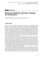
FIGURE 9.1 External view of the cerebral hemisphere. (a) Brain and spinal cord are pro-
tected by many layers including the skin, bone, and special connective tissue layers referred
to as the meninges. (b) Schematic diagram of the protective layers that cover the brain.
(c) Major divisions of the human brain as seen from a midsaggital view.
᭣
206 COMPONENTS OF CELL AND GENE THERAPY FOR NEUROLOGICAL DISORDERS
Posterior view
The peripheral nerves in humans
C1
C2
C3
C4
C5
C6
C7
C8
T1
T2
T3
T4
T5
T6
T7
T8
T9
T10
T11
T12
L1
L2
L3
L4
S1
S2
S3
S4
S5
C1
L5
Brachial plexus
Cauda equina
Lumbosacral
plexus
Coccygeal nerve
Sacral nerves
Lumbar nerves
Thoracic nerves
Spinal cord
Cervical nerves
Sacrum
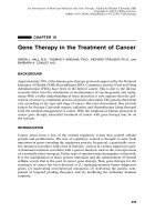
FIGURE 9.2 Brain, spinal cord, and peripheral nerves. There are 31 vertebral bones in the
spinal column that house and protect the spinal cord. Between the vertebrae, spinal (periph-
eral) nerves emerge bilaterally. The individual nerves are made of sensory and motor fibers
that interface the peripheral parts of the body with the central nervous system (brain and
spinal cord).
WHAT GOES WRONG IN NEUROLOGICAL DISORDERS?
Given the vast number and types of neurons and glial cells in the nervous system,
one quickly realizes the potential for several neurological dysfunctions, depending
on the cell type(s) affected. Neuronal degeneration can occur in selected areas of
the brain or neurodegenerative events may affect the entire brain (global neu-
WHAT GOES WRONG IN NEUROLOGICAL DISORDERS? 207
Dendrites
Axon
Oligodendrocyte
(glia)
Myelin
sheath
Synapse
Motor neuron
Axon
Myelin
Oligodendrocyte cell
cytoplasm
Direction of
action potential
Neuron
cell body
Astrocytes
(glia)
FIGURE 9.3 Schematic representation of neurons and glial cells. Neurons are surrounded
by astrocytes that fill the interstices between neuronal cell bodies. Glia outnumber neurons
by at least 10 to 1. Oligodendrocytes wrap around the axon and produce the myelin sheath.
Inset shows how the myelin wraps around segments of the axon.
rodegenerative conditions) as in the case of the neurogenetic lysosomal storage
diseases (LSD) associated with single-gene mutations.
For the majority of neurological disorders, specific classes of neurons in the brain
or spinal cord show selective vulnerability. Depending on the type of neuron/
neurotransmitter affected, changes will occur in behavior, memory, or movement.
In Parkinson’s, neurons located in the substantia nigra of the midbrain that contain
208 COMPONENTS OF CELL AND GENE THERAPY FOR NEUROLOGICAL DISORDERS
Mitochondrion
Presynaptic
membrane
Postsynaptic
membrane
Receptor site
Postsynaptic cell
Axon
Microtubules
Synaptic
vesicle
Synaptic cleft
Dendrite
Channel
K
+
Action
potential
Axon potential
moves down axon
to nerve terminal
Axon
Synaptic
vesicle
Receptor
site
Neurotransmitter
Dendrite
K
+
Depolarization
Synaptic vesicle
releases
neurotransmitter.
Neurotransmitter on
receptor site.
Channel opens.
Reuptake of
neurotransmitter by
presynaptic neuron or
astrocytes
The flow of sodium
ions (Na
+
) and
potassium ions (K
+
)
generates a new
electrical signal
FIGURE 9.4 Components of a synapse. Illustration shows aspects of neurotransmitter
release, receptor interaction, and generation of the electrical signal. All electrical signals
arise from the action of various combinations of ion channel proteins that form aqueous
pores through which ions traverse the membranes. When ion channels are open, ions move
through the channels down their electrochemical gradients. Their net movement across
the membrane constitutes a current that changes the membrane potential and generates an
electrical signal.
the neurotransmitter dopamine undergo accelerated cell death. Loss of these
neurons influences the normal function of the extrapyramidal system in the brain
and results in rigidity and tremor of the limbs. Alzheimer’s isolates the hippocam-
pus and regions of the cerebral cortex due to death of acetylcholine-rich neurons,
causes dementia, and prevents the formation of new memory. Amyotrophic lateral
sclerosis (ALS) damages the motor neurons in the CNS and causes weakness and
spasticity. Alternatively, when oligodendrocytes in the central nervous system are
affected, problems develop with routine motor functions, and sensory deficits
become noticeable in individuals with multiple sclerosis.
The LSD are genetic disorders resulting from mutations in genes that code for
proteins involved with the degradation of normal body compounds that include
lipids, proteins, and carbohydrates. Although most lysosomal disorders result from
defects in genes that code for lysosomal enzymes, some are caused by genes coding
for transport proteins, protective proteins, or enzymes that process the lysosomal
enzymes. Individually, the LSD occur infrequently, but collectively they occur
approximately in 1/5000 births. The accumulation of enzyme substrates in cells
of the CNS characterizes disorders like the mucopolysaccharidoses or GM
1
gangliosidosis.
What triggers selected cell death in the nervous system? In some cases, genetic
causes have been associated with neuronal degeneration. In Huntington’s disease,
a mutation (triplet repeat mutations) in chromosome 4 is linked with the death of
neurons in a region of the brain called the caudate/putamen, a complex of inter-
connected structures tuned to modulate motor activities. The identification of un-
stable triplet repeat mutations represents one of the great discoveries of human
neurogenetics. Genetic linkages discussed later in this chapter have also been deter-
mined for a small percentage of individuals with Alzheimer’s and Parkinson’s.
We have identified various types of cytological and molecular changes in neurons
that are associated with the death of neurons. Research has identified numerous,
specific changes in neurons at risk associated with the prevalent CNS disorders and
also with the aging process. Abnormal accumulations of filaments and altered pro-
teins are recognized as primary features of neurons targeted in neurological dys-
function. The accumulations may occur in the cytoplasm of the neuron or in the
extracellular environment. In certain instances, the pattern of neuronal loss is dic-
tated by how the neurons are connected to one another.Alzheimer’s is an excellent
example of this point. Virtually all the subgroups of neurons lost in Alzheimer’s
are found to be connected to regions of the cerebral cortex that show high levels
of neuritic plaque formation—foci of degenerating processes and twisted arrays of
cytoskeletal elements in the neurons referred to as neurofibrillary tangles.
What sets off the initial changes in neurons that lead to a cascade of cell death
in specific areas and pathways of the nervous system? A number of molecular mech-
anisms at different levels of neuronal function have been proposed. Changes to the
cytoskeleton, oxidative injury, deoxyribonucleic acid (DNA) modifications, changes
in ribonucleic acid (RNA)/protein synthesis, abnormal protein accumulation, toxic-
free radicals, reduced axonal transport, and programmed cell death have been iden-
tified as possible reasons for neurological disease. Several animal models are used
to generate these molecular changes, and, in turn, they help define the possible
etiology of neurodegeneration and provide a way to test gene therapy strategies
for CNS disorders, injury, or aging.
WHAT GOES WRONG IN NEUROLOGICAL DISORDERS? 209
NEUROTROPHIC FACTORS AND GENE THERAPY
Neurotrophic Factors
There are a variety of molecules in the nervous system that are important to the
survival, differentiation, and maintenance of neurons in both the PNS and CNS.
These molecules, referred to as neurotrophic factors (Table 9.1), induce pattern and
synapse formation and create highly specialized neural circuits in the brain. The
factors are secreted from the target innervated by the neurons, taken up at the nerve
terminals, and then transported over long distances to the cell body where they act
to regulate neuronal functioning by a variety of signaling mechanisms (Fig. 9.5).We
now realize that neurotrophic factors bind to cell surface receptor proteins on
the nerve terminals, become internalized (receptor-mediated endocytosis), and
then move toward the cell body by the mechanism of retrograde axonal transport.
Advances in the understanding of the structure of the receptors for neurotrophic
factors indicate that they are similar to the receptors used by traditional growth
factors and cytokines. The expression of the receptors for the neurotrophic factors
is exclusively or predominantly in the nervous system, and, when activated, the
factors display distinctive molecular actions.
Nerve growth factor (NGF) is the prototype member of the neurotrophins, a
family of proteins that have common structural features. It was discovered and char-
acterized in the 1950s by Rita Levi-Montalcini, Stanley Cohen, and Viktor Ham-
burger and was the first molecule to show potent nerve growth promoting activity
on explants of neural tissue maintained in tissue culture. Since the discovery of NGF,
a number of molecules have been identified and added to the expanding list of
substances grouped under the broad umbrella of neurotrophic factors. Common,
well-studied factors are listed in Table 9.1.Responses to the neurotrophins are medi-
ated through receptor tyrosine kinases that belong to the trk family of protoonco-
210 COMPONENTS OF CELL AND GENE THERAPY FOR NEUROLOGICAL DISORDERS
TABLE 9.1 A Listing of Common Neurotrophic Factors
Class Members Receptor Responsive Neurons
Neurotrophins NGF TrkA Forebrain cholinergic neurons
NT-3 TrkC Corticospinal neurons
NT4/5 TrkB Caudate/putamen
BDNF TrkB Substantia nigra
Transforming growth GDNF Ret Substantia nigra neurons
factor b TGF-b Motor neurons
Cytokines CNTF CNTFa Spinal cord motor neurons
LIF gp130/JAK Spinal cord motor neurons
LIFRb/TYK
Insulinlike growth IGF-1 IGF Forebrain cholinergic neurons
factors receptor Forebrain cholinergic neurons
IGF-2
Fibroblast growth bFGF FGF Forebrain cholinergic neurons
factors receptor Spinal cord motor neurons
aFGF
genes. It is now clear that neurotrophic factors can be provided by a number of
sources including glial cells, afferent processes of neurons, muscle, and even by the
extracellular matrix. Numerous biological events including neuronal growth, phe-
notype (neurotransmitter) expression, and programmed cell death have been linked
with retrograde neurotrophic factor signaling. Hence, there are many possible lines
of study to explore the effects of neurotrophic factor gene therapy in relation to
basic neural cell survival and function for the treatment of neurodegenerative
disorders.
From basic research, we have learned that if the brain is injured, these molecules
can be released to play a significant role in the recovery process. In addition to
limiting the loss of neurons, neurotrophic factors can stimulate new outgrowth from
the axons and dendrites, regulate axon branching, modulate neurotransmitter
synthesis, and influence synapse formation. This inherit property of structural and
functional change in neurons in response to environmental cues (like the release
of neurotrophic factors) is referred to as plasticity. Many factors have been shown
to have overlapping effects (primarily on development and survival) on subsets
of neurons in the central and peripheral nervous system. It is now very clear that
any given type of central or peripheral neuron needs a combination of factors,rather
than a single neurotrophic factor to optimize survival and function. Therefore,
decisions must be made regarding the most effective combinations of factors for
the neurons/neurological disorder in question. As discussed later in this chapter,
NEUROTROPHIC FACTORS AND GENE THERAPY 211
Dendrites
Cell body
Axon
Axon terminal Target
Receptor
Ligand (e.g., NGF)
FIGURE 9.5 Retrograde signaling by neurotrophic factors.The neurotrophic factor ligand
(supplied by a target tissue) binds to the receptor on the surface of the axon terminal. This
receptor–ligand complex is then transported along the axon to the cell body. Retrograde
trophic signals have been shown to modulate neuronal growth, survival, death, and the
expression of neurotransmitters.
the logic of combined neurotrophic factor therapy must, however, be balanced
against the increased risk of adverse effects that have surfaced from many clinical
trials.
The identification and characterization of each neurotrophic molecule has been
followed by the establishment of transgenic (knock-out) mice that do not produce
that factor or the associated receptor components to help unravel the physiological
function of these molecules and to assess their contribution to the survival of dif-
ferent neuronal types. It should be pointed out, however, that we do not know if
neurotrophic gene defects in humans are associated with any aspect of neurologi-
cal dysfunction.
Extensive research has focused on the beneficial effects of delivering neu-
rotrophic factors in the animal models of neurodegeneration and this research has
set the foundation for a number of clinical trials (discussed later). The extent of the
nervous system damage, the available concentration of neurotrophic factors, and the
time at which the factor is released are key parameters in relation to the effective-
ness of these molecules to rescue neurons from death. It should be realized that the
precise roles of neurotrophic factors and their therapeutic potential in degenera-
tion disorders remains to be elucidated.
Gene Therapy in Animal Models of Neural Degeneration
At the present time CNS gene therapy initiatives follow in vivo and ex vivo
approaches. Gene transfer by viral vectors is currently the most common and pre-
ferred method of gene delivery to cells of the CNS. The in vivo method involves
direct administration of the virus to the nervous system. For this approach, viral
vectors are injected into specified locations of the brain or spinal cord. In the case
of ex vivo gene transfer, new genes are first introduced into cells in a tissue culture
environment, and then the cells are stereotaxically transplanted into desired regions
of the nervous system.
As gene therapy efforts continue, the list of viral systems continues to grow.The
types of viruses and cells that have been used for gene delivery in the nervous
system are shown in Figure 9.6. Now, viral vectors and cells are used together and
certain combinations show real promise and benefits over the gene and cell replace-
ment procedures used just a few years ago. As each neurotrophic factor is identi-
fied, cells are genetically modified to secrete the factor and then tested in animal
models for effects on neuronal survival and animal behavior (Table 9.2). Some of
the gene therapy models are highlighted here with a special focus on the promising
vectors and the cells used to transfer genes with therapeutic value in the CNS. The
purpose of this section is to provide some examples of the streams of gene therapy
used in the animal models for the neurodegenerative disorders described in this
chapter.
To model Alzheimer’s, animals are used that show cholinergic neuron loss, the
formation of neurofibrillary tangles plaques, or the generation of the amyloid pre-
cursor protein. In mammals, transection of the fimbria-fornix pathway (connection
between the hippocampus and medial septum) produces significant death (approx-
imately 50%) of cholinergic neurons in the medial septum, paralleled by a loss of
cholinergic inputs to the hippocampal formation. If a neurotrophin (e.g., NGF) is
administered, the transection-induced neuronal loss in the medial septum/forebrain
212 COMPONENTS OF CELL AND GENE THERAPY FOR NEUROLOGICAL DISORDERS
region can be minimized. Infusions of NGF in animal models of age-related memory
impairments will also improve the memory-associated tasks.
The possibility of supplying a neurotrophic factor to the brain via genetically
engineered cells was first demonstrated by Fred Gage and co-workers in 1988. The
investigators used a rat fibroblast cell line (208F) that had been modified with a
retrovirus designed to synthesize and secrete NGF. The fibroblasts were implanted
into the brains of rats with fimbria-fornix lesions. The engineered fibroblasts pro-
duced enough active NGF to rescue more than 90% of the cholinergic neurons from
cell death. This work indicated that this approach to ex vivo gene therapy is feasi-
ble in the CNS. Similar neuroprotective effects on medial septal cholinergic neurons
NEUROTROPHIC FACTORS AND GENE THERAPY 213
stem cells
glial cells
myoblasts
fibroblasts
ex vivo
transplantation
in vivo
injection of virus
neurotransmitter
neurotrophic factor
Adeno-associated virus
Retrovirus
AdenovirusHerpes virus
FIGURE 9.6 Viruses and cell types used for experimental gene/graft therapy in the nervous
system.
have been shown with primary fibroblasts, baby hamster kidney (BHK) cells, and
neuroblastoma cells all modified to produce NGF.
In addition to gene therapy with neurotrophic factors, strategies that use regula-
tory proteins of cell death have been examined. Antiapoptotic factors like Bcl-xL
is one of three isoforms of Bcl-x that protects cells from the damaging effect of re-
active oxygen molecules. These antiapoptotic factors are being evaluated by gene
therapy in animal models of neural degeneration (see section on programmed cell
death and neurodegeneration).
The most popular animal model of Parkinson’s is the rat model. Involving intrac-
erebral injections of the catecholamine neurotoxin 6-hydroxydopamine (6-OHDA),
this neurotoxin destroys the dopamine fibers that project from the substantia nigra
to the striatum. This treatment results in a loss of dopamine and causes a circling
behavior in the animals when they are given a dopamine agonist (e.g., amphetamine
or apomorphine) to activate the dopamine receptors.The circling tendencies can be
reduced when the enzyme tyrosine hydroxylase (rate-limiting enzyme for dopamine
production) is made available to neurons in the striatum.Initial ex vivo gene therapy
experiments in consideration of Parkinson’s used cell lines of fibroblasts genetically
modified in culture to express the gene for tyrosine hydroxylase. In this case, the
function of the implanted fibroblasts was monitored by observing reductions in
the circling behavior of the recipient host rats. In addition to fibroblasts, primary
myoblasts and a variety of other cell lines have been modified to synthesize tyro-
sine hydroxylase and have shown to reduce the behavioral impairments in the 6-
OHDA-lesioned rat model. It should also be pointed out that fibroblasts as well as
other non-neuronal cell types do not make connections with the host brain circuitry
but still produce strong functional effects when producing the transgene product.A
primary drawback when using fibroblast cell lines has been the continued expan-
sion of the fibroblast cell mass within the brain.To prevent tumor formation by these
cell lines, the cells can be encapsulated by materials that allow for the exchange of
the transgene product between the cells and the host tissue. Important advances
that use primary cells, stem cells, and cell lines that withdraw from the cell cycle are
214 COMPONENTS OF CELL AND GENE THERAPY FOR NEUROLOGICAL DISORDERS
TABLE 9.2 Rodent Models Used to Study Neurological Disorders
Disorder Model Principal Cell Related Survival Transgenic
Type Affected Trophic Factor Mouse Model
Parkinson’s 6-OHDA Dopamine BDNF, GDNF NURR 1
injection neurons
Alzheimer’s Transection of Cholinergic NGF, NT4/5 APP
fimbria-fornix neurons
pathway
Huntington’s Excitotoxin GABA neurons BDNF, NT4/5, CAG repeat
injection (e.g., CNTF
kainic acid)
ALS Injection of IDPN Motor neurons BDNF, CNTF SOD1
MS EAE Oligodendrocytes CNTF, IL-6 2–5 MBP
now the focus of attention when considering the transplantation of cells into the
nervous system.
Although we do not know why neurons that contain dopamine preferentially die
in Parkinson’s, neurotrophic factors that enhance the survival and function of these
dopamine neurons are the center of attention for gene therapy possibilities with the
hope of preventing the death of these neurons. Promising factors include brain-
derived neurotrophic factor (BDNF), fibroblast growth factor (FGF), and glial-
cell-line-derived neurotrophic factor (GDNF). These three factors show significant
protection of dopaminergic neurons. Primary fibroblasts and fibroblast cell lines
engineered to deliver BDNF by retroviral infection can prevent the degeneration
of dopamine neurons when the fibroblasts are transplanted into the striatum of
animals that model Parkinson’s. In this situation, BDNF is taken up by the nerve
terminals of the dopamine neurons and moved back to the cell body by retrograde
transport. In the cell body, the BDNF activates a cascade of molecular signals that
prevents neuronal death.
GDNF is a member of the transforming growth factor b (TGF-b) family, a large
group of cytokines that play roles in the control of cell proliferation, migration, and
morphogenesis. This molecule, discovered in the culture supernatants of a glial cell
line by Leu-Fen Lin in the laboratory of Frank Collins in 1993 was shown to have
potent effects on the survival of dopamine neurons. Replication-defective aden-
ovirus vectors that encode for GDNF are able to reduce experimentally induced
rotational behavior when injected into the 6-OHDA rat model of Parkinson’s.These
Ad vectors using the Rous sarcoma virus (RSV) promoter to control the GDNF
transgene, however, showed significant reductions in transgene expression levels
after 1 month. Host immune reactions to adenovirus and down-regulation of the
viral promoters are common problems observed with adenoviral injections in the
brain. Next generation Ad vectors will be designed to minimize the immune reac-
tions and extend gene expression. Like other neurotrophic factors, GDNF now
appears to have pharmacological effects on a wide variety of neurons. It is a potent
survival factor for motor neurons in the spinal cord and for Purkinje neurons in the
cerebellum.
Another technique to prevent neuronal degeneration has been to transplant
support cells with fetal neurons. In this situation, referred to as a co-grafting strat-
egy, the support cells assist with the survival of the transplanted neurons. Fibrob-
lasts modified to produce a local supply of FGF helps maintain grafts of fetal
dopamine neurons.The fibroblasts not only help to maintain the population of trans-
planted neurons but also help to reduce the need for large numbers of fetal cells
when dissected from embryonic brains.
In consideration of Huntington’s, encapsulated human fibroblasts made to
secrete ciliary neurotrophic factor (CNTF) can prevent behavioral deficits and stri-
atal degeneration in the rodent model of Huntington’s disease. Experimental gene
therapy in a monkey model of Huntington’s has been evaluated. Monkeys given an
injection of quinolinic acid show features of neurodegeneration that are character-
istic of Huntington’s disease. Researchers at CytoTherapeutics in Rhode Island
engineered baby hamster kidney fibroblasts to secrete CNTF and then enclosed the
cells in polymer capsules before implantation into the striatum. When the capsules
containing the modified fibroblasts were grafted into the monkeys that model Hunt-
NEUROTROPHIC FACTORS AND GENE THERAPY 215
ington’s, the production of CNTF protected several populations of cells including
GABAergic and cholinergic neurons from death.
It should be noted that the vectors are designed to eliminate viral gene expres-
sion to avoid cytotoxic and immunological effects. The exclusion of these genes,
however, often reduces the efficiency and length of transgene expression. Control
of the gene product will be a critical aspect of successful gene therapy in the
CNS. There are intense efforts to develop gene regulatory elements that offer
cell-specific (spatial) expression and/or drug-dependent (temporal) expression
of the desired therapeutic gene. Potential transgene promoter/regulatory elements
to guide neuronal expression include the light neurofilament subunit, a-tubulin,
neuron-specific enolase, and tyrosine hydroxylase. Promoters for glial fibrillary
acidic protein and myelin basic protein have been constructed to drive transgene
expression in astrocytes and oligodendrocytes, respectively. A common inducible
(temporal) transgene system uses tetracycline or tetracycline derivatives as con-
trolled promoters. Transcriptional control of tyrosine hydroxylase, various reporter
genes, and CNTF has been achieved with the inducible tetracycline system in neural
progenitors and in cell lines.The ability to control the genetic elements and the level
of the new transgene via a pharmacological effector such as tetracycline will be very
important in consideration of CNS gene therapy protocols that focus on the deliv-
ery of neurotrophic factors and neurotransmitters.
Exploiting the Properties of HIV for Gene Delivery in the CNS
The power and potential of molecular biology techniques is exemplified through the
creation of very useful gene delivery vectors that are based on potentially harmful
viruses such as the human immunodeficiency virus type 1 (HIV-1). Neurons in the
nervous system reside in a nondividing state and therefore potential virus vectors
for gene therapy must be capable of infecting postmitotic cells. A method devel-
oped by Inder Verma, Luigi Naldini, and Didier Trono at the Salk Institute in La
Jolla, California, took advantage of HIV genome elements to generate recombinant
viruses capable of infecting nondividing cells, including neurons. The HIV virus is
a well-characterized lentivirus that belongs to the retrovirus family. Lentiviruses
(from the Latin word lentus meaning slow) cause slow chronic and progressive
degenerative diseases of the nervous, hematopoietic, musculoskeletal, and immune
systems.
The lentiviruses have powerful gene regulatory systems and the HIV-1 tat-LTR
(long terminal repeats) transactivator–promotor combination is one of the strongest
known. These viruses are the only retroviruses able to integrate into the chromo-
somes of cells that are not mitotically active. This virus was stripped of its ability
to reproduce but used the HIV nuclear import components to guide the inte-
gration of new genes into the nuclei of infected cells. The HIV genetic sequences
that control integration into the target cells plus the elements from two other
viral plasmids were used to produce highly efficient virus vectors that directed
long-term, stable, novel gene expression in neurons. The efficiency of gene transfer
is high and reports indicate that lentiviral vectors injected into the adult rat
brain stably transduce terminally differentiated cells in vivo, without a decrease in
transgene expression or toxicity for at least 6 months in vivo. Furthermore, the
injection of HIV-derived vectors into the nervous system does not set off
216 COMPONENTS OF CELL AND GENE THERAPY FOR NEUROLOGICAL DISORDERS
significant inflammatory or immune responses. The ability to construct HIV-
based viral vectors for efficient and stable gene delivery into nondividing cells is an
important step to increase the applicability of retroviral vectors in human gene
therapy.
Programmed Cell Death and Neurodegeneration
Programmed cell death (PCD), also referred to as apoptosis, occurs during the
development of all animals and is the process where cells activate an intrinsic
death program. Recent attention has been focused on the observations of increased
PCD rates in the major neurological disorders discussed in this chapter.While there
is no definitive evidence that PCD is the key problem in neurological disorders,
there is a rapidly growing body of evidence that PCD is involved with the death
of neurons and glial cells. There are numerous genes that modulate PCD. These
genes and their products show homology throughout the animal kingdom from the
nematode to the primates.The products of the Bcl-2 family of protooncogenes have
been extensively characterized as proteins that regulate cell death. A possible ther-
apeutic approach to preventing neuronal degeneration may be via the modula-
tion of apoptosis by members of the Bcl-2 family, including bcl-xl and bax.In
Alzheimer’s,levels of Bcl-2 protein are significantly higher than aged-matched adult
brain, and this protein is predominantly localized to activated astrocytes rather than
neurons.
Overexpression of bcl-2 in the superoxide dismutase (SOD) transgenic mouse
model of ALS delays the onset of the motor neuron disorder but does affect the
duration of the condition. Bcl-2 has strong antioxidant properties. Thus, overex-
pression of Bcl-2 may prevent the degeneration of motor neurons by inhibiting free
radical mediated damage. Studies of this type suggest the possibility of Bcl-2 gene
therapy for ALS. However, these experiments indicate that potential treatment
should begin before the clinical symptoms of ALS are apparent.
Poor survival of grafted neurons has been a major issue in neural transplanta-
tion. Attempts to increase the survival of grafted neurons have been made by
expressing the Bcl-2 gene in cells before transplantation. This concept has been
tested with a cell line generated from the substantia nigra. When this cell line over-
expresses the Bcl-2 protein in the striatum of 6-OHDA treated rats, enhanced
behavioral improvements are observed in the rat (i.e., reductions in apomorphine-
induced rotation).
In the rodent fimbria-fornix lesion model of cholinergic neuron degeneration,
neuroprotective effects have been demonstrated by the Bcl-xL gene. Expression of
Bcl-xL by lentiviral vectors in this model significantly increases cholinergic neuron
survival in the septal region subsequent to axotomy of the pathway. Studies of this
nature provide evidence that overexpression of antiapototic factors via gene trans-
fer in vivo is sufficient to rescue neuronal populations after axotomy.
A new family of anti-apoptotic proteins called inhibitors of apoptosis (IAP) has
recently been discovered. Human IAP proteins include XIAP, HIAP1, HIAP2,
NAIP, BRUCE, and Survivin. The neuronal apoptosis inhibitory protein (NAIP) is
expressed in neuronal cells.The administration of NAIP with adenoviral vectors has
been shown to reduce the death of hippocampal neurons in cases of ischemia and
rescue motor neurons in laboratory axotomy models.
NEUROTROPHIC FACTORS AND GENE THERAPY 217
NEURAL TRANSPLANTS AND STEM CELLS
Experimental Transplantation to Clinical Application
In the 1970s as the concept of neural transplantation grew, the parameters to max-
imize the survival and function of grafted cells were established. One of the key pio-
neers in the field of transplantation research, Anders Björklund at the University
of Lund, Sweden, has been instrumental in the refinement of cell grafting in the
CNS. Within a span of approximately 20 years, the transplantation of cells into the
brain evolved from the laboratory setting to clinical trials for severe Parkinson’s.
Table 9.3 lists several variables that have been identified as essential to maximize
the survival of cells when grafted into the CNS.
Throughout the 1980s and 1990s, tissues were grafted into the brain to study
aspects of neural cell development and to identify the function of different brain
areas.Animal models of CNS degeneration and injury were refined; the survival and
restorative effects of various neurons and glial cells in the animals were studied
from cellular, molecular and behavioral perspectives. Now, we are in a new era of
establishing the most appropriate cell grafting technologies for application in the
clinic. Unfortunately, the dramatic restorative functional changes seen in certain
animal models of neurological disease were not seen with the transfer of the
grafting techniques to the human situation. Case in point—transplants of fetal
substantia nigra neurons stereotaxically injected into the striatum of Parkinson’s
patients. While the 6-OHDA-lesioned rats (the rodent model of Parkinson’s) with
implants of substantia nigra showed significant and remarkable recovery of some
behavioral impairments, the outcome for individuals with Parkinson’s who received
stereotaxic injections of fetal neurons was not as favorable as the laboratory find-
ings. The results from the initial clinical trials were partly encouraging in that there
were no major side effects from this type of operation. Some of the transplanted
cells in the human striatum show extended survival for years, and for some pa-
tients there was a therapeutically significant reduction in the motor symptoms
(rigidity and bradykinesia). In fact, survival of grafted fetal neurons up to 8 years
has been reported. The modest to moderate improvement seen in some patients
does, however, gradually disappear. There has been considerable variability in the
outcome from patient to patient. To date we cannot predict with certainty that
Parkinson’s patients who are ideal candidates for a transplant will benefit from this
grafting procedure.
One of the primary problems with transplanting neurons into the lab animal and
human brain has been the issue of poor graft survival. In humans only about 5% of
the fetal dopamine neurons survive using the current transplantation protocols.
218 COMPONENTS OF CELL AND GENE THERAPY FOR NEUROLOGICAL DISORDERS
TABLE 9.3 Variables that Encourage Survival of Cells Grafted in the Brain
Fetal cells: survive better than adult cells
Young hosts: are more receptive to grafts
Trophic factor(s): improve cell survival and enhance process growth
Target access: is a key in long-term survival
Vascular supply: is essential for survival
Immune compatibility: reduces the risk of rejections
However, in animals, poor cell survival has been correlated with surprisingly sig-
nificant restoration of behavior. This raises the issue of just how representative
are the animal models of human neurological disorders. Although fetal neurons
have shown the greatest potential in terms of graft survival and clinical efficacy
for Parkinson’s, there are serious concerns associated with the use of human fetal
neurons, namely tissue availability, quality control, and ethics. To circumvent some
aspects of these problems, research has examined neural xenografts for Parkinson’s
and the use of stem or neuronal cells grown in culture. It is now possible to isolate
subpopulations of stem or neuronal progenitor cells from the developing or adult
nervous system, expand the cells in culture, and then use the cells for transplanta-
tion or as vehicles for gene delivery to selected sites of the nervous system. These
cells survive in vitro in media enriched with growth factors and with passage express
a neuronal phenotype. A major advantage of using progenitor cells for transplan-
tation is that they have not been transformed or immortalized and exist naturally
in the brain. Continued collaborative efforts between the basic and the clinical
research sectors using stem or progenitor cells for ex vivo transgene delivery will
be critical to the progression of effective therapy for Parkinson’s and other neu-
rodegenerative conditions.
As previously described, a variety of non-neuronal primary cells and cell
lines have been used largely as a way to deliver an active substance that promotes
survival or growth of neurons.Cells of non-neural origin (e.g., fibroblasts, myoblasts)
do not integrate into the host brain tissue and therefore remain as isolated tissue
masses. These types of cells are foreign to the brain and we do not know the long-
term consequences of these foreign cells within the CNS. The ideal cells
used for cell replacement should be derived from the CNS. Research centered
on cell replacement strategies now focus predominantly on the use of neural stem
cells. Cells that can fully differentiate and integrate in the CNS provide excellent
prospects for therapy and also for the delivery of gene products.
Stem Cells in the Adult Brain
Until just a few years ago, it was generally assumed and believed that the adult brain
was incapable of generating new neurons.Research on a number of fronts has estab-
lished that the adult mammalian brain contains stem cells that can give rise to the
full spectrum of neurons and glial cells. In particular, the subventricular zone, an
important layer that forms during development and persists into adulthood retains
the capacity to generate both neurons and glial cells (Fig. 9.7). Stem cells by
strict definition over the lifetime of the animal must be able to proliferate, show
self-renewal, produce progeny with multilineage characteristics, and divide when
injured. Progenitor cells refer to cells with a more restricted potential than
stem cells, and precursor cells refer to cells within a given developmental pathway.
The presence of neural stem cells in the adult brain has established the possibility
for using the mature brain as a source of precursor cells for transplantation
and helps to establish new therapy directions for neurological injury and disease. In
fact, as our understanding of stem cell neurobiology grows, it may be possible to
control the proliferation and migration of such cells into areas of the nervous system
affected by the diseases discussed in this chapter. The notion of self-repair in the
brain is now visible at the basic research level.With eloquent neuroanatomical tech-
NEURAL TRANSPLANTS AND STEM CELLS 219
niques, Sanjay Magavi, Blair Leavitt, and Jeffrey Macklis of the Children’s Hospi-
tal/Harvard Medical School have shown that stem cells in the adult mouse brain
can migrate and replace neurons that undergo apoptosis in the neocortex. More-
over, these newly generated neurons had also made connections to their appropri-
ate target.
Multipotent stem cell proliferation and differentiation can be regulated by neu-
rotrophic factors. For example, epidermal growth factor (EGF) can induce the pro-
liferation of stem cells from embryonic and adult CNS tissue in vitro. When growth
factors are added in sequence to neural stem cells, they regulate whether the cells
will acquire neuronal or glial characteristics.The addition of basic fibroblast growth
factor to progenitor cells derived from EGF responsive stem cells produces neu-
ronal progenitors.
One sector of gene therapy research focuses on a neural-stem-cell-based strat-
egy. There is hope that progenitor or stem cells will play the critical role in effec-
tive CNS gene therapy. With the capability of differentiating along multiple cell
lineages, stem cells may be very effective for the delivery of therapeutic gene prod-
ucts throughout the brain or spinal cord.The potential of combining progenitor cells
with CNS gene therapy was demonstrated by Evan Snyder, Rosanne Taylor, and
John Wolfe in 1995.They demonstrated that neural stem cells, engineered to secrete
the enzyme b-glucuronidase (GUSb) could deliver therapeutic levels of GUSb
sufficient to enhance the life span of mice modeled for a neurogenetic LSD—
220 COMPONENTS OF CELL AND GENE THERAPY FOR NEUROLOGICAL DISORDERS
Embryonic or adult
nervous system
Multipotent
stem cell
Progenitor cells
Glial precursor cells
AstrocytesMature neuron Oligodendrocytes
Neuronal precursor cells
EGF
EGF, bFGF
bFGF
BDNF
FIGURE 9.7 Theoretical model for the generation of neurons and glial cells from stem cells
in the brain. The potential growth factors governing the commitment and differentiation of
the neuronal lineage are indicated.
mucopolysacchaidoses type VII (MPSVII). The enzyme deficiency in this mouse
model causes lysosomal accumulations of undegraded glycosominoglycans in the
brain and other tissues that results in fatal degenerative changes. Fibroblasts trans-
duced by a retrovirus encoding GUSb have also been successful in clearing the
lysosomal lesions in this model. The ability to clear the lysosomal distentions
from neurons and glial cells by gene therapy is an important advance because most
patients are not diagnosed with LSD until the lesions are advanced enough to affect
phenotype or developmental milestones. Similar therapeutic paradigms are also
being evaluated for other inherited neurogenetic diseases that are characterized
by an absence of discrete gene products. Engineered cells and progenitors are also
being grafted into mouse models of hexosaminadase deficiencies causing Tay-Sachs
and Sandhoff disease.
Oncogene Transfer to Neural Cells
A variety of methods have been developed to generate cell lines from primary cells
and developmental neurobiologists have used specially constructed retrovirus
vectors to establish cell lines from the developing CNS.Clones of stem cells or prog-
enitor cells are used extensively to study aspects of differentiation along neuronal
and glial lineages. These types of progenitor cell lines have been useful in the iden-
tification of molecules and neurotrophic factors that initiate and modulate differ-
entiation at specific developmental time points. Stage-specific lines of neurons or
glial cells have been established with retrovirus vectors containing oncogenes such
as the simian virus 40 (SV40) large tumor T antigen, neu, and the myc family.
The myc family of protooncogenes consist of a number of well-characterized
members including c-myc,N-myc, and L-myc. The myc gene was originally identi-
fied as the oncogene of the MC29 avian leukemia virus. This retrovirus induces a
number of carcinomas in addition to the leukemic disorder myelocytomatosis (myc)
in birds and can transform primary cells in tissue culture.
The transformation of cells from the developing nervous system with a retrovirus
expressing v-myc have revealed extraordinary characteristics. In culture, progenitor
cells immortalized with the v-myc oncogene divide continuously. However, when
removed from the culture environment and transplanted back into the nervous
system of laboratory animals, these v-myc-immortalized cells withdraw from the cell
cycle and undergo terminal differentiation. In addition, certain neural progenitor
cells generated with v-myc not only stop dividing in the animals’ brain, but the
cells also undergo site-specific differentiation. A well-characterized clonal cell line
(termed C17.2) with stem cell features will acquire glial characteristics or neuronal
features when situated in the white matter or gray matter, respectively. The C17.2
cells will also differentiate into the appropriate neuronal phenotype and express the
neurotransmitter specific to the transplant region. Several hundred grafts of neural
cells carrying the v-myc gene have been studied in laboratory animals in numerous
regions of the central and peripheral nervous system, and not a single graft has
shown continued proliferation (tumor growth). Hence, the cells with this oncogene
fall into a special category with highly desired characteristics in consideration of cell
replacement strategies for therapeutic restoration of nervous system function. At
this time, the precise mechanism(s) that override the expression of the v-myc onco-
gene product and pull the cells from mitotic cycling are not known.
NEURAL TRANSPLANTS AND STEM CELLS 221
CLINICAL NEURODEGENERATIVE CONDITIONS
Alzheimer’s
In the strictest sense, the conditions of Alzheimer’s and also Parkinson’s should be
defined as disorders rather than diseases, since no etiological agents have been iden-
tified at this time. Alzheimer’s represents the single greatest cause of mental dete-
rioration in older people, affecting approximately 4 million in the United States and
300,000 in Canada. Men and women are affected almost equally.The German physi-
cian Alois Alzheimer first described this condition in 1907 as a case presentation of
a 51-year-old woman whose symptoms included depression, hallucinations, demen-
tia, and, upon postmortem examination, a “paucity of cells in the cerebral cortex
. . . and clumps of filaments between the nerve cells.”
Alzheimer’s is a progressive, degenerative condition of the brain, usually associ-
ated with advancing age. Although the majority of individuals are in their sixties,
Alzheimer’s can develop at a younger age. No matter when a person is affected, the
condition is always progressive and degenerative. Formerly self-reliant people even-
tually become dependent upon others for routine daily activities.
The first indication of Alzheimer’s are subtle changes in behavior. Difficulty with
short-term memory then becomes apparent. Adjustments to new places or situa-
tions may prove to be stressful. Learning, making decisions, or executing tasks
becomes problematic. Eventually, emotional control becomes more and more
difficult.
Although there are a number of promising clues, the definitive cause of
Alzheimer’s has not been determined. Scientists recognize that there are two forms
of Alzheimer’s—familial and sporadic.The familial (sometimes referred to as early-
onset Alzheimer’s) stream is known to be entirely inherited. These autosomal-
dominant inheritance patterns are linked to specific mutations in the genes encod-
ing presenilin 1 (PS1), presenilin 2 (PS2), and the amyloid precursor protein (APP).
Mutations at all three of these loci lead to increased production of the amyloid
polypeptide Ab42. This peptide is derived from APP and spans the transmembrane
region of cells.Abnormal phosphorylation events lead to the deposition of Ab42 in
the neuropil and blood vessel walls and may be the initiating factor in Alzheimer’s.
It is estimated that 10 to 20% of cases belong to the familial group. It progresses
faster than the sporadic, late-onset form of the disorder, which generally develops
after age 65.The late-onset forms have been associated with the presence of APOE-
z4 alleles. APOE is a serum protein that mediates cholesterol storage, transport,
and metabolism. It appears that the APOE allele type does not predict risk of
Alzheimer’s but influences the age at which the disease is likely to occur.
In Alzheimer’s, axons and dendrites in the brain neurophil degenerate and
disrupt the normal passage of signals between cells. These focal areas of degenera-
tion (senile plaques) have specific cytological characteristics. The plaques are com-
posed of degenerating neuronal processes associated with extracellular deposits of
amyloid peptides. These foci tend to recruit astrocytes and microglia. In addition,
changes also occur inside the neurons, leading to cytoskeletal disruption and the
accumulation of abnormal filament proteins in twisted arrays called neurofibrillary
tangles. Tangles consist predominantly of abnormal phosphorylated forms of
tau—a protein that binds to microtubules as part of the neuronal cytoskeleton.
222 COMPONENTS OF CELL AND GENE THERAPY FOR NEUROLOGICAL DISORDERS
The severity of mental deterioration has been correlated with a high density of
neuritic plaques and neurofibrillary tangles in the cortical areas of the brain.
Acetylcholine and somatostatin are the principal neurotransmitters that are
depleted in Alzheimer’s.
There is strong evidence implicating cholinergic neurons as the mediators of
memory loss in Alzheimer’s. The illness results from selective damage of specific
neuronal circuits in the neocortex, hippocampus, and basal forebrain cholinergic
system. In fact, the extent of the cholinergic deficit correlates with the degree of
memory impairment and the loss of cholinergic function appears to be one of the
earliest changes. Nerve growth factor has a potent influence on the survival of
cholinergic neurons, and NGF administration prevents cholinergic neuron atrophy
during normal aging and in cases of experimental injury. These observations have
provided part of the rationale for NGF therapy of Alzheimer’s. This chapter
describes experiments applying gene therapy to the animal models of Alzheimer’s
and Parkinson’s as well as related clinical trials.
Parkinson’s
In 1817, the British physician James Parkinson published a study entitled An Essay
on the Shaking Palsy. In this work, he outlined the major symptoms of the disorder
that would later bear his name.
Parkinson’s runs a lifetime incidence of about 2% and an estimated one million
people in the United States have this neurodegenerative disorder. It generally
affects men and women 40 years of age or older. Symptoms appear slowly and in
no particular order. In fact, many years may pass before early symptoms progress
to the point where they interfere with normal activities. The four major hallmarks
or symptoms are debilitating rigidity, resting tremor, bradykinesia or akinesia (slow-
ness or lack of movement), and postural instability demonstrated by poor balance.
Parkinson’s is caused by the progressive deterioration of a small area in the mid-
brain called the substantia nigra.This region contains neurons that produce the neu-
rotransmitter dopamine. Dopamine is transported through the axons that terminate
in the striatum—a large structure consisting of the caudate nucleus and the
putamen. This structure is part of the basal nuclei and is involved in complex mus-
cular activities such as postural adjustments, locomotion, and balance. The striatum
may also be viewed as responsible for inhibiting unwanted movements and per-
mitting selected actions. As neurons in the substantia nigra die, less dopamine is
transported to the striatum. Other groups of neurons connected with the striatum
may also die. Eventually a low threshold level of dopamine leads to the neurologi-
cal symptoms (Fig. 9.8). There is muscle stiffness and difficulty with bending the
extremities. Walking patterns change and the gait will often assume a shuffling
pattern. There is freezing of movement when the movement is stopped and often
the inability to resume motion. The finger-thumb rubbing (pill-rolling tremor) may
be present. Changes in facial expression are described as a “masklike” appearance.
Speech becomes slow and very low, with a monotone quality. There is also a loss of
fine motor skills and hand writing takes on distinctive features.
A pattern of familial aggregation for the autosomal dominance and inheritance
of early-onset Parkinsons’ has been established, and a susceptible gene associated
with this group has been located on the long arm (q) of chromosome 4 at band 21
CLINICAL NEURODEGENERATIVE CONDITIONS 223
(4q21). A mutation in the a-synuclein gene (a substitution of alanine to threonine
at position 53), which codes for a presynaptic nerve terminal protein, was identified
to be at fault in a large Italian family in 1997 by Mihael Polymeropoulos and co-
workers at the National Human Genome Research Institute in Bethesda, Maryland.
A number of additional defective genes including Parkin, PARK3, UCH-LI, and
2p13 have also been identified in certain family pedigrees.
Current treatment for Parkinson’s is aimed at controlling the symptoms. The
primary pharmacological therapy is based on increasing dopamine levels in the
brain by supplying the precursor l-DOPA and disabling the side effects by
the co-administration of a peripheral DOPA-decarboxylase inhibitor. Combined
l-DOPA/carbidopa medication is the primary method to alleviate akinesia and
rigidity in the early to middle stages of Parkinson’s.Basic research and gene therapy
initiatives are directed at preventing the loss of neurons that synthesize dopamine
(possibly by supplying a neurotrophic factor) or by engineering cells to increase the
dopamine concentration in the striatum.
Modern imaging techniques and an improved understanding of basal ganglia
224 COMPONENTS OF CELL AND GENE THERAPY FOR NEUROLOGICAL DISORDERS
Motor cortex
Somatosensory
cortex
Premotor
cortex
Corticostriate
fibers
Putamen
Pallidus:
external
Globus
internal
Subthalamic
nucleus
Thalamus
Compacta Reticulata
Substantia nigra:
FIGURE 9.8 Circuits of the basal ganglia. A variety of reciprocal connections are made
between neurons joining the substantia nigra with the striatum (putamen). Dopamine made
in the substantia nigra is transported to the putamen (arrow). Death of substantia nigra
neurons results in reduced levels of dopamine transported to the putamen and causes the
neurological symptoms of Parkinson’s.
function and organization has revitalized the surgical treatments for Parkinson’s.
Magnetic resonance imaging and electrophysiologically monitoring during surgery
permits detailed localization within the brain. Common procedures include the pal-
lidotomy and thalamic deep brain stimulation.The presence of high-frequency stim-
ulation through electrodes placed deep in the brain appears to produce a functional
lesion in the desired target area (deep brain stimulation). One of the main applica-
tions of neurosurgery is the control of l-DOPA induced dyskinesia by electrical
ablation of the posterior ventral globus pallidus (pallidotomy).
Huntington’s
In 1872, George Huntington described a disease that he, his father, and his grand-
father had observed in several generations of their patients. Huntington’s disease
(HD) is a hereditary neurodegenerative condition that results in a pattern of
cumulative damage to the basal ganglia. HD is expressed in a dominant manner
and affects about 5 in every 100,000 individuals. It is estimated that 30,000 persons
have HD in the United States. However, 150,000 individuals are at a 50% risk
of inheriting the disease from an affected parent. It usually develops in a subtle
fashion in the fourth to fifth decade of life and gradually worsens over a course of
10 to 20 years until death. The hallmark feature is distinctive choreic (dancelike)
movements. The motor symptoms develop gradually, initially characterized by
involuntary movements. Uncontrolled movements increase until the patient is
confined to a bed or wheelchair. Aspects of cognitive loss and psychiatric distur-
bances also surface. The movement symptoms appear in the form of clumsiness,
stiffness, and trouble with walking. Aspects of dementia include a decline in
memory, concentration, and problem solving. If psychiatric symptoms appear, there
are episodes of depression, instability, and even personality changes associated
with mood swings.At the neuropathological level, there is a selective loss of neurons
that is most aggressive in the striatum (caudate and putamen regions). Specific
sets of cholinergic, GABA, and substance P neurons die and leave the dopamine
afferent terminals in the striatum relatively intact. Nerve cell death (up to 90%)
in the striatum is thought to cause the chorea. Areas of astroglial propliferation
are also evident. The marked atrophy of the striatum and enlargement of the
ventricles is readily visible by computed axial tomography (CAT) scans and nuclear
magnetic resonance (NMR) imaging. There is no specific therapy or treatment
for this disease.
Although the genetic defect causing Huntington’s was assigned to chromosome
4 in 1983, it took 10 additional years of intense research to identify the gene in ques-
tion. This gene produces the protein termed huntingtin. The Huntington’s Disease
Collaborative Research Group showed that a section of the gene contains CAG
nucleotides that repeat several times causing an elongated polyglutamine tract
in the mutant huntingtin protein. There is an inverse relationship between the
increased number of CAG repeats in the gene and the age of onset of the clinical
symptoms. More than 50 CAG repeats are associated with the most extreme forms
of juvenile Huntington’s. Individuals with more than 40 repeats will develop
Huntington’s. No one with fewer than 30 repeats will develop Huntington’s. The
function of this trinucleotide sequence has not been identified. Despite the selec-
CLINICAL NEURODEGENERATIVE CONDITIONS 225
tive neuronal cell death, the transcripts for the mutated gene are widely expressed
in brain and non-nervous system tissues. The gene has been implicated as a tran-
scription factor to regulate the expression of other genes. Because HD is dominant,
most HD patients carry one copy of the expanded triplet gene and one normal copy
of the gene. Therefore, each of their children has a 50/50 chance of receiving the
gene and a 50/50 chance of inheriting the condition.
Amyotrophic Lateral Sclerosis
Amyotrophic lateral sclerosis (ALS) is also called motor neuron disease. Since the
1930s, this disease has been widely referred to as Lou Gehrig’s disease. The inci-
dence of ALS in the United States is 1 to 3 per 100,000. In this condition, there is
a system degeneration of the upper and lower motor neurons in the brain and spinal
cord. Lower motor neurons constitute the large neurons in the anterior horn of the
spinal cord that connects with the skeletal (voluntary) muscles of the body. The
upper motor neurons refer to the pyramidal neurons in the cerebral cortex that
interact and modulate the activity of the lower motor neurons. Neurons affected
usually show accumulations of phosphorylated neurofilaments in swollen proximal
regions of axons and in cell bodies. There are signs of axonal degeneration leading
to a reduction in the number of motor neurons in the spinal cord and brain stem
nuclei.A loss in the number of pyramidal neurons in the brain motor cortex is asso-
ciated with degeneration of the corticospinal pathways (responsible for voluntary
movement). This condition is very progressive, resulting in muscle weakness and an
atrophy of muscle mass due to the degenerating neurons.
ALS occurs sporadically in 90% of the cases. In 10% of patients, a family history
link can be found.Mutations of the copper–zinc superoxide dismutase (SOD1) gene,
mapped to chromosome 21, have been associated with ALS in approximately 20%
of the patients with the familial links. The SOD1 are a group of enzymes that cat-
alyze the conversion of the radical ·O
2
to hydrogen peroxide and oxygen. These
enzymes provide cellular defense against the radical ·O
2
and its toxic derivatives.
The cause of ALS is not known and there is no known cure. Life expectancy from
the time of diagnosis is about 2 to 5 years, but there is a wide range because some
patients have prolonged survival. ALS is recognized and classified on clinical
grounds since no definitive diagnostic test is currently available.
This condition presents in different ways, depending on the muscles initially
affected. Symptoms may include stumbling, a loss of dexterity and strength in the
hands, or difficulty in swallowing. With progression, muscle twitching and cramping
become frequent. The degeneration of the neuromuscular components may be
present for some time before the symptoms cause real concern. In the majority
of cases, all voluntary muscles become affected, leaving the patient completely
paralyzed.
Multiple Sclerosis
Multiple sclerosis (MS) is a chronic disorder of the CNS involving decreased nerve
functioning.About 350,000 Americans have MS, with women affected twice as often
as men. MS usually starts between the ages of 15 and 50 with the average age of
onset at 30. The risk of MS varies for different geographic areas and tends to
226 COMPONENTS OF CELL AND GENE THERAPY FOR NEUROLOGICAL DISORDERS
increase as one lives farther north or south of the equator. There are several types
of MS, but most patients (85%) initially have relapsing remitting disease, with
abrupt onset of neurological problems that later dissipate.
All forms of MS are associated with inflammation in the CNS that is accompa-
nied by areas of demyelination. Multiple, randomly scattered lesions (referred to as
plaques), representing sites of myelin destruction,accumulate in the brain and spinal
cord and cause a variety of neurological problems. When the myelin is damaged,
neurological transmission may be slowed or blocked completely, leading to dimin-
ished or lost function. During an attack, the neurological symptoms may last for
days, weeks, or months.The initial symptom is often blurred or double vision. Some
individuals can also experience blindness. Nearly all MS patients experience numb-
ness and muscle weakness in the limbs and difficulty with coordination and balance.
These symptoms can be severe enough to impair walking and standing. Speech dif-
ficulty, fatigue, and dizziness are commonly present. The symptoms may be mild or
severe and may appear in various combinations depending on the affected area(s)
of the CNS.
Although genetic and environmental factors are known to contribute to MS, the
cause of MS is unknown. Although MS is not inherited, the condition is more likely
to be present if there is a close relative with the disorder. There is strong evidence
that MS is linked to the immune system and that the patient’s own immune system
attacks the CNS. In MS, the main targets of the misguided immune system appear
to be myelin and oligodendrocytes. Astrocytes contribute to the scar tissue in the
plaques throughout the brain and spinal cord. The mediator of the autoimmune
attack is the patients’ T lymphocytes—a type of white blood cell derived from the
thymus gland that normally responds to infection and offers long-term immunity.
The abnormal autoimmune response involves activation of helper T cells and cyto-
toxic T cells, with a corresponding decrease in suppressor T-cell activity (see
Chapters 11 and 12 for immune cell functions). Experimental autoimmune
encephalitis (EAE) is an inflammatory immune disease of the CNS that serves as
a model for MS. EAE is produced in animals by immunization with myelin proteins.
Animal studies are now guiding the evolution of experimental gene therapies to
delay, control, or prevent MS, and a number of promising immunotherapies are cur-
rently being evaluated for future use in MS. Local delivery of interleukins (IL-4, IL-
10) by retroviral transduction or transfection of T lymphocytes has been shown to
delay the onset and reduce the severity of EAE in mice immunized with myelin
basic protein.
CLINICAL NEURODEGENERATIVE CONDITIONS 227
TABLE 9.4 Clinical Trial Examples with Neurotrophic
Factor Administration
Disorder Neurotrophic Factor
Alzheimer’s NGF
ALS BDNF
Parkinson’s GDNF
ALS CNTF
Diabetic neuropathy NGF