Đề ôn thi thử môn hóa (676)
Bạn đang xem bản rút gọn của tài liệu. Xem và tải ngay bản đầy đủ của tài liệu tại đây (182.21 KB, 5 trang )
CHAPTER 54 Mechanical Ventilation and Respiratory Care
resolved by setting the trigger sensitivity low enough to capture
the patient’s efforts but not result in autotriggering (see following discussion).
Delayed triggering: Delayed triggering is defined as a lag from sensing
the trigger to delivering the mechanical breath (see Fig. 54.9).
This is usually intrinsic to the trigger sensitivity and electronic
response of the ventilator.
Autotriggering: When a breath is delivered neither in response to a
scheduled event nor triggered by a patient effort, it is referred to
as autotriggering (see Fig. 54.9). It is best detected with esophageal
monitoring, in which a breath is delivered in the absence of a diaphragmatic contraction. Ventilator factors associated with autotriggering are a low triggering threshold, circuit leak, leak around
endotracheal tube, and water in the circuit. Patient-related factors
include cardiac oscillation and low respiratory drive.
Double triggering: When two consecutive inspirations occur
within an interval of less than half of the mean inspiratory
time, it is referred to as double triggering (see Fig. 54.9). The
usual reason for double triggering is a mismatch, with the
neural inspiratory signal being longer than the machine’s inspiratory time. This results in the second breath being triggered
after the first breath has been delivered. Double triggering can
also occur when a pressure-supported breath has a high termination criterion. When these breaths are stacked, the peak
inspiratory pressure may be increased by the second breath.
While missed trigger events are due to the low patient effort
relative to the trigger threshold, double triggering is usually
due to excessive patient demand or effort. Double triggering
may also occur with sighs or coughing.
Reverse triggering: Reverse triggering occurs when the ventilator
delivers a breath not triggered by the patient (usually a timetriggered breath) and the distension of the lungs causes the diaphragm to contract, triggering a spontaneous breath. In this
setting, the ventilator may sense the pressure reduction in the
circuit and prolong the flow into the patient, delivering larger
than intended tidal volumes. If the ventilator does not recognize
the pull of flow into the patient, it may prematurely terminate
the breath while the patient is still trying to inhale.
Asynchrony During Inspiration
(Flow Asynchrony)
Once a breath is initiated, the flow of gas into the patient must
match the needs of the patient, providing a “physiologic” breath.
The most common form of flow asynchrony occurs when the
patient’s demand exceeds the delivered flow.37 The pressure-time
curve in ventilator graphics display can be useful in assessing flow
asynchrony. A commonly witnessed pattern for inadequate flow is
an M-shaped flow pattern as the patient maximizes the available
flow, slightly exhales, and then inhales again to the maximal available flow (see Fig. 54.9). Less common is a delivered flow that is
in excess of the patient’s needs, resulting in larger Vt.
Asynchrony During Cycling
Cycling asynchrony occurs when the patient’s desire to exhale and
ventilator cycling criteria are mismatched.37 Premature cycling
refers to the ventilator terminating inspiration while the patient is
still maintaining an inspiratory effort. This most commonly occurs in the presence of a leak in the ventilator circuit or around
the endotracheal tube, which will often lead to air hunger. Delayed cycling refers to the prolongation of inspiration beyond the
637
start of the patient’s expiration. This may be associated with adverse effects such as increasing hyperinflation and intrinsic PEEP
due to a shortened expiratory time as well as patient discomfort.38
Use of Neuromuscular Blockade
Neuromuscular blockers are often used as adjuncts to mechanical
ventilation. Although spontaneous breathing should be encouraged whenever possible, respiratory muscle paralysis may become
necessary at times. The indications for the use of neuromuscular
blocking agents during mechanical ventilation include (1) unmanageable asynchrony between the patient and ventilator;
(2) entities in which there is a need to decrease oxygen demand of
skeletal muscles, especially in patients with hemodynamic instability or severe heart failure; or (3) prevention of coughing, especially
in patients with intracranial hypertension or after complex
abdominal or airway surgery. The benefit of neuromuscular blockade early in the disease course of adults with severe ARDS remains
a matter of debate.39,40 Neuromuscular blockade may also be
considered in patients when ventilation is to be controlled such
that a specific targeted minute ventilation is delivered. Prolonged
neuromuscular blockade should be avoided, as it tends to promote muscle atrophy and debility. This, in turn, will generally
prolong weaning from mechanical ventilation.
High-Frequency Ventilation
High-frequency ventilation (HFV) refers to diverse modes of
ventilation generally characterized by a supraphysiologic ventilatory frequency and Vt less than or equal to physiologic dead
space. Five distinct methods of HFV are recognized: highfrequency positive pressure ventilation (HFPPV), high-frequency
jet ventilation (HFJV), high-frequency oscillatory ventilation
(HFOV), high-frequency percussive ventilation (HFPV), and
high-frequency chest wall oscillation (HFCWO). Only HFJV,
HFOV, and HFPV have been extensively used clinically. A version of HFCWO (vest therapy) is used for airway clearance (see
pulmonary hygiene section that follows) but not primarily for
ventilation.
The principal theoretical advantage for HFV lies in the ability
to ventilate effectively with minimal alveolar stretch. This may be
coupled with a high mean airway pressure to improve lung expansion and oxygenation or a low mean airway pressure to reduce
intrathoracic pressure for patients with air leak syndromes or passive pulmonary blood flow. HFV (particularly HFJV) has been
shown to support adequate gas exchange with severe pulmonary
interstitial emphysema (PIE) in neonates and in the setting of
bronchopleural fistula.41 HFOV use in adults has recently declined owing to data demonstrating questionable utility and potential harm.42 Nevertheless, HFOV remains in use in many
neonatal and pediatric ICUs and is the subject of an ongoing
clinical trial in severe PARDS.34
Mechanism of Gas Flow in High-Frequency
Ventilation in the Normal Lung
The exact primary mechanism of gas transport in HFV remains
unclear, but it is likely that HFV combines multiple forms of gas
transport.43,44 Mechanisms involved in gas transport during HFV
include accelerated axial dispersion, increased collateral flow
through pores of Kohn, intersegmental gas mixing or pendelluft
phenomenon, Taylor dispersion, asymmetric gas flow profiles,
638
S E C T I O N V Pediatric Critical Care: Pulmonary
and gas mixing within the airway due to the nonlinear pressurediameter relationship of the bronchi.
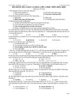
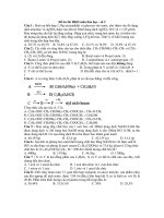
High-Frequency Jet Ventilation
HFJV refers to delivery of inspiratory gases through a jet injector
at a high velocity into the trachea with interruption of the flow
stream at a rate of 100 to 400 cycles/min (Fig. 54.10) The primary
indication for HFJV is air leak syndrome, but HFJV may be used
successfully in patients with bronchiolitis, congenital diaphragmatic hernia, congenital heart disease, and other disease states.
The main controls in HFJV are the driving pressure, inspiratory time, and rate. The driving pressure is usually coupled with
CO2
CO2
CO2
• Fig. 54.10 High-frequency jet ventilation. CO2, Carbon dioxide.
PEEP (provided through a separate bias flow circuit) to achieve a
desired mean airway pressure and chest wall oscillation and is then
adjusted according to the level of lung expansion and gas exchange. The typical rate is 320 to 480 breaths/min, although rates
as low as 240 breaths/min can be used for those patients needing
a longer expiratory time. The optimal rate is dependent on the
child’s size and disease process. Tidal volume increases with higher
driving pressure and increased inspiratory time. In certain circumstances, conventional ventilation can be combined with HFJV to
provide “sigh breaths” of approximately 6 to 10 mL/kg to prevent
atelectasis and maintain lung volumes during HFJV. It should be
noted that the need for these sigh breaths can often be eliminated
with the use of an adequately high mean airway pressure. Gas trapping can be minimized with an inspiratory time close to 20%,
longer expiratory time (i.e., lower rate) and lower driving pressures.
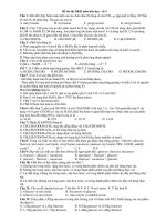
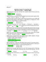
High-Frequency Oscillatory Ventilation
HFOV refers to ventilation by oscillatory flow with alternating
positive and negative pressures in the airway with Vt 1 to 3 mL/
kg and frequency 5 to 15 Hz. HFOV is most commonly used as
a rescue therapy for restrictive lung diseases, such as ARDS, that
are refractory to conventional ventilation.43
Oscillations are produced by a diaphragm connected to a piston (Fig. 54.11). The main controls in HFOV are mean airway
pressure, oscillatory pressure amplitude (power), frequency, and
inspiratory time. Mean airway pressure determines lung volume
and is the primary determinant of oxygenation along with Fio2.
Oscillatory pressure amplitude is the change in pressure around
the mean airway pressure produced by forward and backward
displacement of the piston. These pressures are attenuated in the
distal airways owing to the impedance of the endotracheal tube
and proximal airways. Vt is determined by the amplitude and
duration of each stroke. Oscillatory amplitude is a primary determinant of Vt and therefore minute ventilation as well. Frequency
is usually set in the range of 5 to 12 Hz. For a given amplitude, a
lower frequency will increase Vt and improve minute ventilation
but will also result in less attenuation of pressures along the
airways; the larger effective alveolar Vt is therefore less lung
protective. Inspiratory time is generally set at 33% of the total
cycle time. A higher inspiratory time will also result in a larger
effective tidal volume but can greatly increase the risk of gas
trapping.
Bias
flow
To patient
Magnetically driven piston
Expiratory
valve
• Fig. 54.11 High-frequency oscillatory ventilation.
CHAPTER 54 Mechanical Ventilation and Respiratory Care
639
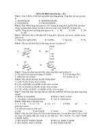
Convective pressure rise
VDR
Pressure
Pulsatile flow rate
PEEP
Oscillatory PEEP
Time
• Fig. 54.12 High-frequency percussive ventilation.
High-Frequency Percussive Ventilation
HFPV is most commonly used by burn centers for inhalation
injury, as it combines aspects of HFJV and conventional ventilation and adds the secretion mobilization benefits of percussive
ventilation. It may also be used for ARDS, plastic bronchitis, and
other disease states.45
In HFPV, gas flow is pneumatically driven and delivers subphysiologic Vt at high rates (up to 500 breaths/min) using the
volume diffusion respirator. High- and low-pressure circuits attach to a system called the Phasitron, which is a sliding Venturi
that acts as both an inspiratory and expiratory valve. There are
seven control variables: (1) peak inspiratory pressure, (2) PEEP,
(3) CPAP, (4) inspiratory time, (5) expiratory time, (6) percussive
frequency, and (7) rate. The Vt delivery is a product of the peak
inspiratory pressure setting with subtidal volumes produced by
the oscillatory function. During inspiration, lung volumes are
progressively increased in a controlled, stepwise fashion by repetitively diminishing subtidal volume deliveries until an oscillatory
plateau is entered and maintained (Fig. 54.12). At the end of inspiration, the lung is allowed to empty passively (with continued
oscillations) until the preset expiratory baseline is reached.
Adverse Effects of Mechanical Ventilation
The success of mechanical ventilation hinges on the balance of
beneficial and deleterious effects. The beneficial effects in the lung
are related to improvements in pulmonary mechanics and gas
exchange, which are often seen immediately. Ventilator-induced
lung injury may not be immediately appreciated but is closely tied
to meaningful clinical outcomes; short-term improvement in gas
exchange will typically not lead to improved outcomes if achieved
with toxic ventilator settings.
Airway Injury
Oropharyngeal and nasopharyngeal injuries secondary to the endotracheal tube are uncommon but may include ulceration of the
ala nasi from pressure necrosis following prolonged nasotracheal
intubation, or ulceration may occur at the angles of the mouth
from tight taping of orotracheal tubes. Palatal grooves and traumatic cleft palate can occur in infants. Laryngeal injury may extend from minor swelling to ulceration of the mucosa of the vocal
cords and aryepiglottic folds. Similarly, injuries in the subglottic
region may extend from minor swelling to major ulceration, and
healing of severe injuries may lead to scarring or granuloma formation with airway obstruction. The majority of the subglottic
tracheal lesions are due to compression of the tracheal mucosa by
the endotracheal tube. High-pressure cuffs, low cardiac output
state, upper respiratory tract infection, duration of intubation,
and head-neck movement all increase the risk of tracheal injury.
However, these injuries have become less common with modern
endotracheal tubes.46 Airway injury can also result from suction
catheters. Necrotizing tracheobronchitis is a severe form of airway
injury seen in patients on mechanical ventilation, which is characterized by extensive ulceration and mucosal damage. The sequelae of tracheal injuries include tracheal stenosis, tracheomalacia, tracheoesophageal fistula, and tracheoinnominate artery
fistula.
Injury to the airway can be prevented by attention to several
details. The endotracheal tube should be of the proper size and
should be inflated with less than 20 cm H2O to avoid pressure
necrosis in the adjacent tracheal mucosa. Excessive pressure on the
skin should be avoided while taping the endotracheal tube. Excessive patient movement should be prevented by targeted sedation.
Suctioning should be gentle, preferably with a catheter with multiple side holes, and suction catheters should not be routinely
advanced beyond the tip of the endotracheal tube.
Effects on the Lung
Adverse effects of mechanical ventilation on the lung may be due
to the following factors: (1) high airway pressures, (2) overdistention of the alveoli, (3) cyclic closing and reopening of alveoli,
(4) inflammation and cytokine exposure, (5) altered mucociliary
clearance, (6) impaired lung water clearance, and (7) oxygen toxicity. In many cases, it may be difficult to discern the contribution
of lung injury from the ventilator from the pathologic effects of
the underlying disease process(es). Pulmonary barotrauma is a general term that encompasses many entities of parenchymal injury.
Increased airway pressure may cause hyperinflation of the alveoli,
increased alveolar dead space, impaired venous return, compressed
alveolar vessels, and risk for air leak syndromes. Alveolar rupture
from overdistended alveoli is the most clinically apparent manifestation of pulmonary barotrauma. Air leak may occur from the lung
into the pleura (pneumothorax), interstitium (PIE), mediastinum
(pneumomediastinum), pericardium (pneumopericardium),
640
S E C T I O N V Pediatric Critical Care: Pulmonary
peritoneal cavity (pneumoperitoneum), and subcutaneous tissue
(subcutaneous emphysema). Even though the term implies high
airway pressures as the main mechanism of parenchymal injury,
pulmonary barotrauma is often multifactorial. The physiologic
consequences of extra-alveolar air may range from no adverse effect to life-threatening cardiorespiratory compromise. A pneumothorax may be small and inconsequential or may be large and
under tension, necessitating immediate evacuation of the pleural
air. PIE may decrease lung compliance and increase pulmonary
vascular resistance. Pneumomediastinum will typically track along
fascial planes either cephalad to produce subcutaneous emphysema or caudad to produce pneumoperitoneum or pneumoretroperitoneum. Pneumomediastinum rarely requires evacuation and
will self-resolve once the impetus for air leak has been halted.
Pneumopericardium can range from a minimally inconsequential
amount of air to life-threatening cardiac tamponade. Cardiovascular compromise is an indication for immediate evacuation of
pericardial air.
An uncommon but important form of pulmonary air leak is a
bronchopleural fistula, in which a fistulous track develops between the bronchus and pleural space. This results in an almost
continuous flow of air from the airway into the pleural space. The
fistula flow is wasted ventilation and may result in hypercarbia.
Attempts to increase minute ventilation by increasing Vt will
only serve to increase the fistula flow by increasing the pressure
gradient across the fistula. If attempts to decrease airway pressures
with conventional ventilation are not possible without compromising gas exchange, a trial of HFV may be considered.
Barotrauma can be minimized by avoiding factors that predispose to pulmonary air leakage. The principal factors that can be
controlled are airway pressures and lung volumes. As long as acceptable gas exchange is maintained, every effort should be made
to reduce airway pressures to a minimum. Hyperinflation must be
avoided. When the lung disease is severe, deliberate hypercarbia
may be tolerated provided that the arterial pH is acceptable.13
Inspired oxygen concentration should be maintained at nontoxic
levels (usually ,0.50). Ventilator-induced lung injury is described
in more detail in Chapter 48.
Effects on the Circulatory System
The cardiovascular effects of positive intrathoracic pressure are
complex and depend on many factors, including the underlying
lung disease etiology, uniformity of lung disease, transmission of
airway pressure to the pleural space, and lung volume. Positive
intrathoracic pressure impedes right ventricular filling by decreasing the pressure gradient for systemic venous return. Positive airway pressure can also increase pulmonary vascular resistance when
the airway pressure significantly exceeds FRC. Positive intrathoracic pressure has also been shown to decrease left ventricular afterload. The net effect is a combination of all effects mentioned
earlier and the reflex cardiovascular adjustments that accompany
these changes. See Chapter 32 for more details on cardiopulmonary interactions.
Specialty Gases
Inhaled Nitric Oxide
Nitric oxide is a potent vasodilator; inhaled nitric oxide (iNO)
produces selective pulmonary vasodilation in any segment of ventilated lung. Indications for iNO include primary pulmonary
hypertension, pulmonary hypertension after repair of congenital
heart disease, congenital diaphragmatic hernia, and isolated right
heart failure. Randomized controlled studies have shown that
iNO safely improves arterial oxygen levels in babies with pulmonary hypertension and decreases the need for extracorporeal
membrane oxygenation (ECMO) therapy.47 In multicenter studies in children and adults with ARDS,48,49 there were no differences in ventilator-free days and no effect on mortality between
treatment groups. Physiologically, iNO preferentially vasodilates
capillaries in well-ventilated alveoli, improving V/Q mismatch
and oxygenation, but not affecting the underlying disease pathology. As such, iNO may be used as a bridge to a different therapy
for ARDS (e.g., HFV or ECMO) but should not be employed
routinely. Not all patients respond to iNO; after a trial period,
iNO should be continued only in those patients who show a
clinical response. Nitric oxide also serves as an oxidizing agent to
convert hemoglobin to methemoglobin; therefore, methemoglobin levels should be monitored during its administration.
Helium-Oxygen Mixture
Helium-oxygen (heliox) mixture has a much lower density as
compared with oxygen-nitrogen mixture. This results in reduced
resistance to breathing during turbulent flow states and makes
low-resistance laminar flow more likely.50 On the basis of the
physics of airflow and the properties of heliox, the following
behaviors can be predicted with its use: (1) heliox will result
in a higher flow when transairway pressures are held constant,
(2) heliox will result in a lower airway pressure when airflow is
constant, (3) density-dependent flow meters will underestimate
flow, (4) heliox can decrease the degree of air trapping and hyperinflation associated with lower airway obstruction, (5) heliox can
decrease the work of breathing, and (6) heliox can result in better
deposition of aerosols.51 Helium is usually administered through
a tight-fitting face mask, high-flow nasal cannula, or invasively
through a heliox-compatible ventilator.
Several studies have shown that heliox administration improves symptoms and relieves the respiratory distress associated
with upper airway obstruction due to viral croup, subglottic narrowing, and postextubation stridor.52,53 Studies of heliox in children with asthma demonstrate improved delivery of aerosolized
bronchodilators with the addition of heliox.51,54 Most clinical
studies also show improvement in clinical scores of respiratory
distress, relief of wheezing, and faster resolution of symptoms, but
fail to show consistent improvement in other clinical outcomes,
such as admission rates or length of stay.51,54 Heliox has also been
used in acute bronchiolitis in infants, with improvement in the
clinical score and work of breathing.55
Altering Pulmonary Vascular Resistance With
Adjusted Inspired Oxygen and Carbon Dioxide
Concentrations
Low alveolar oxygen tension increases pulmonary vascular resistance (hypoxic pulmonary vasoconstriction), while high alveolar
oxygen tension decreases pulmonary vascular resistance. In states
of increased pulmonary vascular resistance, such as pulmonary
hypertension, increasing the Fio2 can be a powerful vasodilator as
long as the vascular bed has normal reactivity. Similarly, with
certain types of “mixing” congenital heart lesions, such as hypoplastic left heart syndrome, it is critical to control pulmonary
CHAPTER 54 Mechanical Ventilation and Respiratory Care
blood flow and prevent pulmonary overcirculation. One approach
is to decrease the Fio2 to less than 0.21 with a blending of room
air with nitrogen. Several studies have shown that hypoxic gas
mixtures can be used both preoperatively and postoperatively to
balance the pulmonary and systemic circulations, but this approach has fallen out of favor due to concerns for combining hypoxia with a baseline state of reduced oxygen delivery. Another
approach, especially in patients undergoing mechanical ventilation either preoperatively or postoperatively, is to increase the
fraction of inspired CO2 concentration (Fico2).56 Increased Fico2
and the concomitant increase in Paco2 also increases pulmonary
vascular resistance and limits pulmonary blood flow. One of the
difficulties with a boost in Fico2 is increased spontaneous ventilatory drive due to an increased Paco2. This increases the work of
breathing and, with marginal cardiac reserve, may impose undue
strain on the heart. Therefore, neuromuscular blockade and total
ventilatory support may be necessary with increased Fico2 to
avoid an increased workload on the heart.
Respiratory Care During
Mechanical Ventilation
Pulmonary Hygiene
The primary objectives of airway clearance therapy are to prevent
and treat atelectasis from mechanical obstruction of airways and
remove toxic substances, including infective materials, proteolytic
enzymes, and other mediators of inflammation. The most effective method of clearing secretions is a combination of changing
body position and vigorous coughing by the patient. If the patient
is unable to cough effectively, chest physiotherapy with or without
active suctioning of the trachea may be beneficial. Handheld mechanical devices may be used in lieu of a caretaker’s hands for
chest percussion or vibration. Hyperoxygenation prior to endotracheal suctioning can help mitigate associated desaturations and
potential hemodynamic changes.
Chest physiotherapy refers to a variety of respiratory maneuvers performed to aid in the clearance of airway secretions and
promotion of lung expansion, including (1) postural drainage,
(2) chest percussion and chest vibration, and (3) deep breathing
exercises. The efficacy of chest physiotherapy in intubated patients
is unclear.57 Several devices have also been used as an adjunct to
standard chest physiotherapy: the intrapulmonary percussive ventilator (IPV); mechanical insufflator-exsufflator (CoughAssist,
Philips Respironics); FLUTTER (Axcan Pharma) mucus clearance device and Acapella devices; intermittent positive pressure
breathing (IPPB); mechanical percussors; and percussive vest devices. The IPV device delivers high-flow jets of air to the airways
by a pneumatic flow interrupter at a rate of 100 to 300 cycles/min
through a mouthpiece. The patient controls variables such as inspiratory time, peak pressure, and delivery rates. IPV has been
shown to be beneficial for secretion clearance (particularly for
cystic fibrosis patients) and improvement in atelectasis in
intubated patients.58,59 CoughAssist is a portable, electric mechanical insufflation-exsufflation device that attempts to simulate
a cough by using a blower and valve to alternately apply a positive
and then a negative pressure to a patient’s airway to assist the
patient in clearing retained bronchopulmonary secretions. This
approach has been shown to be of particular benefit in patients
with neuromuscular weakness.60 The FLUTTER and Acapella
devices are small, handheld devices that provide positive expiratory
641
pressure (PEP). Exhaling through the device creates oscillations in
the airway, resulting in loosening of mucus. Other PEP devices
are used with a small volume nebulizer and function in conjunction with medication delivery. There is no clear evidence that PEP
is more or less effective than other forms of physiotherapy. IPPB
devices use pressure to passively fill the lungs in conjunction with
a patient’s breath and may also nebulize inhaled medication. A
high-frequency chest wall vibrating/oscillating vest device has
been shown to mobilize secretions in patients with cystic fibrosis
and is commonly used as an adjunct airway clearance device in
children with a reduced ability to clear secretions due to neuromuscular abnormalities.60,61
Humidification Systems
During spontaneous breathing, inspired air is warmed and almost
completely humidified as it passes through the upper airways. The
use of an endotracheal or tracheostomy tube bypasses the natural
warming and humidifying functions of the upper airway, leaving
the mucosal surface below the artificial airway to provide both
humidification and heat to the inspired air. This may adversely
affect mucociliary clearance, and delivered dry air may result in
irritated, friable mucosa. Also, if the inspired gases are not
warmed to the body temperature, insensible water loss in the lung
is increased.
Humidifiers can be classified into those that provide only humidity and those that provide both heat and humidity. The nonheated designs are pass-over or blow-by, bubble, and jet humidifiers. In the clinical setting, the amount of humidity provided by
these simple humidifiers is about the same and is determined by
time of contact with the gas and water, temperature of both the
gas and water, and surface area of contact of the gas-water interface. The efficiency of humidification increases as the time of
contact and/or surface area of contact increases. Heating the gas
prior to humidification allows for higher relative humidity.
Aerosol Therapy
Aerosolized drug administration is used for the delivery of medications, including b2-agonists, atropine, ipratropium bromide,
cromolyn sodium, antiviral agents, corticosteroids, antibiotics,
surfactant, pentamidine, and mucolytics. Aerosolization increases
the therapeutic index of the drug by delivering it directly to the
site of action while minimizing systemic side effects. The factors
that affect deposition of aerosol particles are gravity, viscosity of
the gas, kinetic activity of the particles, particle inertia, physical
nature of the particle, temperature and humidity of the aerosol,
and the ventilatory pattern. Compared with adults, deposition of
aerosolized particles in infants and children is poor because of the
small airway caliber, relatively greater airway resistance, high respiratory rate with a short inspiratory time, increased chest wall
compliance, ineffective coordination effort, and inconsistent
breath-holding maneuvers.
Four types of aerosol delivery systems are available for clinical
use: jet or pneumatic nebulizers, ultrasonic nebulizers, metereddose inhalers, and dry-powder inhalers.62 A jet nebulizer uses the
Bernoulli principle to create an aerosol. The size of the particle
depends on the jet flow rate and size of the capillary tube. Baffles
placed in the path of the aerosols tend to remove larger particles,
allowing delivery of smaller particles to the patient. A pneumatic
nebulizer creates an aerosol using the same principle as the jet
nebulizer but may use the main gas flow or a side stream nebulizer.