Đề ôn thi thử môn hóa (688)
Bạn đang xem bản rút gọn của tài liệu. Xem và tải ngay bản đầy đủ của tài liệu tại đây (262.34 KB, 5 trang )
682
S E C T I O N V Pediatric Critical Care: Pulmonary
Freedom from bronchiolitis obliterans
syndrome (%)
100
75
P = .0089
50
25
1994–2003 (N = 314)
2004–6/2016 (N = 528)
0
0
1
2
3
4
5
6
7
8
9
Years
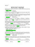
• Fig. 57.4 Pediatric
lung transplants freedom from bronchiolitis obliterans syndrome (transplantation
performed January 1994 to June 2016).
• BOX 57.2 Currently Accepted Characteristics of
the Ideal Donor for Pediatric Recipients
Age ,55 y
ABO compatibility
No HLA antibody sensitization by recipient
Clear chest radiograph
Pao2 . 300 on Fio2 5 1.0, PEEP 5 5 cm H2O
Tobacco history ,20 pack-years
Absence of chest trauma
No evidence of aspiration/sepsis
No prior cardiopulmonary surgery
Sputum Gram stain—absence of organisms and PMNs
Absence of purulent secretions at bronchoscopy
Fio2, Fraction of inspired oxygen; HLA, human leukocyte antigen; Pao2, partial pressure of arterial oxygen;
PEEP, positive end-expiratory pressure; PMN, polymorphonuclear neutrophils.
for pediatric candidates have not been established. However, it is
generally accepted that the ideal lung donor for children should
be a nonsmoker of the same size (chest dimension measured from
the diaphragm to the apex of the lung) and blood type or ABO
compatible. Other than these basic criteria, the evaluation is relatively subjective and occurs at the time of retrieval. The general
tenets followed include that the donor should have no significant
history of lung disease, including asthma. There should be no
pulmonary trauma or infections, gas exchange should not be impaired, and ischemic time should be minimal. Pediatric lung
transplant centers may apply more stringent criteria if the candidate in question is reasonably stable. Donor offers from younger
donors may be more desirable in some cases.
Depending on the need of the candidate, nonideal donors, also
called extended or marginal donors, may be accepted. There is
limited data to either support or prohibit the use of lungs from a
nonideal donor. There is no evidence that a marginal donor will
have any effect on either immediate or long-term morbidity or
mortality except in egregious cases of the diagnosis of bronchopneumonia, the presence of purulent lower airway disease in the
donor, or injury from contusions. It is shown that lungs obtained
from donors older than 65 years who have a significant smoking
history are at risk for developing malignancy in the setting of immunosuppression. As well, lungs from older donors have worse
long-term graft survival.18
Many factors stemming from donor cause of death and subsequent donor maintenance in the ICU can contribute to donor
lung injury. The most common causes include ventilator-induced
lung injury, atelectasis, oxygen toxicity, and volume overload. In
addition, after brain death, a systemic inflammatory response
known as a cytokine storm occurs. This predisposes to the development of lung injury that is similar to acute respiratory distress
syndrome, (ARDS). A different type of cytokine storm that occurs following brain death is a catecholamine storm. In an attempt to protect cerebral perfusion during brain death, the body
will release a large amount of catecholamines. This surge of catecholamines causes significant systemic hypertension, which results in elevated left-sided heart pressures and consequent interstitial edema and can sometimes cause alveolar hemorrhage, resulting
in neurogenic pulmonary edema. This generally precludes the use
of the lungs for transplantation because of poor oxygenation.
However, neurogenic pulmonary edema is a leaky capillary syndrome that is fully recoverable. This is one area in which removal
of the lung from the inflammatory milieu of the brain-dead donor
and a period of time for recovery and removal of extravascular
water with ex vivo lung perfusion (EVLP) has had a significant
impact on donor lung utilization.
The process for donor lung preservation begins at the time of
declaration of death and extends until the lungs are reperfused in
the recipient. Prior to retrieval, fluids are managed to maintain
euvolemia and barotrauma must be avoided. A 1-g bolus of methylprednisolone is given to the donor to mitigate brain death–
induced systemic inflammation.19 At the time of retrieval, the lungs
are prepared for transport, flushed vigorously with a preservation
solution, and inflated with oxygen. An extracellular-type flush
preservation solution with low potassium, coupled with glucose
and dextran, has been established as best practice for prolonged
cold preservation.20 Prostaglandin E1 (PGE1) is a vasodilator given
before the dextran flush to reduce pulmonary vascular resistance
and achieve a more complete flush. PGE1 also has antiinflammatory
CHAPTER 57 Pediatric Lung Transplantation
properties useful for lung preservation and prevention of reperfusion injury.21 A retrograde flush is subsequently performed with
the same solution, again, to improve the homogeneity of the
flush.22 The lungs are inflated with 50% oxygen before their removal from the body in order to maintain the alveolar structure
and to provide oxygen for metabolism.23
A novel strategy of perioperative lung preservation is being
developed by the Toronto Lung Transplant Group. Using their
technique, termed ex vivo lung perfusion (EVLP), lungs are continuously perfused anterograde with an acellular perfusate and
ventilated with room air with an ICU ventilator at normothermia.24 A hypoxic air mix is bubbled into the perfusate to deoxygenate and add carbon dioxide (CO2) to the perfusate. This
method, performed by a separate surgical team, allows for at least
12 hours of donor lung preservation. EVLP allows the team to
further evaluate the donor lungs as to their suitability for transplantation. More important, the lungs can be treated actively to
improve their performance.25 Marginal lungs can be resuscitated
and rehabilitated using EVLP, expanding the donor pool.26
Surgical Approach
Bilateral sequential lung transplantation is the most frequently
performed lung transplantation procedure in children and is performed most often via median sternotomy. The main stem bronchi and left and right pulmonary arteries are connected via endto-end anastomoses. Two pulmonary veins with intact atrial
connections are harvested from each donor lung. Each left atrial
patch is sewn onto the recipient heart. This surgical approach
minimizes cardiopulmonary bypass time, which reduces related
complications.27–29
Though combined heart-lung transplantation had initially
been a favored surgical approach, improved surgical techniques as
well as the profound scarcity of donor organs have led to a dramatic decrease in the frequency of heart-lung transplantation.
Moreover, right-sided heart failure associated with pulmonary
hypertension resolves following lung transplantation, which has
obviated the need for heart and lung transplantation for primary
pulmonary hypertension except in instances of severe, irreversible
right heart failure.30,31 There is no difference in survival between
patients who undergo bilateral sequential lung transplantation
compared with those who undergo heart-lung transplantation.32
Lung transplantation alone maximizes the distribution of organs
from a single donor, benefiting more children.
In the 1990s, living donor lobar lung transplantation was developed as a strategy for transplantation in order to decrease waiting time of severely ill children awaiting lung transplantation, but
with the adoption of a new lung allocation score in 2005 and
improved peritransplant strategies, wait list deaths have decreased.33 The relative efficacy of the new lung allocation scoring
system, combined with the technical and ethical challenges associated with living lobar transplantation, have prevented wider
adoption of the procedure in the United States.34
Presurgical Management in the Intensive
Care Unit
Compared with the early era of lung transplantation, the number
of patients receiving a transplant from the ICU and with mechanical respiratory support has increased recently. Thus, the incidence of bridging severe respiratory failure to lung transplant
683
with ambulatory VV ECMO and mechanical ventilation is increasing. The risk of dying within 1 year of transplantation increases by 58% if the patient was bridged to transplant in the
intensive care unit (ICU) with mechanical ventilation (MV) support before lung transplantation.35 Intubated patients who require
heavy sedation and ventilation with high airway pressures are especially prone to ventilator-induced injury and ICU-related complications, including extrapulmonary organ failure. ICU-related
complications—such as pressure ulcers, vascular complications,
nosocomial infections, delirium, critical illness polyneuropathy/
myopathy, and airway colonization—will increase wait list mortality and mortality after transplant. Candidates for lung transplant on MV support may have uncertain neurologic status. Thus,
an approach with “awake” ventilation, even if supported with VV
ECMO, is often pursued to obtain better short-term outcomes.
There have been substantial improvements in extracorporeal life
support (ECLS) technology and many centers are increasingly using
these devices. Bridge-to-recovery and bridge-to-transplant are the two
basic indications for ECMO support. While the adult experience is
quickly expanding, the pediatric literature is limited. A retrospective
evaluation of the United Network for Organ Sharing database of
pediatric transplantations between 2000 and 2013 in the United
States determined that a small percentage (2.9%) of patients were
bridged to transplant with ECMO and there was no statistically significant increase in hazard for death.36 Major advances in ECLS included use of heparin-coated circuits, development of polymethylpentene oxygenator membranes, introduction of centrifugal pumps,
dual-lumen cannulas (important for small adults and the pediatric
population), and miniaturized systems. For these reasons, VV ECMO
as a bridge to transplant is considered carefully for a small percentage
of critically ill children awaiting lung transplantation.
Postsurgical Management
Immediate postoperative care is focused on respiratory and hemodynamic management. In the perioperative period, pulmonary
care emphasizes reestablishment of functional residual capacity.
Mechanical ventilation is generally necessary for less than 48 hours
but may be prolonged in the event of primary graft dysfunction.
There is a wide variation in MV strategies among lung transplant
centers. In general, lung protective approaches using low tidal
volumes based on recipient’s characteristics are preferred. However, in a retrospective study on patients receiving a transplant
between 2010 and 2013 among three transplant centers, low tidal
volume ventilation was not shown to have an effect on length of
ICU stay, forced expiratory volume (FEV1) at 3 months postsurgery, or survival to 6 months. Conversely, poor outcomes have
been associated with injudicious use of higher-pressure ventilation
strategies.37 To minimize hyperoxic-related injury to the lungs,
the fraction of inspired oxygen is maintained at less than 60%
while maintaining systemic arterial saturation at 94% to 95%.
Ventilator strategy uses 5 to 7 mL/kg tidal volumes and an inspiratory plateau pressure of less than 30 cm H2O. Sufficient positive
end expiratory pressure is used to fully recruit and maintain the
functional residual capacity of the newly transplanted lungs.
Once the patient is extubated, aggressive tracheobronchial toilet,
chest physiotherapy, and bronchoalveolar lavage can mobilize secretions to ensure patency of the airways.
Hemodynamic status must be closely monitored though data on
hemodynamic management are limited. Vascular permeability and
myocardial function may be adversely affected by cardiopulmonary
bypass, necessitating inotropic support in the perioperative
684
S E C T I O N V Pediatric Critical Care: Pulmonary
period. Usually, restrictive fluid support (0.9–1.0 3 maintenance)
is encouraged. Central venous pressure monitoring is beneficial in
order to optimize cardiac output.38 Central venous pressure alone
may be unreliable to guide volume status. Hemodynamic instability may be exacerbated by diminished intravascular volume. Early
recognition of compromised renal function is essential, as the
prescription of all medications excreted and metabolized by the
kidneys will need to be promptly altered. Additionally, clinical
and ultrasound observations may be helpful.
The most common causes of hypotension in the immediate posttransplantation period include hypovolemia from overly aggressive
diuresis, systemic inflammatory response syndrome from surgical
insult causing low systemic vascular resistance, medication-induced
hypotension (including sedatives/analgesics), lung hyperinflation,
hemorrhage, tamponade, or heart failure.39,40 Management should
be causally determined, generally requiring a combination of fluid
volume management, transfusion of blood products, administration
of vasopressors or inotropes, correction of bleeding diatheses, chest
tube drainage, and, when indicated, surgical revision.
Recipients may experience early severe graft dysfunction as a result
of lung injury incurred during or prior to organ harvest. The occurrence of primary graft dysfunction (PGD) is between 10% and 35%
of all patients. The clinical presentation of PGD is entirely consistent
with ARDS as manifested by elevated alveolar-arteriolar gradient,
compromised pulmonary compliance, poor ventilation and perfusion matching, and impaired diffusion.41 PGD refers to acute respiratory failure defined by reduced oxygenation index and pulmonary
infiltrates within 72 hours of lung transplantation (Table 57.2).42 In
most patients, a mild and transient course is observed, but 10% to
20% of patients will be affected by a severe form (partial pressure of
arterial oxygen/fraction of inspired oxygen [Pao2/Fio2] ,200). Secondary causes of hypoxemia—such as volume overload, pneumonia,
acute rejection, atelectasis, or pulmonary venous outflow obstruction—
should be excluded. Severe PGD is associated with high hospital
mortality rates of 30% to 40%, prolonged ICU stay, and impaired
long-term graft function and survival. It is the leading cause of mortality in the perioperative period. In a multicenter study from 10 US
centers, increased oxygen fraction levels at the time of graft reperfusion was associated with increased risk of subsequent PGD. Severe
PGD is associated with donors with any smoking history, PAH, the
use of cardiopulmonary bypass, large-volume blood product transfusion, elevated pulmonary arterial pressures, or obesity.43 Improved
surgical techniques and organ perfusate have diminished the severity
of early graft dysfunction over the last decade.
TABLE
Grading of Primary Graft Dysfunction
57.2
Grade
Po2/Fio2
Radiographic Changes
Grade 0
.300
Absent
Grade 1
.300
Present
Grade 2
200–300
Present
Grade 3
,200
Present
Fio2, Fraction of inspired oxygen; Po2, partial pressure of oxygen.
Modified from Christie J, Carby M, Bag R, et al. Report of the ISHLT Working Group on
Primary Lung Graft Dysfunction part II: definition. A consensus statement of the International
Society for Heart and Lung Transplantation. J Heart Lung Transplant. 2005;24(10):
1454-1459.
Treatment of PGD is primarily supportive. Most grafts will
recover under careful ventilator management, diuretics, and pressor support as well as high-dose corticosteroid pulses. Inhaled
nitric oxide (iNO) has been shown to improve oxygenation in the
presence of acute graft dysfunction, likely as a result of enhanced
ventilation and perfusion matching. A few observational trials
suggest that use of iNO in patients with PGD may result in a
better outcome. Despite these limited data, iNO has been used as
salvage therapy for severe allograft dysfunction following transplantation. It may be useful in patients with refractory hypoxemia
posttransplantation.44,45 ECMO has been successfully employed
as a therapeutic modality.46
The postoperative course can be complicated by technical
problems associated with the surgery. At many centers, the
patency of the airway anastomoses is routinely assessed within
24 hours by direct visualization with flexible bronchoscopy. While
the vascular anastomoses are more difficult to assess, arterial
anastomoses are generally amenable to inspection with nuclear
medicine studies. In order to assess the venous anastomoses,
transesophageal echocardiography may be necessary.47,48
Vocal cord paresis or diaphragmatic paresis can complicate virtually any major thoracic surgery, both of which derive from surgical injuries to the respective nerve. Vocal cord paralysis or paresis
results from injury to the recurrent laryngeal nerve, and phrenic
nerve injury leads to diaphragmatic paralysis or paresis. However,
the clinical symptoms entailed by these issues generally are not
apparent until after extubation. The likelihood of phrenic nerve
injury is increased in patients who have had prior thoracic surgery.
Most of these injuries resolve within several weeks of surgery, but
serious consideration should be given to early diaphragmatic plication, as the risk of infection in the lung affected by the paretic
hemidiaphragm is quite high.49 Vocal cord function may be temporarily compromised following removal of an endotracheal tube
even in the absence of true injury. Thus, definitive evaluation for it
should be deferred to at least 72 hours after extubation.
In cases of respiratory failure after extubation, noninvasive ventilation may be an option to prevent reintubation.50 In immunosuppressed patients with acute respiratory failure, early initiation of
noninvasive ventilation was associated with significant reductions
in reintubation and an improved likelihood of survival to hospital
discharge.51 For patients with profound hypoxemia, high-flow oxygen
through a nasal cannula is an increasingly applied option.52
Immunosuppression
The long-term success of lung transplantation is achieved with the use
of immunosuppressive drugs that inhibit rejection of the lung allograft. Immunosuppression strategies in lung transplantation generally consist of a triple-drug maintenance regimen composed of a
calcineurin inhibitor, T-cell antiproliferative, and corticosteroids. In
the United States, this regimen is most often tacrolimus, mycophenolate mofetil/mycophenolic acid, and prednisone. Approximately 60%
of pediatric lung transplant centers use an induction regimen in the
perioperative time period, though data do not support the impression
that induction confers either a survival benefit or reduction in the
incidence of CLAD.53,54
Immunobiology
Acute cellular rejection (ACR) of the transplanted lungs occurs in
almost one-third of children within the first year of lung transplantation.2 Alloreactivity toward the graft is likely augmented by
CHAPTER 57 Pediatric Lung Transplantation
685
local innate immune activation in various situations, such as preexisting inflammatory processes in the donor, tissue injury related
to ischemia and reperfusion injury at the time of implantation,
and posttransplantation infections. As well, the airways are continuously exposed to the environment via inhalational toxins,
pathogens, allergens, and irritating organic and inorganic particles, all of which stimulate the protective response of the innate
immune system. Episodes of ACR entail activation of the innate
and adaptive immune responses, resulting in recruitment of alloreactive CD41 and CD81 T lymphocytes to the lung allograft,
which magnify further recruitment of neutrophils, eosinophils,
B lymphocytes, macrophages, and natural killer (NK) cells, causing
lung injury.55,56 On the other hand, tolerance can be facilitated by
regulatory Foxp31 CD41 T cells, central memory CD81 T cells,
and NK cells.57,58
Rejection
The clinical manifestations of ACR include fever, dyspnea, and
hypoxia. Chest radiograph findings are relatively nonspecific but
often include perihilar infiltrates and effusions. Airflow obstruction may be detected with spirometry. The patient must be evaluated for both infection and rejection when these signs are
detected. This requires bronchoscopy to obtain bronchoalveolar
lavage samples and transbronchial biopsies for histologic evidence
and grading.59 At least five pieces of alveolated tissue are required
for the highest level of confidence to determine the presence and
grade of severity of ACR, if present. Since ACR occurs in the
majority of patients in the first year following lung transplantation and clinical signs of graft dysfunction are often not present,
surveillance bronchoscopies are performed on a predetermined
schedule in the first year posttransplantation to monitor the lung
allograft for ACR. The pathologist examines the transbronchial
biopsy specimens for the presence ACR, lymphocytic bronchiolitis, and for evidence of chronic rejection. The A grade designation describes ACR, referring solely to the extent and distribution
of the mononuclear cells that form as perivascular cuffs, and includes evaluation for extension of the process beyond the vascular
adventitia into adjacent alveolar septae.60 The B designation applies to the presence and severity grade of lymphocytic inflammation surrounding small airways. The grade reflects the intensity of
the inflammatory infiltrates surrounding bronchioles. The C designation applies to whether fibrotic changes consistent with either
obliterative bronchiolitis (luminal obliteration of the small airways
with fibrosis) or RAS (interstitial fibrosis) is present (Fig. 57.5). Of
note, transbronchial biopsies are miniscule in size, and bronchioles often are not present for histologic examination. Thus airway
disease itself may not be reportable. As well, technical issues regarding tissue preservation can be induced by crush artifact from
the forceps. Because of this, an “ungradable” category in lymphocytic bronchiolitis is designated for biopsies limited by those and
other sampling problems. Histologic evaluation of the specimens
leads to the assignment of a grade: A0 indicates the absence of
rejection and grade A4 indicates severe rejection.61
Treatment for ACR is initiated with high-dose intravenous
methylprednisolone and bronchoscopy is repeated 2 to 4 weeks later to
assess for resolution of abnormal histopathology. For refractory
ACR, optimization of the oral immunosuppression regimen concomitant with more aggressive treatment with monoclonal antibodies—
such as alemtuzumab (CD52 receptor antagonist), basiliximab
(CD25 a-chain antagonist), other biologicals—or photopheresis
may be considered.
• Fig. 57.5 Histopathology of obliterative bronchiolitis.
Antibody-Mediated Rejection
The many consequences of immunologic injury to the lung from
the development of donor-specific antibodies (DSAs) include
persistent or recurrent episodes of ACR of all grades, lymphocytic
bronchiolitis, and all subtypes of CLAD.61–63 The term antibodymediated rejection (AMR) describes the production of damaging
DSA targeted against the allograft by recipient immune cells.
HLA molecules are the major transplant antigens that can cause
AMR. DSAs largely target HLA molecules. The presence of graft
dysfunction, complement deposition at the alveolar capillary
membrane, detection of circulating DSAs, and histopathologic
changes (capillaritis) are considered sufficient evidence of AMR.64
However, it is unusual that all of these criteria are met in lung
transplant recipients and, if present, are detected after severe, irreversible damage has been conferred to the allograft. AMR is
often refractory to therapy, resulting in graft failure and death.
A 2012 ISHLT consensus statement includes a much broader
set of histologic findings felt to be consistent with AMR if, in addition, donor-specific HLA antibodies are present and capillary
complement deposition positivity is present in at least 50% of
interstitial capillaries.65 This document was updated in 2016.66
There is no consensus on treatment of humoral rejection. Pulse
steroids, plasmapheresis, intravenous immunoglobulin, and B cell–
directed therapy (Cytoxan or rituximab) are used, often in combination.67 The proteasome inhibitor bortezomib (targets plasma
cells) and the complement inhibitor eculizumab are also considered
in the treatment of AMR.68,69
The lung transplant community has made important headway
in recognizing cases of AMR, but substantial challenges remain in
standardization in the diagnosis of and determining the most
optimal therapeutic options for pulmonary AMR.
Chronic Lung Allograft Dysfunction
Broadly, the term CLAD has been adopted to include manifestations of graft dysfunction that can occur due to a variety of immunologic or nonimmunologic allograft insults (Fig. 57.6).70
While there is a clear relationship between ACR and eventual
development of CLAD, other etiologies that are associated with
chronic graft dysfunction must be investigated and ameliorated.
686
S E C T I O N V Pediatric Critical Care: Pulmonary
CLAD Not Due to Chronic Rejection
• Allograft related
°
°
°
°
°
°
ARAD
Follicular bronchiolitis
Refractory acute cellular rejection
Infection/colonization
Antibody-mediated rejection
Anastomotic stenosis
• Allograft related
RAS
BOS
• Non–allograft related
°
°
°
°
Pleural disease
Diaphragm dysfunction
Neuromuscular dysfunction
Other
Acute Lung Allograft Dysfunction
CLAD
ALAD
°
°
°
°
°
°
°
°
°
Acute rejection (cellular/humoral)
Lymphocytic bronchiolitis
Infection
Anastomotic abnormality
ARDS
ABPA
Pulmonary embolism
Pneumothorax
Other
• Non–allograft related
° Pleural disease
° Measurement error
• Fig. 57.6 Etiology of allograft syndromes. ABPA, Allergic bronchopulmonary aspergillosis; ALAD, acute
lung allograft dysfunction; ARAD, azithromycin-responsive allograft dysfunction; ARDS, acute respiratory distress syndrome; BOS, bronchiolitis obliterans syndrome; CLAD, chronic lung allograft
dysfunction; RAS, restrictive allograft syndrome.
CLAD is a diagnosis of exclusion. CLAD tends to be a nonuniform process; thus, TBBx sampling is not likely to identify fibrosis early in the course posttransplantation. Thus, CLAD diagnosis
per se does not hinge on histopathologic evidence but rather on
the development of composite findings of histopathologic, radiologic, and measured changes in allograft function.
Historically, chronic lung graft dysfunction was thought to
present only as BOS, in which the progressive development of
obliterative bronchiolitis led to a fall in the FEV1 and, ultimately,
to graft loss. However, in 2005, a novel subtype of BOS was first
described as a distinct entity.71 Since then, different phenotypes of
CLAD with distinct prognostic significance have been described.
The entity described by Pakhale et al.71 is now known as restrictive
allograft syndrome (RAS). RAS is described histologically as fibrosis occurring predominantly in the peripheral lung tissue rather
than in small airways, resulting in a decline in total lung capacity
in addition to a decline in FEV1.
CLAD phenotypes are characterized with a combination of
pulmonary function and imaging with chest CT. Of CLAD patients, 80% will have no identifiable cause for chronic graft failure, yet identifiable causes of graft dysfunction must be pursued
and treated, such as ACR, AMR, chronic infection, obesity, gastroesophageal reflux disease, aspiration, and chronic inflammation. Distinct phenotypes of CLAD have individual prognostic
significance. For example, neutrophilic allograft syndrome can be
slowed or arrested with a prolonged course of oral azithromycin,
whereas RAS is more rapidly progressive and associated with a
worse long-term outcome and low survival.72
Treatment Options for CLAD
There is no maintenance immunosuppression protocol proved to
be superior for preventing CLAD nor has any advantage been
demonstrated with the use of an induction regimen at the time of
transplantation. Intensifying the immunosuppressive treatment
generally has little effect in patients with established BOS or RAS.
Current therapy for the BOS subset of CLAD—if not associated
with AMR, infection, or inflammation—is limited to optimizing
immunosuppressant levels and maintaining good pulmonary toilet
to prevent postobstructive pneumonia and chronic inflammation.
Most practitioners will substitute tacrolimus for cyclosporine,
begin a trial of azithromycin for a minimum duration of 3 months,
or proceed to fundoplication of the gastroesophageal junction if
gastroesophageal reflux has been refractory to medical treatment.
Unfortunately, this approach tends to have limited success. Photopheresis, particularly in the setting of recurrent ACR or plasmapheresis in the setting of AMR, may be of utility in abrogating
rapid deterioration. Failing these approaches, retransplantation is
recommended in selected cases.73–75 As to treatment options for
RAS, no formal treatment guidelines exist. Pirfenidone is a synthetic molecule that has recently been approved for the treatment
of idiopathic pulmonary fibrosis (IPF) in Europe, Canada, Japan,
South Korea, and the United States. In vivo and in vitro studies
have shown a potent antifibrotic effect of pirfenidone, which inhibits the synthesis of transforming growth factor-b (TGF-b) and
tumor necrosis factor-a (TNF-a), leading to a reduction in fibroblast proliferation and collagen synthesis and thus a slower decline
in lung function in animal models of fibrosis and in IPF patients.76 Lung transplant recipients have been treated in single
case series reports. The treatment is currently experimental, but
the case reports have demonstrated some beneficial effects (i.e.,
mild improvement of interstitial changes and lung function) with
pirfenidone.77,78
Similarly, nintedanib is a drug indicated for the treatment of
IPF that targets multiple receptor tyrosine kinases and nonreceptor
tyrosine kinases that stimulate fibroblast growth factor receptor,
platelet-derived growth factor receptor, and vascular endothelial
growth factor receptor. These receptors have been implicated in
IPF pathogenesis. Nintedanib binds competitively to the adenosine triphosphate binding pocket of these receptors and blocks the
intracellular signaling crucial for the proliferation, migration, and
transformation of fibroblasts representing essential mechanisms of
the IPF pathology.79
Extracorporeal photopheresis (ECP) has emerged as an effective option, having been used successfully in cutaneous T-cell
lymphoma and graft-versus-host disease.80 ECP induces psoralenmediated deoxyribonucleic acid cross-linking and results in apoptosis of lymphoid cells, including NK and T cells. These apoptotic
lymphocytes are phagocytosed and eliminated upon reinfusion by
immature dendritic cells, which subsequently undergo maturation and present antigenic peptides. The first successful application of ECP in lung transplant recipients was reported in 1995.81
Experimental models and human studies have demonstrated
ECP-associated modulation of dendritic cells, alteration of