Đề ôn thi thử môn hóa (691)
Bạn đang xem bản rút gọn của tài liệu. Xem và tải ngay bản đầy đủ của tài liệu tại đây (154.04 KB, 5 trang )
692.e2
O
Choline
N+
HO
O
L-Tyrosine
CH3
OH
CH3
CoA
S
O2
Tetrahydrobiopterin
CH3
AcetylCoA
CoA
chAT
Acetylcholine
N+
O
A
O
HO
CH3
O
Tyrosine hydroxylase
Dihydrobiopterin
H2O
SH
H3C
NH2
HO
CH3
L-DOPA
OH
CH3
NH2
HO
CH3
DOPA decarboxylase
O
L-Tryptophan
OH
NH2
HN
HO
Dopamine
O2
Tetrahydrobiopterin
Dihydrobiopterin
H2O
Tryptophan
hydroxylase
O2
Ascorbic acid
HO
OH
OH
HO
NH2
HN
Dopamine β-hydroxylase
Dehydroascorbic
acid
H2O
O
5-HTP
NH2
HO
Norepinephrine
NH2
HO
5-HTP
decarboxylase
S-adenosylmethionine
Phenylethanolamine
N-methyltransferase
HO
Homocysteine
OH
Serotonin
HO
C
HN
Epinephrine
NH2
N
HO
B
H
H
O
C
[CH2]2
C
COO–
NH3
H2N
Glutamine
[CH2]2
C
α-Ketoglutarate
D
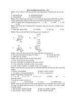
• eFig. 58.1
H
–OOC
C
[CH2]2
NH3
O
–OOC
Glutamate
COO–
CH3
COO–
Glutamic acid
decarboxylase
–OOC
[CH2]3
GABA
Neurotransmitter structure and synthesis. (A) Acetylcholine. (B) Catecholamines. It should be
noted that synthesis of norepinephrine and epinephrine requires dopamine as a precursor. (C) Serotonin,
an amine synthesized from tryptophan. (D) Glutamate and g-aminobutyric acid, amino acid derivatives.
NH3
CHAPTER 58 Structure, Function, and Development of the Nervous System
into epinephrine by an enzyme phentolamine N-methyltransferase, which is found only in adrenergic neurons. Thus, all neurons
that synthesize catecholamines contain TH and dopa decarboxylase, but only noradrenergic and adrenergic neurons contain
the synthetic enzymes required to produce norepinephrine and
epinephrine, respectively.
Catecholamine-using neurons reside primarily in the brainstem. Dopaminergic neurons in humans are located in two mesencephalic nuclei: the substantia nigra and its medial neighbor,
the ventral tegmental area. Dopaminergic neurons in the substantia nigra project primarily to the basal ganglia, where they are
involved with initiation of voluntary movement. Ventral tegmental neurons send dopaminergic fibers to the amygdala and the
cerebral cortex and participate in regulation of emotion, reward,
and addiction. Brainstem noradrenergic neurons are located in
the locus ceruleus and in the reticular formation. They project
widely to the thalamus and cortex, as well as to the spinal cord,
and play a significant role in arousal and vigilance. Adrenergic
CNS neurons are located in the ventrolateral medulla and participate in temperature regulation via their projections to the
hypothalamus.
Unlike ACh, which is cleared from the synapse by hydrolysis,
catecholamines are cleared from the synaptic cleft by reuptake
into the axonal terminal. Once inside the cell, catecholamines are
either repackaged into vesicles or destroyed by monoamine oxidase (MAO). Pharmacologic manipulation of synaptic catecholamine concentrations plays a therapeutic role in the management
of several disorders, such as depression and attention deficithyperactivity disorder (ADHD). Furthermore, recreational drugs
affecting catecholamine concentrations at the synapse continue to
gain popularity and to grow in number. Therapeutic uses include
treatment of severe depression with MAO inhibitors, which inhibit catecholamine breakdown, and treatment of ADHD with
amphetamines, which interfere with dopamine transport and
increase dopamine concentrations. Recreational drugs include
amphetamine and its analogs, along with cocaine, a selective norepinephrine transporter blocker. Excess catecholamine levels at
the synapse result in sensations of euphoria, increased energy
levels, improved focus, anxiety, paranoia, and jitteriness. Notably,
hypertensive crises leading to myocardial infarction and stroke may
occur with use of cocaine, amphetamines, and MAO inhibitors.
Serotonin
Serotonin is an amine neurotransmitter synthesized from the
amino acid tryptophan in a two-step process (eFig. 58.1C). First,
tryptophan is hydroxylated by tryptophan hydroxylase to form
5-hydroxytryptophan (5-HTP). 5-HTP is then decarboxylated
by 5-HTP decarboxylase to form serotonin, also known as
5-hydroxytryptamine (5-HT). After 5-HT is released at the synapse,
it is cleared by a specific serotonin reuptake transporter. Serotonergic
neurons are located in the rostral and caudal raphe nuclei in the
brainstem. Rostral raphe neurons innervate the cerebral cortex,
including the limbic system, where serotonin levels help regulate
mood and attention. Caudal raphe neurons project to the brainstem and spinal cord, where they are involved in regulation of
general arousal and pain perception, respectively. Importantly,
dysfunction of the serotonergic pathways originating in the raphe
nuclei has been linked with sudden infant death syndrome
(SIDS).15 Additionally, serotonin levels play a key role in depression, giving rise to an entire class of drugs in clinical use called
selective serotonin reuptake inhibitors (SSRIs). SSRI abuse or
overdose is rare but may result in patients presenting with the
693
potentially life-threatening “serotonin syndrome,” characterized
by hypertension, tachycardia, mental status changes, myoclonus,
and severe hyperthermia. The latter may lead to shock, rhabdomyolysis, renal failure, and death. The serotonin syndrome is
particularly likely to occur when SSRIs and MAO inhibitors, inadvertently or intentionally, are taken together. Treatment includes serotonin antagonists, blood pressure control with either
adrenergic antagonists or agonists as clinically indicated, and temperature control with benzodiazepines and neuromuscular blockade.
Amino Acids
Neurotransmitters derived from common amino acids include
glutamate, g-aminobutyric acid (GABA), and glycine. These are
among the most widely distributed neurotransmitters in the
CNS. Glutamate and glycine exist as amino acids in all cells,
where they are used as protein building blocks. Glutamatergic and
glycinergic neurons have the additional capacity to package glutamate and glycine, respectively, into synaptic vesicles and release
them at the synapse. GABA must be synthesized from glutamate
via an additional reaction catalyzed by an enzyme glutamic acid
decarboxylase (GAD; eFig. 58.1D). Only GABA-ergic neurons
contain GAD.
Glutamate is generally an excitatory neurotransmitter, whereas
GABA and glycine are inhibitory. Excitatory glutamatergic neurons exert their influence both locally and over a long distance,
depending on the shape of their axons. Inhibitory neurons, on the
other hand, tend to exert local inhibitory control over neuronal
circuitry either in the brain (GABA) or spinal cord (glycine). A
major exception is cerebellar Purkinje cells, which are GABAergic but project over long distances to the brainstem, thalamus,
and cerebral cortex (see later discussion).
Adenosine, Peptides, and Nitric Oxide
In addition to the “classic” neurotransmitters described earlier, a
number of substances have been documented to mediate or
modulate information transfer between neurons. These include
ATP and adenosine, which, at the synapse, is a metabolite of ATP
released in the synaptic vesicle. ATP modulates neuronal excitability such that energy may be conserved during times of ATP
depletion. Adenosine functions as a neurotransmitter in the autonomic nervous system (ANS), in the basal ganglia, and at some
cortical synapses. It also modulates the respiratory rate, and adenosine antagonists, such as caffeine, are used to treat apnea and
bradycardia of prematurity. In addition to ATP and adenosine, a
number of peptides can be released in synaptic vesicles, including
substance P, vasoactive intestinal peptide (VIP), endogenous opioids, and endogenous cannabinoids. These peptides are involved
in pain sensation and perception (substance P and opioids),
modulation of vascular tone (VIP), and as yet uncharacterized
processes (cannabinoids).
Finally, several gaseous molecules function as neurotransmitters. These include nitric oxide (NO), carbon monoxide (CO), and
possibly hydrogen sulfide. NO, the most thoroughly studied of the
gaseous neurotransmitters, is produced by brainstem neurons in
the nucleus tractus solitarius, where it interacts with a-amino3-hydroxyl-5-methyl-4-isoxazole-propionate (AMPA)–type and
N-methyl-d-aspartate (NMDA)–type glutamate receptors and
regulates cardiovascular function.16 Hydrogen sulfide is produced
from the amino acid cysteine and may influence cellular redox
state and glutamatergic transmission.17 Intriguingly, hydrogen
sulfide induces a suspended animation-like state in animals18 and
may be protective after resuscitation from cardiac arrest.19
694
S E C T I O N V I Pediatric Critical Care: Neurologic
Neurotransmitter Receptors
Nicotinic Acetylcholine Receptors
nAChRs are ligand-gated ion channels, related structurally and
functionally to GABAA channels and a subset of serotonin receptors. Five transmembrane subunits comprise the nAChR and form
a central pore that allows ionic currents to pass. There are two subunit classes, a and b, with multiple members in each class. The
nAChR is generally a heteromer, but homomer channels have
been described. Each nAChR binds two ACh molecules, with
affinity for ACh and nicotine dependent on subunit composition.
The receptor exists in three distinct states: closed, open, and desensitized. In the closed position, no ionic current passes through the
central core. When an agonist, such as ACh or nicotine, binds the
nAChR, the receptor opens, becoming permeable to monovalent and
divalent cations. After a short period of time, the receptor spontaneously closes. On continued exposure to an agonist, however, the
nAChR assumes a permanently closed, or desensitized, conformation.20 As discussed earlier, nAChRs mediate neuromuscular coupling
at the NMJ. In addition, nAChRs are widely distributed in the CNS,
where they perform a variety of functions depending on subunit
composition and location.
In the CNS, unlike at the NMJ, nAChRs are permeable to
both Na1 and Ca21. In neurons, however, nAChR-evoked Ca21
current exceeds the Na1 current twofold to tenfold. The greater
Ca21 permeability indicates that nAChRs mostly modulate synaptic transmission and release of other neurotransmitters. Indeed,
although direct nAChR-dependent responses have been observed
in the hippocampus and developing visual cortex, overwhelming
evidence points to nicotinic receptors playing the role of modulator in the CNS. Their wide distribution in the CNS—with locations presynaptically, postsynaptically, and extrasynaptically—further supports that role. Activation of presynaptic nAChRs
enhances release of ACh, dopamine, glutamate, and GABA. Coupling of enhanced glutamate release with nAChR-dependent increase in intracellular Ca21 suggests that nAChRs participate in
synaptic plasticity during learning. Postsynaptic and extrasynaptic
nAChRs regulate excitability and signal propagation in neuronal
circuits. In the hippocampus, for example, nAChR activation
leads to increased release of GABA from inhibitory interneurons,
which decreases the excitability of hippocampal pyramidal neurons. Nicotinic receptors also interact with the dopaminergic
system in regulating neuronal circuitry in the basal ganglia and
limbic system. Thus, nicotinic receptors have been implicated in
not only learning and memory but also regulation of addiction
and reward. Furthermore, loss of cholinergic neurons represents
one of the distinguishing neuropathologic features of Alzheimer
disease, and cholinesterase inhibitors are widely used to improve
cognition and memory in Alzheimer patients.20 In pediatrics, a
mutation in nAChR is responsible for a specific clinical epilepsy
phenotype, called autosomal-dominant nocturnal frontal lobe epilepsy.21 Seizure onset usually occurs around 12 years of age in
otherwise healthy children. Seizures originate in the frontal lobe
and occur predominantly during non-REM sleep.22 The mutant
nAChR is more sensitive to ACh than the wild-type receptor, suggesting that cholinergic medications should be avoided in these
patients.
Muscarinic Acetylcholine Receptors
Muscarinic ACh receptors (mAChRs) comprise a group of
metabotropic receptors that link ACh exposure at the surface with
G protein activation inside the cell. There are five distinct
subtypes of mAChRs, designated M1 through M5. These subtypes
are divided into two broad classes on the basis of the identity of
the G protein with which they interact. M2 and M4 mAChRs
couple with Gi proteins, inhibit adenylyl cyclase activity, and reduce intracellular cAMP levels. M1, M3, and M5 receptor subtypes couple with Gq proteins and increase intracellular Ca21
levels via activation of phospholipase C.23 In the CNS, the M1
mAChR is the most abundant subtype, located on neurons in the
cortex, thalamus, and striatum.24 mAChRs are also present in the
PNS in the sweat glands and organs of lacrimation and salivation,
as well as in the heart, where they mediate the parasympathetic
control of heart rate and contractility. mAChRs thus mediate
many of the systemic effects of organophosphate exposure and
nerve gas poisoning.25
Glutamate Receptors
Glutamate is the major excitatory neurotransmitter in the CNS.
In mammals, it depolarizes postsynaptic neurons by binding to
three types of ionotropic glutamate receptor (iGluR), each of
which is characterized by different affinities for synthetic analogs,
different selectivity to ions, and different time course of the current that is permitted to pass through the cell membrane. The
three types of iGluR are the AMPA receptor, NMDA receptor,
and kainate receptor. All three channel types are widely present
throughout the CNS, with AMPA and NMDA channels mediating the bulk of the excitatory transmission. At present, the function of the kainate channel remains to be clearly defined.
AMPA and NMDA receptors differ from each other with respect to ion permeability and time course of ion flow through the
channel. On binding of glutamate, AMPA receptors open their
pores, which are permeable to Na1 and K1 ions. At the negative
resting potential of the neuronal cell membrane, Na1, driven by
the electrochemical gradient, flows into the cell and causes a
large, fast depolarization. AMPA receptors are generally impermeable to Ca21, although more recent findings indicate that
Ca21-permeable AMPA receptors do exist and may significantly
contribute to pathology observed in amyotrophic lateral sclerosis
(ALS) and stroke.26 In contrast, NMDA receptors are universally
permeable to Ca21, as well as to Na1 and K1, generating a slow,
inward depolarizing current. NMDA receptors possess a unique
property, however, that allows them to pass current only when
the neuronal membrane is already depolarized. This property,
termed voltage dependence, stems from Mg21 ions blocking the
entry pore of the NMDA channel at negative membrane potentials even in the presence of glutamate. As the neuron depolarizes
further, mostly due to current flow via the AMPA receptor, the
Mg21 block is relieved and Ca21, as well as Na1, flows into
the cell. Once inside the cell, Ca21 ions mediate a multitude
of effects, from modifying protein phosphorylation and gene
expression to overt excitotoxicity and cell death. Thus, NMDA
receptors are thought to function as coincidence detectors, linking events at the cell membrane (e.g., AMPA receptor–mediated
depolarization, with long-term changes in synaptic strength and
gene expression in the neuron).
In addition to directly modulating current flow via the ionotropic channels, glutamate modulates effector molecules within
the neuron by binding to a diversity of G protein–linked metabotropic glutamate receptors (mGluRs). Eight mammalian subtypes
of mGluR are divided into three categories on the basis of sequence homology and coupling to secondary effector systems.27
Group I mGluRs (mGluR1 and mGluR5) are localized at the
edge of the postsynaptic neuronal membrane and are positively
CHAPTER 58 Structure, Function, and Development of the Nervous System
coupled with phospholipase C (PLC) via the Gq protein. Group II
and group III mGluRs are located on the edge of the presynaptic
neuronal membrane and are negatively coupled with adenylyl cyclase (AC) via the Gi protein. All three groups are activated only
when excess glutamate spills out of the synaptic cleft and diffuses
toward mGluRs located at the periphery of the synaptic membrane.
Differential secondary messenger coupling and synaptic localization of the three mGluR groups point to their divergent roles
in regulating neuronal function. Binding of glutamate to group I
mGluRs leads to activation of PLC, which releases two secondary
messengers—diacylglycerol (DAG) and inositol triphosphate
(IP3)—from the membrane phospholipid phosphoinositol
1,4,5-bisphosphate (PIP2). DAG activates protein kinase C in the
neuronal membrane, whereas PIP2 diffuses toward its receptor on
the internal cell membrane and triggers a massive Ca21 release
into the cytoplasm. Downstream events lead to (1) modulation
of K1 currents, resulting in increased neuronal excitability; and
(2) potentiation of glutamate-dependent current at NMDA
receptors specific to the synapses at which excess glutamate release
has occurred. Thus, group I mGluRs participate in activitydependent strengthening of synaptic connections and play a
significant role in learning and memory.
In contrast, group III, and probably group II, mGluRs on
presynaptic neurons provide a negative feedback loop by inhibiting glutamate release. When glutamate binds to group III
mGluRs, an inhibitory G protein (Gi) is activated. It then functions to decrease AC-mediated production of cAMP. A decrease in
cAMP leads to lower Ca21 concentrations at the presynaptic neuronal membrane and decreased synaptic vesicle fusion. The net
effect is a decrease in the amount of glutamate released at the
synapse and a reduction in synaptic transmission. Recently,
mGluRs have emerged as a major therapeutic target due to the
multitude of effects that they exert on synaptic transmission.
Their extrasynaptic location presumably will allow newly developed pharmaceutical agents to maximize therapeutic value and
minimize unwanted side effects.28
GABAA and GABAB Receptors
GABA is the major inhibitory neurotransmitter in the brain. Like
glutamate and ACh, it binds two distinct classes of GABA receptors; GABAA receptors are ionotropic and GABAB receptors are
metabotropic. Both receptor classes are involved in regulation of
physiologic and pathologic states, and pharmacologic manipulation of GABA receptors plays a major role in the management of
pediatric critical illness.
GABAA receptors are chloride channels. At the normal resting
membrane potential, opening of the GABAA receptor allows chloride ions to flow into the cell down their electrochemical gradient.
Influx of negatively charged Cl2 ions results in hyperpolarization
of the cell membrane. In neurons, membrane hyperpolarization
decreases the probability that the neuron will reach threshold and
fire an action potential. Thus, on an individual cell level, GABA
decreases neuronal activity via the GABAA receptor.
GABAA receptors are heteropentamers, similar in structure to
the nAChRs. At least eight subunit classes exist in mammals, including humans. Each class consists of several members, allowing
for a staggering 150,000 possible subunit combinations to create
one functional GABAA receptor. Only 500 combinations are
known to exist, and most receptors contain a varying complement
of the a, b, and g subunits.29 Most GABAA receptors cluster
at postsynaptic densities; such clustering appears to depend on
the presence of the g subunit. However, a subset of the GABAA
695
receptors—in particular, those containing the d subunit—localize
to extrasynaptic sites, mediate tonic levels of inhibition in
the brain, and may underlie the pathophysiology of absence
seizures.30
The pharmacology of the GABAA receptor is of particular relevance in critical care because the two classes of first-line anticonvulsants and anxiolytics in clinical practice—the benzodiazepines
and barbiturates—allosterically modulate the GABAA receptor.
GABA itself binds the receptor at the junction of the a and b
subunits. Benzodiazepines bind the GABAA receptor at a different
site, classically between the a and g2 subunits, and, in the presence of GABA, increase the frequency with which the chloride
channel opens. Barbiturates, in contrast, bind at yet a different
site and increase the duration of the open state in the presence of
GABA. Thus, both benzodiazepines and barbiturates increase the
efficacy of endogenous GABA in hyperpolarizing the cell membrane. A major difference between the two drug classes is that at
increasing concentrations, barbiturates, but not benzodiazepines,
become direct GABA agonists and can open GABAA channels
independent of endogenous GABA release. Hence, barbiturates
have a significantly narrower safety window compared with
benzodiazepines.
Additional GABAA receptor ligands of clinical importance include (1) general inhalational anesthetics, which are thought to
modulate tonic inhibition via the d subunit–containing receptors;
(2) alcohol, although its mechanistic action is poorly understood;
and (3) flumazenil, a competitive benzodiazepine antagonist used
clinically to reverse benzodiazepine overdose.
GABAA receptor mutations contribute to several known disease states in humans. Two different point mutations on chromosome 5, both affecting the g2 subunit, are associated with development of febrile seizures and generalized epilepsy, as well as with
a link between the two conditions.31,32 In patients with temporal
lobe epilepsy, GABAA receptor expression is altered in hippocampal neurons33 and GABA-evoked responses actually depolarize,
rather than hyperpolarize, neurons in excised tissue (see also the
Developmental Processes section later in this chapter).34 GABAA
receptor dysfunction is also thought to contribute to anxiety,
panic disorder, schizophrenia, and sleep disturbances.35 Thus,
pharmacologic modulation of GABAA receptor function represents an active area of research and novel drug development.
GABAB receptors are heterodimeric proteins that activate G
protein–coupled second messenger systems upon interaction with
GABA at the membrane.36 The GABAB1 subunit binds GABA at
the cell membrane, while the GABAB2 subunit interacts with the
G protein on the cytosolic surface. GABAB receptors are widely
distributed throughout the CNS but are particularly abundant in
the thalamus, cerebellum, and hippocampus. They can be located
both presynaptically and postsynaptically. Presynaptic GABAB
receptors allow G protein–coupled Ca21 influx into the cell and
lead to feedback inhibition of transmitter release. Postsynaptic
GABAB receptors function as K1 channels by allowing a slow, G
protein–dependent K1 current to leak out of the cell and lead to
cell hyperpolarization (because positive ions have left the inside of
the cell membrane).37
Baclofen is the only pharmacologic GABAB receptor ligand in
current clinical use. It directly activates the GABAB receptor and,
through its action in the spinal cord, leads to reduction in muscle
tone. Thus, it is used primarily to relieve spasticity after CNS injury. When administered systemically, it has a significant side-effect profile, including hypotension and bradycardia.36 Systemic
side effects have been minimized with intrathecal administration
696
S E C T I O N V I Pediatric Critical Care: Neurologic
via an indwelling catheter and pump.38,39 Indeed, intrathecal
baclofen infusion has emerged as an alternative to the more invasive dorsal rhizotomy in the treatment of spasticity refractory to
medical therapy in pediatric patients.40 Although intrathecal baclofen delivery systems are effective, they have a 10% to 20%
failure rate over time. When an intrathecal baclofen pump fails,
patients can develop baclofen withdrawal symptoms characterized
by agitation, increasing spasticity and dystonia, hypertension,
tachycardia, hyperthermia, and potentially death.40 Thus, baclofen withdrawal should be considered in the differential diagnosis of an agitated child with an indwelling baclofen pump.
Major Anatomic Organization
of the Nervous System
The nervous system in mammals is organized along an evolutionarily conserved axis. It can be divided anatomically and functionally into the central and peripheral nervous systems. Broadly
speaking, the CNS consists of the spinal cord and brain inside the
skull. All other components, such as nerves after they leave the
spinal canal or exit the brain, as well as autonomic ganglia in
the body, comprise the PNS. The general subdivisions are shown
in eFig. 58.2. The following sections focus on well-defined functions of these subdivisions as well as on their clinical relevance in
pediatric critical care medicine.
Central Nervous System
Spinal Cord
The spinal cord is organized into segments delineated by the exiting spinal nerves. In humans, there are 31 segments: 8 cervical, 12
thoracic, 5 lumbar, 5 sacral, and 1 coccygeal.41 The first seven
cervical nerves exit the spinal canal above the corresponding vertebra, that is, the C1 nerve exits between the occiput and the C1
vertebra (the atlas). Because in humans there are only seven cervical vertebrae and eight cervical nerves, the last cervical nerve, C8,
exits the spinal canal between C7 and T1. From T1 on, each corresponding spinal nerve exits the spinal cord below its corresponding vertebra, that is, the T12 nerve exits between T12 and
L1. Early in fetal life, the spinal cord extends throughout the entire length of the spinal canal. Beginning in gestation week 12, the
growth rate of the vertebrae exceeds that of the spinal cord, such
that by birth in humans, the spinal cord ends at L3. During postnatal development, further differential growth occurs; in adults,
the spinal cord ends more rostrally, between L1 and L2. The nerve
roots continue to exit the spinal canal through their corresponding foramina, such that the caudal spinal roots extend past the
end of the spinal cord toward their exit points and form the cauda
equina. The end of the spinal cord forms an important landmark
during development because the lumbar puncture must be performed below the spinal cord in order to avoid severe injury.
Hence, in infants, the preferred location of the lumbar puncture
is between L4 and L5 vertebrae, with an alternate site between L3
and L4. In adults, it is safe to perform the lumbar puncture between L3 and L4, with both the L2–L3 and the L4–L5 intervertebral spaces as alternative sites.
Spinal cord lesions result in two general subsets of neurologic
deficits: those caused by interruption of ascending information
flow toward the brain and those caused by interruption of descending brain control of the spinal cord circuitry and the PNS.
Thus, complete spinal cord transection leads to loss of sensation
and muscular paralysis below the level of the lesion due to injury
to the ascending sensory pathways and descending motor pathways, respectively. Immediately after the transection, paralysis is
flaccid, with loss of deep tendon reflexes, characteristic of spinal
shock (not to be confused with neurogenic shock; see later discussion). After a period of time, paralysis becomes spastic, with increased muscle tone and hyperactive deep tendon reflexes due to
disrupted inhibitory control of spinal cord circuitry by the brain’s
motor centers.
Medulla
The medulla extends rostrally from the spinal cord in the rough
shape of an ice cream scoop.41 The closed “handle” portion contains an enclosed central canal contiguous with that in the spinal
cord. The open “scoop” portion is located rostrally where the
central canal opens into the fourth ventricle. The medulla gives
origin to four cranial nerves (CN): the glossopharyngeal (CN IX),
vagus (CN X), accessory (CN XI), and hypoglossal (CN XII). It
also contains decussations (crossings) of two major fiber tracts.
The postsynaptic fibers from the nuclei gracilis and cuneatus,
which carry tactile information from the lower and upper parts of
the body, respectively, cross the midline in the caudal medulla,
giving rise to the sensory decussation. The pyramidal tracts, which
contain fibers descending from the motor cortex into the spinal
cord, decussate slightly more rostrally, giving rise to the pyramidal
or motor decussation. These decussations are responsible for the
fact that the left half of the brain controls and senses the right half
of the body and vice versa. Thus, in general, damage to brain
structures above the decussation gives rise to contralateral symptoms, whereas damage below the decussation results in ipsilateral
symptoms (except for the cerebellum, as discussed later). Finally,
the medulla contains the brain’s respiratory control center, which
is of paramount importance in determining brain death (see later
discussion). In the absence of neuromuscular blockade, complete
apnea in response to rising Paco2 results only when the medulla
has been extensively injured.
Although extensive damage to the medullary structures is often
quickly fatal due to ensuing apnea, more localized injury produces
a number of recognizable syndromes. Wallenberg, or lateral medullary, syndrome occurs when the territory supplied by posterior
inferior cerebellar artery has been compromised and consists of
loss of temperature and pain sensation on the contralateral side of
the body and ipsilateral side of the face. Additionally, Horner
syndrome and varying degrees of vertigo, dysphagia, and dysarthria can be present. The medial medullary syndrome (Dejerine,
or inferior alternating, syndrome) results from injury to the territory supplied by the anterior spinal artery or, occasionally, the
vertebral artery. It is characterized by weakness or complete
hemiplegia and loss of tactile and vibratory perception on the
contralateral side of the body, together with preservation of temperature and pain sensation in the body and full sensation in the
face. Medullary injury also occurs as a life-threatening, albeit rare,
complication of tonsillectomy and adenoidectomy.42
Pons
The pons extends from the medulla to the midbrain and is readily
recognized by the massive, bulbous structure with horizontally
oriented fibers on its ventral surface that gives rise to its name
(from the Latin, bridge). The pons begins at the pontine-medullary junction, characterized by a groove from which the abducens
(CN VI), facial (CN VII), and vestibulocochlear (CN VIII)
nerves emerge. It extends rostrally to the point of emergence of