Đề ôn thi thử môn hóa (786)
Bạn đang xem bản rút gọn của tài liệu. Xem và tải ngay bản đầy đủ của tài liệu tại đây (205.99 KB, 5 trang )
CHAPTER 84 Endocrine Emergencies 1009
Alanine
Glycolysis
ALT
Pyruvate
LDH
Lactate
Pyruvate
Pyruvate carboxylase
Oxaloacetate
Malate dehydrogenase
Fatty acid
oxidation
Malate
Mitochondria
Malate
shuttle
Cytoplasm
GLUCOSE
Malate
Triglycerides
Glucose-6-phosphatase
+
Fatty acids
Glucose 6phosphate
Oxaloacetate
Epinephrine
growth
PEPCK
hormone Phosphoenolpyruvate
Glycerol 3phosphate
DHAP
Fructose 6phosphate
Phosphoenolpyruvate
Glyceraldehyde 3-phosphate
+ Glucagon
Fructose 1,6-biphosphatase
Fructose 1,6biphosphate
– Insulin
+
Glucagon
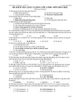
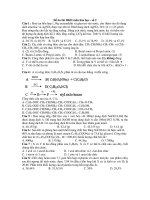
• Fig. 84.4 Schematic
overview of gluconeogenesis. Pyruvate is generated from glycolysis, lactate, or
alanine through pyruvate kinase, lactate dehydrogenase (LDH), and alanine aminotransferase (ALT), respectively. Pyruvate enters the mitochondria freely and is converted into oxaloacetate, which is then
converted into malate to enter the malate shuttle and cross the mitochondrial membrane into the cytoplasm. In the cytoplasm, it is again converted into oxaloacetate and, through phosphoenolpyruvate carboxykinase (PEPCK), is converted into phosphoenolpyruvate that serves as substrate for a series of enzymatic-driven reactions and is finally converted into glucose. It should be noted that the rate-limiting step
of gluconeogenesis is the conversion of fructose biphosphate into fructose 6-phosphate and is regulated
by the actions of glucagon (stimulates) and insulin (inhibits) on fructose 1,6-biphosphatase. Triglycerides
also contribute to gluconeogenesis by their breakdown into fatty acids and glycerol 3-phosphate. The
latter is then transformed into dihydroacetone phosphate and then into fructose 1,6-biphosphate to follow
the rest of the gluconeogenic pathway and result in the generation of glucose.
receive antifungal and antibiotic medications in large volumes of
dextrose-containing fluids). Corticosteroids can increase the risk
of SHG, especially when given in bolus doses. Thiazide diuretics
are also associated with the occurrence of SHG. Calcineurin inhibitors, such as tacrolimus and cyclosporine, can result in SHG
and posttransplant diabetes due to decreased insulin biosynthesis
and release.
Nutritional support practices strongly influence SHG. Critically ill children are frequently prescribed parenteral nutrition
(PN). Provision of excess carbohydrate calories in PN can result in
SHG, which has been associated with increased risk of mortality
in critically ill pediatric patients.140 Overfeeding is common in
critically ill children, regardless of whether PN or enteral nutrition
is employed. Studies have shown that commonly used predictive
equations to calculate caloric needs are inferior to targeted indirect
calorimetry and frequently result in overprescription of calories.141
Mechanisms of Stress Hyperglycemia
Adverse Outcomes
Postulated mechanisms by which SHG causes harm include direct
cellular damage and alterations of essential organ function. In
diabetic patients, four main molecular mechanisms have been
identified in glucose-mediated complications: (1) increased polyol
pathway flux, (2) increased advanced glycation end-product formation, (3) activation of protein kinase C isoforms, and (4) increased hexosamine pathway flux. Each of the four pathogenic
mechanisms reflects a hyperglycemia-induced process, namely,
overproduction of superoxide anion by the mitochondrial electron-transport chain.142 Diabetes-related injury develops over
years, but some common mechanisms parallel acute stress-related
hyperglycemia. During SHG, overexpression of insulin-independent transporters (glucose transporter types 1 to 3 [GLUT-1 to
1010
S E C T I O N V I I I Pediatric Critical Care: Metabolic and Endocrine
GLUT-3]) results in glucose overload and toxicity essentially in
every organ.143 Cells damaged by hyperglycemia are primarily
those unable to effectively control their intracellular glucose
concentration—notably, neuronal, capillary endothelium, and
renal mesangial cells. Glucose overload results in excessive glycolysis
and oxidative phosphorylation with increased production of reactive oxygen species such as superoxide anion, the same toxic end
product involved in the injury pathway for diabetic patients. Reactive oxygen species cause mitochondrial dysfunction and altered
metabolism with subsequent apoptosis and cellular and organ
system failure in the critically ill child. Glucose overload can also
lead to glycation, the reaction of glucose with the amine group of
proteins, which may impair the function of these proteins.
Hyperglycemia is a risk factor for infection in acute illness.112
The relative bacterial overgrowth witnessed in hyperglycemia may
be partly due to altered host defenses. Acute, short-term hyperglycemia impairs macrophage activity, reduces polymorphonuclear
leukocyte chemotaxis and bactericidal capacity, and alters complement fixation in critically ill patients.144 SHG affects all major
components of innate immunity and impairs the ability of the
host to combat infection.144 Furthermore, hyperglycemia is associated with poor gut motility, a factor that may be important in
bacterial overgrowth and translocation. A raised blood glucose
level is also recognized as being proinflammatory and pro-oxidant.
Mononuclear cells isolated from healthy volunteers exhibited
higher levels of NF-kB binding activity, raised reactive oxygen
species, and increased levels of TNF-a mRNA following exposure
to hyperglycemia.145
Hyperglycemia also results in a hypercoagulable state partly
through the increased expression of tissue factor, which is both
pro-coagulant and proinflammatory.146 SHG is implicated in
other abnormalities, such as endothelial dysfunction and alteration in vascular smooth muscle tone, commonly observed during
critical illness.147 Likewise, hyperglycemia has been associated
with deleterious effects on the nervous system. Underlying mechanisms in critical illness remain largely speculative and are often
extrapolated from knowledge in diabetic patients. Hyperglycemia-induced blood-brain barrier permeability, oxidative stress,
and microglia activation may play a role and compromise neurons
and glial cell integrity.148
Clinical Trials Examining Management of Critical
Illness Hyperglycemia
In 2001, a single-center adult study reported reduction of hospital
mortality by more than 30% using a tight glycemic control
(TGC) protocol.149 The effect was attributed to the actual glycemic control rather than the infused insulin dose. These impressive
results brought the issue of SHG to clinical attention and fostered
considerable discussion. Previously seen as an aspect of a normal
stress response, physicians now started viewing SHG differently,
considering it to be a major cause or contributor to pathophysiology that must be aggressively addressed and treated. The appeal of
such straightforward intervention was too great to resist.150 Subsequent studies failed to reproduce these results, yet guidelines
generated by professional societies initially recommended TGC
for adult critically ill patients.151,152 A large international randomized multicenter study involving more than 6000 adult patients153
and many other smaller studies reported that TGC increased
mortality and risk for severe hypoglycemia among adults in the
ICU. In the setting of such consistent negative results, guidelines
were revised and currently recommend using a higher glucose
threshold for initiation of insulin therapy at 150 mg/dL, with the
goal of keeping blood glucose ,180 mg/dL, focusing on close
monitoring and safety margins to avoid hypoglycemia and minimize glucose variability.154 While TGC became the standard of
care for adults, the pediatric critical care community was hesitant
to adopt any guidelines or consistent standard approach.155 There
are, however, several prospective randomized clinical trials to
guide practice.
In very-low-birth-weight neonates, a multicenter trial of insulin with continuous 20% dextrose infusion was terminated prematurely for concerns of futility and potential harm associated
with hypoglycemia.156 This study took a proactive approach to
glycemic control in that the treatment group received insulin with
glucose infusion regardless of their blood glucose level before the
intervention. Considering the study population and the fact that
the study was not designed to treat hyperglycemia, it is difficult to
compare the results with any other pediatric study.
Five randomized prospective studies have examined the association between TGC and clinical outcomes in critically ill children. The first study, published in 2009, was derived from a single
center in Belgium. The study enrolled a mixed medical/surgical
patient population, although 75% of the subjects had undergone
cardiac surgery.157 The study targeted age-adjusted glycemic range
for infants and children, 50 to 80 mg/dL in children less than
1 year old, and 70 to 100 mg/dL in the remainder. Results showed
improved short-term outcome, including mortality, in the TGC
group despite the fact that the TGC group had severe hypoglycemia (,40 mg/dL) at unacceptable rates (25% overall and 44% in
neonates).
A two-center prospective randomized trial published in 2012
enrolled 980 children below age 3 years who underwent cardiac
surgery with cardiopulmonary bypass. The authors reported that
TGC (target range, 80–110 mg/dL) could be achieved with a low
hypoglycemia rate. However, the study found no clinical benefit
for TGC in terms of infection rate, mortality, LOS, or measures
of organ failure when compared with standard care.158 A post hoc
analysis of this study demonstrated that TGC may, in fact, lower
the rate of infection in children older than 60 days of age at the
time of cardiac surgery when compared with standard care.159 In
a secondary analysis of this trial, insulin appeared to have no discernible impact on skeletal muscle degradation.160
The concern for the impact of hypoglycemia on neurocognitive long-term outcome was addressed by both investigator teams.
Four-year neurocognitive follow-up in the single-center study ascertained that insulin-induced hypoglycemia caused by TGC was
not associated with worse neurocognitive outcome.161 However,
the outcomes of both treatment groups were similar to the few
patients who developed moderate or severe hypoglycemia in the
two-center, cardiac-only trial. The group that had no hypoglycemia, as reported by continuous glucose monitoring, had a markedly better neurocognitive outcome than the other three groups,
raising the possibility that the group in the first trial with no hypoglycemia detected may have had undetected hypoglycemia
leading to the moderately impaired outcomes. Subsequent studies
have confirmed the dangers of hypoglycemia in this population.162 Taken together, these data suggest that, in order to ensure
optimal outcome, hypoglycemia should be assiduously avoided.
In 2014, a large multicenter randomized trial involving
13 centers in the United Kingdom reported that TGC in critically
ill children had no significant effect on major clinical outcomes
(number of days alive and free from mechanical ventilation at
30 days after enrollment), but patients in the TGC arm had lower
CHAPTER 84 Endocrine Emergencies
need for renal replacement therapy and reduced 12-month health
care costs. These effects were mostly notable in the noncardiac
patient population.163
In 2010, a single-center study focusing on pediatric patients
with severe burns concluded that intensive insulin therapy significantly decreased infections and sepsis and improved organ
function by decreasing inflammation.164
After five randomized clinical trials of TGC to low versus
high target ranges, this area of critical care therapeutics has become one of the most well studied in the field. The end result in
general pediatric ICU and cardiac ICU patients, although initial
promising findings were noted in the original single-center trial,
is that low targets produce little to no benefit yet increase hypoglycemia, which is becoming increasingly associated with harm.
Consensus has evolved that insulin infusion should be initiated
when glucose levels reach 150 mg/dL, with the goal of keeping
blood glucose less than 180 mg/dL. In the burn population,
which has repeatedly been shown to be physiologically different
from other critically ill children, the single-center study that has
been completed stands in support of targeting a low range of 80
to 110 mg/dL.
Glucose Measurement
Until recently, intermittent blood glucose levels (using point of
care, blood gas analyzer, or central laboratory measurement) were
the only means of blood glucose monitoring. Accuracy is probably the most important metric in selecting the best glycemic
management device for critically ill children, but rapidity/turnaround time, cost, and sample volume are also important factors.165 Intermittent measurements are limited by the workload
associated with the sampling process and with the potential that
“between measurements” events will be missed. A simulation
study modeling adult patients on TGC protocol demonstrated
that increasing the frequency of glucose measurements reduced
the adverse impact of glucose measurement imprecision on glycemic control.166 A mathematical simulation in a cohort of critically ill patients suggested that glycemic control is more optimal
with a blood glucose measurement interval of no longer than
1 hour, with further benefit obtained with use of a measurement
interval of 15 minutes. These findings have important implications for the development of glycemic control standards and future studies.167 With growing interest in glycemic control and the
possible beneficial effect of frequent glucose measurements, continuous glucose monitoring systems have been developed. Although termed continuous, current systems still sample glucose
intermittently with a measurement interval of a few milliseconds
up to 15 minutes. The Clinical and Laboratory Standard Institute
uses 15 minutes as the cutoff for definition of continuous measurement.168
One of the key advantages of continuous glucose monitoring is the ability to identify and display trends in blood glucose
measurements. High-quality continuous glucose monitoring
devices enable clinicians to assess the complexity of the glycemic signal—how one point in time changes relative to neighboring measurements.169 Continuous glucose monitoring using
subcutaneous sensors measuring interstitial fluid has been validated in the pediatric population,170,171 and sensor performance
has improved exponentially over the past decade.172 Although
not yet approved by the US Food and Drug Administration for
use in the inpatient setting, we expect such approvals to be
forthcoming.
1011
Hypoglycemia
Although the determination of which glucose levels represent
hypoglycemia is controversial, a glucose level less than 40 mg/dL is
generally accepted to represent severe hypoglycemia. However, this
concentration is well below the level at which counterregulatory
responses occur. As plasma glucose levels reach 80 to 85 mg/dL,
insulin secretion decreases, and as levels approximate 65 mg/dL,
glucagon, epinephrine, cortisol, and GH are released.173 In addition, a decrease in mental efficiency may be seen when levels fall
below 50 to 60 mg/dL. Because a delay in the recognition and
management of hypoglycemia may lead to long-term neurologic
sequelae,174 it is important to make a distinction between the
laboratory diagnosis of hypoglycemia (,40 to 50 mg/dL) and an
interventional threshold at which therapies to raise serum glucose
should be applied. Setting the interventional threshold at a level
similar to that which elicits counterregulatory responses seems
appropriate; as such, treatment should be offered for hypoglycemia when levels fall below 60 mg/dL to prevent complications,
especially in young children. An even higher interventional
threshold (,70 mg/dL) is warranted for children who are at
increased risk of hypoglycemia.
Clinical Manifestations
Diaphoresis, tremor, tachycardia, anxiety, weakness, hunger,
nausea, and vomiting are all autonomic manifestations caused
by the adrenergic stress response that occurs with a rapid decline in blood glucose levels. Other symptoms associated with
hypoglycemia are a result of a deficiency of the brain’s primary
energy substrate, which are known as neuroglycopenic symptoms. These symptoms include headache, visual disturbances,
lethargy, restlessness, irritability, dysarthria, confusion, somnolence, stupor, coma, hypothermia, seizures, and motor and
sensory disturbances. The glycemic ranges at which these symptoms manifest vary, as critically ill patients cannot recognize or
communicate symptoms. The picture is further masked by sedation and analgesia.
Pathogenesis
Imaging studies of infants who sustained neonatal hypoglycemic
brain injury display diffuse cortical and subcortical white matter
damage that is most prominent in the parietal and occipital lobes.
This pattern differs from the neuroimaging features of other neonatal insults, including hypoxic-ischemic encephalopathy.175 Interestingly, this pattern does not resemble the glucose uptake
pattern of neonatal brains by positron emission tomography,
which may indicate that neuronal damage is not simply due to
cerebral deprivation of its primary substrate for energy production. Evidence indicates that hypoglycemia activates receptors for
excitatory amino acids within the brain and causes cell depolarization, with subsequent cellular edema and apoptosis.176
Fasting Adaptation
Consumption of glucose is largely dependent on the brain-tobody ratio. This phenomenon explains the reduced fasting tolerance of infants whose glucose utilization rate (approximately
6 mg/kg per minute) is significantly higher than that of older
children and adults (1 to 2 mg/kg per minute). This reality places
younger patients at increased risk of hypoglycemia. In addition,
1012
S E C T I O N V I I I Pediatric Critical Care: Metabolic and Endocrine
their ability to maintain euglycemia through glycogenolysis and
gluconeogenesis is reduced because glycogen stores and muscle
bulk are small, thus reducing the pool of available gluconeogenic
substrates. Within the brain, astrocytes, but not neurons, are capable of storing glycogen. The brain contains less than 1 mmol/
kg of free glucose reserve. Fasting tolerance increases rapidly in the
first days of life. Neonates may fast up to 18 hours after 1 week of
age. By 1 year, a 24-hour fast is tolerated; by 5 years, a child may
fast for up to 36 hours without experiencing hypoglycemia.
Understanding fasting physiology is crucial to the logic and
methodologic approach required for diagnosing the etiology of
hypoglycemia. Normally in the postabsorptive state, metabolism
is governed primarily by counterregulatory hormones. In the first
4 hours of a fast in infants or in the first 8 hours in older children,
glucagon is released and euglycemia is maintained primarily by
glycogenolysis. Following glycogen store depletion, gluconeogenesis gains importance in the maintenance of normal glucose levels
(see Fig. 84.4).
Muscle provides amino acids, particularly alanine and glutamine, as gluconeogenic substrates. Glycerol 3-phosphate derived
from triglyceride hydrolysis is also a gluconeogenic precursor.
Fatty acids resulting from triglyceride hydrolysis are transported
to the liver, where they are oxidized to generate acetyl CoA and
ketones. The latter may then be used as alternative fuel by skeletal
and cardiac muscle to help ensure availability of glucose to the
brain and to erythrocytes that are strictly dependent on glucose
for energy production. The brain may also use ketones as an alternative fuel source, but it does so only during a prolonged fast.
Hypoglycemia that occurs early during fasting may indicate
hormonal imbalance or a primary disorder of glycogenolysis. Disorders of gluconeogenesis (see also Chapter 81) will not manifest
during early fast. They become apparent only after glycogen stores
have been depleted; hence, typically, they present later in infancy
once feeding intervals become increasingly prolonged. The same
is true for fatty oxidation disorders. These disorders generally require a more prolonged fast to manifest, nearing 12 to 18 hours
in infants and 18 to 24 hours in older children. However, the
most common cause of childhood hypoglycemia is ketotic hypoglycemia.177 This illness most frequently occurs in toddlers and
preschoolers and is uncommon after 8 to 9 years. It is typically
triggered by intercurrent infection and caloric restriction, both
common events in the PICU. A defect in protein catabolism,
transamination, or amino acid efflux from skeletal muscle, as well
as impaired autonomic regulation of epinephrine secretion, has
been postulated.
Hypoglycemia has been observed in association with a variety of
critical illness diagnoses, including sepsis, congestive heart failure,
renal failure, liver failure, and pancreatitis, and it has been associated with increased mortality among critically ill children.178,179
Critically ill patients are at risk of hypoglycemia not only because
of their underlying illness but also because of factors unique to their
hospitalization, such as muscular atrophy from prolonged immobilization and gluconeogenic substrate depletion, undernutrition often resulting from the limitation of caloric intake because of fluid
restriction, increased glucose consumption, AI, loss of IV access or
inadvertent disconnection of infusion lines, or iatrogenic factors
related to drugs and therapies, including the practice of TGC.
Hypoglycemia Treatment
After obtaining the “critical” blood/urine samples, administration
of 2 mL/kg of 10% dextrose water solution (or an equivalent dose
of dextrose) is indicated for patients with hypoglycemia. Subsequently, an IV maintenance fluid regimen should be considered
to provide a glucose infusion rate of 6 to 8 mg/kg per minute.
Serum glucose should be rechecked 15 minutes after the initial
bolus; if hypoglycemia persists, a repeat bolus of 2 to 4 mL/kg of
10% dextrose water (or an equivalent dose of dextrose) should be
administered and the glucose infusion rate increased by 25% to
50%. If the volume of fluid required to maintain glucose concentrations greater than 70 mg/dL is excessive, a higher dextrose
concentration should be used. Glucagon (0.03 mg/kg for patients
,30 kg, or 1 mg for patients .30 kg) can reverse hypoglycemia
in patients with adequate glycogen stores and normal glycogenolytic pathways. Definitive treatment will depend on the underlying etiology.
In summary, hypoglycemia is a manifestation of iatrogenic,
intentional, or accidental drug ingestion or administration or the
manifestation of an underlying disorder. All critically ill patients
with hypoglycemia should raise a high index of suspicion because
many defects that cause hypoglycemia remain silent until an intercurrent illness or stress overwhelms the compensatory capacity
of the individual. Unless certitude of the etiology of the hypoglycemia exists before therapy, a “critical” blood/urine sample should
be obtained to guide diagnosis and further management. Prompt
recognition and treatment are necessary to prevent neurologic
injury. A multidisciplinary approach, including endocrine and/or
metabolism consultation, is often necessary.
Alterations of Thyroid Hormone
In Critical Illness
Thyroid Biochemistry
Thyroid-stimulating hormone (TSH) derived from the anterior
pituitary is a pleotropic hormone that modulates all aspects of thyroid hormone synthesis.180 TSH action within the thyroid follicular
cells facilitates the sodium iodide symporter, resulting in (1) enhanced iodine concentration in the thyroid gland; (2) increased
synthesis of thyroglobulin, the site of tyrosine residues destined for
iodination; and (3) activated thyroid peroxidase, which catalyzes
iodination of tyrosine residues as well as tyrosine coupling. It is
important to note that autoantibodies may bind to TSH receptors
and stimulate a response similar to TSH, resulting in a hyperthyroid state. Leptin is likely to mediate an important role in the regulation of the thyroid axis, as suggested by the close correlation between the circadian rhythm of leptin secretion and TSH.181
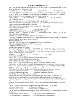
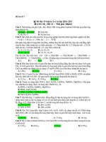
An overview of thyroid hormone biosynthesis and secretion is
provided in Fig. 84.5.34 In this schematic diagram, iodide is transported into the thyroid follicular cell by the action of the sodiumiodide symporter (NIS). Subsequently, this iodide diffuses passively
through the iodide channel termed pendrin (P). Thyroglobulin
(TG) is synthesized within the rough endoplasmic reticulum (ER)
and subsequently packaged by the Golgi apparatus into thyroglobulin secretory vesicles that are released into the follicular cell
lumen. Thyroid oxidase (TO) produces hydrogen peroxide that is
subsequently used by thyroid peroxidase (TPO) to oxidize iodide to
iodine, which subsequently reacts with the tyrosine residues within
thyroglobulin to produce monoiodotyrosine (MIT) and diiodotyrosine (DIT) residues within the thyroglobulin peptide.
Thyroid peroxidase also catalyzes coupling of adjacent iodotyrosines to form thyroxine (T4) and lesser amounts of triiodothyronine (T3). Secretion of thyroxine from the thyroid follicular cell
CHAPTER 84 Endocrine Emergencies
DIT
DIT
DIT
1013
Follicular
lumen
DIT
MIT
MIT
DIT
TG H2O2
T4
P
TO
T4
DIT
TPO
TG
vesicle
DIT
MIT
T4
Endosome
E.R.
Golgi
MIT PF
DIT T4
MIT
Nucleus
DIT
Lysosome
T4
ITDI
I–
3Na+
NIS
I–
2Na+
ATP
2K+
T4
• Fig. 84.5 Thyroid
hormone biosynthesis and secretion. ATP, Adenosine triphosphate; DIT, diiodotyrosine; ER, endoplasmic reticulum; H2O2, hydrogen peroxide; I2, iodide ion; ITDI, iodotyrosine deiodinase;
MIT, monoiodotyrosine; Na1, sodium ion; NIS, sodium-iodide symporter; P, pendrin; P1, potassium ion;
PF, peptide fragments; T3, triiodothyronine; T4, thyroxine; TG, thyroglobulin; TO, thyroid oxidase;
TPO, thyroid peroxidase. (From Goodman HM. Basic Medical Endocrinology. Philadelphia: Elsevier;
2009.)
begins with thyroglobulin phagocytosis with a subsequent fusion
of thyroglobulin endosomes containing proteolytic enzymes capable of digesting thyroglobulin to peptide fragments (PF), as well
as MIT, DIT, and T4. While T4 is released from the cell at the
basal membrane, both MIT and DIT are deiodinated by iodotyrosine deiodinase (ITDI) and subsequently recycled (Fig. 84.6).
T4 is transported to peripheral tissues via transport hormones
T4-binding globulin, transthyretin, and albumin. Because all the
T4 transport proteins are moderately sized, T4 is not filtered by
the kidney.
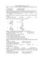
In peripheral tissues, T4 is metabolized to T3 and reverse T3
(rT3) by the action of various isoforms of iodothyrosine deiodinases.182 Transcription and translation of this enzyme are highly
dependent on cytokine stimulation. Selenocysteine residues characterize the active site of this iodine cleavage enzyme. As indicated
in Fig. 84.6, if monodeiodination occurs on the outer tyrosine
ring, the product is T3, and if the monodeiodination occurs on
the inner tyrosine ring, the resultant product is rT3.34
In peripheral tissues, T3 binds to thyroid hormone receptors
that subsequently undergo homodimerization with other thyroid
hormone receptors or heterodimerization with retinoid receptors.
Thyroid hormone receptors can bind to specific nucleotide
sequences termed thyroid responsive elements within promoter
O
HO
C
C
COOH
NH2
Thyroxine
3,5,3´,5´-Tetraiodothyronine (T4)
O
HO
C
C
COOH
NH2
3,5,3´-Triiodothyronine (T3)
O
HO
C
C
COOH
NH2
3,5´,3´-Triiodothyronine
reverse T3 (rT3)
• Fig. 84.6 Thyroid
hormone chemical structures. (From Goodman HM.
Basic Medical Endocrinology. Philadelphia: Elsevier; 2009.)