Một số tư liệu hình ảnh về bệnh vi rút và vi khuẩn ở gia súc
Bạn đang xem bản rút gọn của tài liệu. Xem và tải ngay bản đầy đủ của tài liệu tại đây (3.95 MB, 39 trang )
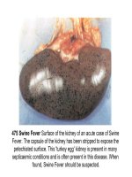
475 Swine Fever Surface of the kidney of an acute case of Swine
Fever. The capsule of the kidney has been stripped to expose the
petechiated surface. This 'turkey egg' kidney is present in many
septicaemic conditions and is often present in this disease. When
found, Swine Fever should be suspected.
476 Swine Fever Haemorrhages in the pelvis of the kidney
strongly suggestive of the disease.
477 Swine Fever
The bladder of an
acute case
of Swine Fever
opened to show the
petech-
iation of the cystic
mucosa.
478 African Swine Fever Larynx from a case of African Swine
Fever. There is oedema and congestion but no petechiae.
.
479 African Swine Fever Haemorrhages in the lung.
480 African Swine Fever Auricle of the heart with haemorrhages.
There are no haemorrhages on the ventricular muscle in this case.
481 African Swine Fever Liver of a pig with acute African Swine
Fever. Note the subcapsular haemorrhages, the distended and
oedematous gall bladder and the haemorrhagic hepatic lymph
node. Haemorrhage in the hepatic lymph node is one of the most
consistent findings in the atypical and chronic forms of the
disease.
482 African Swine Fever Haemorrhagic lymph nodes may
resemble blood clots. There are few, if any, other diseases In
which this type of lesion can be found.
483 African
Swine Fever
Kidney showing
severe
haemorrhage
into the pelvis.
The petechiated
'turkey-egg'
kidney of Swine
Fever is less
common in
African Swine
Fever.
484 Swine Vesicular Disease Early lesions on the coronary
bands, 24 hours post-infection.
485 Swine Vesicular Disease Coronary band lesions. at least five
days post-infection.
486 Swine Vesicular Disease Separation at the coronary band at
nine days plus.
487 Swine Vesicular Disease Marked claw separation and a skin
lesion above the supernumerary digit. Seen 13 days post-
infection.
488 Swine Vesicular Disease Late lesions with under-running of
the horn in two locations on the same foot. This type of lesion
appears to be caused by successive waves of infection.
489 Swine Vesicular Disease Note the shallow ulcers on the
snout and one on the lower lip
.
490 Foot and Mouth Disease A clinically- affected pig. Note the
posture. The animal is severely lame, and would normally be lying
down but has been made to stand. Affected pigs resent being
disturbed.
491 Foot and Mouth Disease Vesicle on the snout of a pig
affected with Foot and Mouth Disease. These vesicles rupture very
quickly but fluid from them is the best source of virus for diagnosis.
492 Foot and Mouth Disease An early case, the vesicles on the
nose having ruptured recently leaving shallow ulcers.
493 Foot and Mouth Disease Lesions on the tongue; ruptured
and unruptured vesicles.
494 Foot and Mouth
Disease Unruptured
vesicles along the
coronary band in an
early case.
495 Foot and Mouth Disease The vesicles on the feet have
ruptured recently leaving raw, shallow ulcers.
496 Foot and
Mouth Disease
Note the marked
separation at the
coronary band.
497 Foot and Mouth Disease Marked separation occurring at the
coronary band and severe heel lesions.
498 Foot and Mouth Disease Thimbling of the horn occurring in
older lesions.
499 Vesicular Exanthema Snout of a pig with primary lesions of
Vesicular Exanthema. Note the ragged area resulting from a ruptured
vesicle.


