Chấn thương cơ xương potx
Bạn đang xem bản rút gọn của tài liệu. Xem và tải ngay bản đầy đủ của tài liệu tại đây (144.57 KB, 9 trang )
Acute Compartment
Syndrome in Lower
Extremity Musculoskeletal
Trauma
Abstract
Acute compartment syndrome is a potentially devastating
condition in which the pressure within an osseofascial
compartment rises to a level that decreases the perfusion gradient
across tissue capillary beds, leading to cellular anoxia, muscle
ischemia, and death. A variety of injuries and medical conditions
may initiate acute compartment syndrome, including fractures,
contusions, bleeding disorders, burns, trauma, postischemic
swelling, and gunshot wounds. Diagnosis is primarily clinical,
supplemented by compartment pressure measurements. Certain
anesthetic techniques, such as nerve blocks and other forms of
regional and epidural anesthesia, reportedly contribute to a delay in
diagnosis. Basic science data suggest that the ischemic threshold of
normal muscle is reached when pressure within the compartment
is elevated to 20 mm Hg below the diastolic pressure or 30 mm Hg
below the mean arterial blood pressure. On diagnosis of impending
or true compartment syndrome, immediate measures must be
taken. Complete fasciotomy of all compartments involved is
required to reliably normalize compartment pressures and restore
perfusion to the affected tissues. Recognizing compartment
syndromes requires having and maintaining a high index of
suspicion, performing serial examinations in patients at risk, and
carefully documenting changes over time.
T
he importance of timely diagno-
sis and management of compart-
ment syndrome was recently empha-
sized in a review of the medical-legal
aspects of this condition.
1
McQueen
et al
2
studied 164 patients (149 men,
15 women) with acute traumatic
compartment syndrome. The inci-
dence of compartment syndrome was
7.3 per 100,000 in men (average age,
30 years) and 0.7 per 100,000 in
women (average age, 44 years).
2
The
most common cause of acute com-
partment syndrome in this series was
fracture (69%); fracture of the tibial
diaphysis was most frequent (36%),
followed by distal radius fractures
(9.8%). Soft-tissue injury without
fracture was the second most com-
mon cause (23.2%), with 10% of
these occurring in patients taking an-
ticoagulants or with a bleeding disor-
der. The incidence of compartment
syndrome associated with high- and
Steven A. Olson, MD, and
Robert R. Glasgow, MD
Dr. Olson is Associate Professor,
Division of Orthopaedic Surgery, Duke
University, Durham, NC. Dr. Glasgow is
Orthopaedic Surgeon, Division of
Orthopaedic Surgery, Royal Alexander
Hospital, Edmonton, AB, Canada.
None of the following authors or the
departments with which they are
affiliated has received anything of value
from or owns stock in a commercial
company or institution related directly or
indirectly to the subject of this article:
Dr. Olson and Dr. Glasgow.
Reprint requests: Dr. Olson, Duke
University, Box 3389, Durham, NC
27710.
J Am Acad Orthop Surg 2005;13:436-
444
Copyright 2005 by the American
Academy of Orthopaedic Surgeons.
436 Journal of the American Academy of Orthopaedic Surgeons
low-energy injuries is nearly equal.
The presence of open wounds does
not mean that compartments are de-
compressed; compartment syndrome
is seen after open fractures.
2-4
Etiology
A variety of injuries and medical
conditions may initiate acute com-
partment syndrome (Table 1). Frac-
tures; contusions; bleeding disor-
ders; burns; trauma; postischemic
swelling; tight casts, dressings, or
external wrappings; and gunshot
wounds are some of the most fre-
quent causes.
2-12
Anatomic struc-
tures, including epimysium, fascia,
and skin, may limit the potential
size of a compartment. Therefore,
closure of incisions or defects in
these structures should not be done
acutely when the patient is at risk
for compartment syndrome. Ther-
mal injuries, especially circumferen-
tial third-degree burns, can cause an
acute compartment syndrome by
forming inelastic constrictions, es-
chars, and massive edema which, in
combination, result in ischemia to
neurovascular and muscular struc-
tures.
2,5,7,13
Circumferential wraps,
such as casting material or cast pad-
ding, can lead to restriction of com-
partment expansion and increased
compartmental pressure.
8,13,14
Re-
leasing all circumferential dressings,
splitting casts, and cutting casting
material results in a marked decrease
in intracompartmental pressure.
Pneumatic antishock garments
have been associated with lower ex-
tremity compartment syndromes.
Templeman et al
9
reported on a pa-
tient who developed bilateral com-
partment syndromes in uninjured
extremities after wearing a pneu-
matic antishock garment. However,
inflation pressures <50 mm Hg in
these garments have been used for
long periods of time (eg, 48 hours for
pelvic fractures) without adverse se-
quelae.
9
Traction, ankle joint position, and
limb positioning have been shown to
affect compar tment volume and
pressure and to contribute to the
formation of compartment syn-
dromes.
3,8,10,11,13,15,16
Traction causes
the fascia to tighten and constrict
the limb, decreasing the compart-
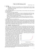
ment volume. Shakespeare and
Henderson
15
described compartmen-
tal pressure changes during calca-
neal traction for tibial fractures.
Pressure in the anterior and deep
posterior compartments rose linear-
ly with increasing traction, up to 9.1
kg (Figure 1). The pressure did not
fall during the time the traction was
applied. For each increase of 1 kg in
longitudinal traction, the compart-
ment pressure within the deep pos-
terior compartment increased by
more than 5%; pressure in the ante-
rior compartment increased by <2%.
Intramuscular pressure is lowest in
the anterior compartment with the
ankle in the neutral to dorsiflexed
position; it is lowest in the deep pos-
terior compartment when the ankle
is in the plantarflexed position.
13
Longitudinal calcaneal traction
tends to dorsiflex the ankle and in-
crease the pressure in the deep pos-
terior compartment more than in
the anterior compartment. After cast
application, the pressure in both the
anterior and deep posterior compart-
ments increases three- to seven-fold,
depending on the position of the an-
kle.
13
Ankle plantar flexion of 0° to
37° is the most protective position
for minimizing the combined risks
of anterior and posterior compart-
ment syndromes.
13
Table 1
Causes of Compartment Syndrome
Fracture
Soft-tissue trauma without fracture
Intracompartmental bleeding
Tight casts, dressings, or external
wrappings
Thermal injury, burn eschar
Extravasation of intravenous infusion
Venous obstruction
Reperfusion injury following
prolonged ischemia
Penetrating trauma
Figure 1
Change in compartment pressure (percent) with increasing calcaneal pin traction
(kg) in patients with tibial shaft fractures. (Adapted with permission from
Shakespeare DT, Henderson NJ: Compartmental pressure changes during
calcaneal traction in tibial fractures. J Bone Joint Surg Br 1982;64:498-499.)
Steven A. Olson, MD, and Robert R. Glasgow, MD
Volume 13, Number 7, November 2005 437
Compartment syndromes have
been described with prolonged use
of the Lloyd-Davies (lithotomy) po-
sition with flexion, elevation, and
abduction of the well leg during in-
tramedullary nailing of femoral frac-
tures.
11,13
The combined effects of
direct compression on the leg, com-
pressive circumferential bindings or
stockings, sequential inflatable de-
vices, and relative elevation of the
limb contribute to increased com-
partment pressure, decreased com-
partment volume, and decreased
blood flow, leading to the formation
of compartment syndromes.
11
Many authors have discussed ele-
vated compartment pressures asso-
ciated with intramedullary nailing
of tibial fractures.
4,16-20
The etiology
of acute compartment syndrome as-
sociated with intramedullary fixa-
tion is multifactorial: tissue damage
secondary to the injury causes swell-
ing, traction decreases the volume of
the compartments, reaming forces
blood and marrow into the compart-
ments, and limb supports may cause
outflow constriction.
16
Moed and
Strom,
18
using a canine model, found
that pressure changes during ream-
ing were transient, returning to base-
line or lower after the reamer was
removed from the intramedullary
canal. After nail insertion, the pres-
sure remained elevated in the an-
terolateral compartment and was
transiently elevated in the posterior
compartment. Mawhinney et al
20
showed that peak pressures were
reached after the first two reaming
cycles.
Several authors have recom-
mended using an unreamed nail in
tibial fractures with associated
compartment syndrome, or in pa-
tients without compartment syn-
drome who have elevated compart-
ment pressures, in order to minimize
pressure elevation during the pro-
cedure.
16-19
Tornetta and French
16
reported on anterior compartment
pressures during unreamed tibial
nailing without traction. Eight of 20
patients had transient compartment
pressure elevation ≥40 mm Hg (max-
imum, 58 mm Hg); all pressures
returned to below 20 mm Hg by the
end of the procedure. The authors
concluded that patients with a tibial
fracture who demonstrate signs and
symptoms of an acute compartment
syndrome on presentation should
undergo a four-compartment fasci-
otomy before intramedullary nail-
ing, and that pressure elevations
during nailing should be minimized
by avoiding prolonged traction.
McQueen and Court-Brown
4
used
continuous compartment pressure
monitoring during tibial nailing in a
prospective study of 116 patients.
Use of reamed versus unreamed nail-
ing had no effect on the incidence of
compartment syndrome. Tibial nail-
ing with or without prior canal ream-
ing is a safe method of managing
tibial shaft fractures at risk for com-
partment syndrome. Prolonged fixed
traction should be avoided to the ex-
tent possible.
Pathophysiology of
Ischemia
The pathophysiologic mechanism
that causes compartment syn-
dromes is increased tissue pressure
and the resulting development of is-
chemia, which leads to irreversible
muscle damage. Cellular anoxia is
the final common pathway of all of
the varieties of compartment syn-
drome. However, the interaction be-
tween increased compartment pres-
sure, blood pressure, and blood flow
are incompletely understood. It was
originally suggested that there was a
threshold compartment pressure
above which irreversible changes
would occur.
21
More recent evidence
indicates that the absolute differ-
ence between compartment pressure
and blood pressure is the critical
variable.
21-26
To avoid collapsing of
the veins, the pressure inside the
veins cannot be less than that of the
surrounding tissue.
7,27
An increase i n
compartment pressure results in an
increase in venous pressure, leading
to a decrease in the arteriovenous
gradient.
7
Change in the local vascu-
lar resistance can accommodate for
some of the reduction in the arterio-
venous gradient; however, this
change becomes ineffective with in-
creasing tissue pressure. Compart-
ment syndrome occurs when the lo-
cal arteriovenous gradient does not
allow sufficient blood flow to meet
the metabolic demands of the tis-
sue.
7
Vascular tone, blood pressure,
duration of pressure elevation, and
metabolic demands of the tissue are
important in determining whether a
compartment syndrome will oc-
cur.
7
Muscle ischemia can lead to re-
lease of myoglobin from damaged
muscle cells. During reperfusion,
myoglobin is released into the cir-
culation with other inflammatory
and toxic metabolites. Myoglobin-
uria, metabolic acidosis, and hyper-
kalemia can lead to renal failure,
shock, hypother mia, and cardiac
arrhythmias and/or failure. The de-
velopment and extent of these sys-
temic effects depends on the sever-
ity and duration of compromised
tissue perfusion and the size and
number of muscle compartments
involved.
7
By using objective, noninvasive
techniques, experimental and clini-
cal investigators have determined
the changes in muscle blood flow
that occur during compartment syn-
drome.
24
Induced compartment syn-
dromes in dogs revealed three histo-
logic regions of muscle injury.
24
In
skeletal muscle, the central portion
of the muscle becomes ischemic
first. The surrounding zone of mus-
cle tissue then shows evidence of
partial ischemic injury with in-
creased tissue edema and swelling.
The peripheral layers of muscle are
the last to be affected, often remain-
ing normal in incomplete compart-
ment syndromes. Microangiograms
showed an abundance of epimysial
vessels with occlusion of central
penetrating branches in specimens
from severe cases.
24
Acute Compartment Syndrome in Lower Extremity Musculoskeletal Trauma
438 Journal of the American Academy of Orthopaedic Surgeons
Using autologous plasma infusion
in a canine compartment syndrome
model, Heckman et al
21
studied the
ischemic threshold of muscle by in-
ducing elevated pressures for 8
hours. Irreversible histologic chang-
es, including focal muscle infarction
and fibrosis, were documented in all
compartments subjected to tissue
pressures within 10 mm Hg of dia-
stolic pressure. None of the animals
with a difference in perfusion pres-
sure >30 mm Hg from mean arterial
and >20 mm Hg from diastolic pres-
sure demonstrated any evidence of
irreversible changes, although occa-
sional cells underwent myofibrillar
degeneration. Mean compartment
pressures of 59 mm Hg with ade-
quate perfusion pressure were toler-
ated for 8 hours without evidence of
infarction. The authors concluded
that the ischemic threshold of skel-
etal muscle, beyond which irrevers-
ible tissue damage occurs after 8
hours, is directly related to the dif-
ference between the compartment
and mean arterial or diastolic pres-
sures. The critical tissue pressure
differentials were ≤30 mm Hg from
mean arterial pressure and ≤20 mm
Hg from diastolic blood pressure.
21
Matava et al
22
performed a similar
study in canines and also found that
the threshold for muscle necrosis
was 20 mm Hg less than the diastol-
ic pressure. These findings support
the hypothesis that tissue damage is
directly related to absolute differ-
ence between compartment pressure
and blood pressure and that this dif-
ference is a variable that affects not
only microvascular perfusion but
also the onset of tissue damage.
Bernot et al
25
observed that mus-
cle subjected to ischemia before
compartment pressurization had a
lower threshold for metabolic deteri-
oration than did nontraumatized
muscle. Hypoxic metabolic changes
occurred in the postischemic limbs
in all compartments with a perfu-
sion pressure (∆P) <40 mm Hg. The
metabolic deterioration observed
was more rapid and severe as the ∆P
value diminished toward a value of
10 mm Hg. Normal limbs did not
become metabolically compromised
until the ∆P value declined to <30
mm Hg. Postischemic muscle is
more easily and much more rapidly
compromised metabolically by in-
creased interstitial pressure than is
normal muscle.
25
Vollmar et al
26
used a skinfold
chamber to examine vessel response
to increased pressure in hamsters.
Venules exhibited early reduction in
size proportional to external pres-
sure. No similar change was observed
in arterioles. This study suggests that
impaired venous drainage with cap-
illary stasis but without arteriolar
constriction is a significant patho-
physiologic component in the devel-
opment of compartment syndrome.
Diagnosis
History and Physical
Examination
Critical to recognizing compart-
ment syndrome is having and main-
taining a high index of suspicion and
performing serial examinations in
patients at risk to document chang-
es over time.
2,5-7,12
Patient history is
important for determining the me-
chanism of injury and whether there
are associated risk factors for devel-
oping compartment syndrome.
6
The
classic clinical diagnosis encompass-
es the six Ps: pain, pressure, pulse-
lessness, paralysis, paresthesia, and
pallor.
12
Pain out of proportion to the in-
jury, aggravated by passive stretching
of muscle groups in the correspond-
ing compartment, is one of the earli-
est and most sensitive clinical signs
of compartment syndrome. However ,
pain may be an unreliable indicator
and may be absent in an established
compartment syndrome.
3
Pain per-
ception may be diminished or absent
in the obtunded patient, thus requir-
ing additional diagnostic methods.
7
The absence of pain in a compart-
ment syndrome is often caused by a
superimposed central or peripheral
neural deficit. However, McQueen
and Court-Brown
4
reported a patient
in whom a compartment syndrome
was diagnosed by increased compart-
ment pressures before the onset of
signs or symptoms.
Pressure or firmness in the com-
partment, a direct manifestation of
increased intracompartmental pres-
sure, is the earliest and may be the
only objective finding of early com-
partment syndrome. Peripheral puls-
es are palpable and, unless a major
arterial injury is present, capillary
refill is routinely present. Only rare-
ly is the compartment pressure ele-
vated sufficiently to occlude arterial
pressure.
7
Paresis is difficult to interpret and
may be caused by muscle ischemia,
nerve ischemia, guarding secondary
to pain, o r a combination of all three.
True paralysis is a late finding that is
caused by prolonged nerve compres-
sion or irreversible muscle damage.
Paresthesia is an early sign of com-
partment syndrome that, without
treatment, progresses to hypesthesia
and anesthesia. Sensory symptoms
and signs are often the first indica-
tion of nerve ischemia.
3
Matava et
al
22
have shown that peripheral
nerve tissue is actually more sensi-
tive to an ischemic event than mus-
cle, with nerve function ceasing af-
ter 75 minutes of total ischemia.
The duration and degree of pressure
elevation leading to irreversible
nerve injury secondary to compres-
sion is uncertain.
22
Typically , abnor-
mal neurologic findings are associat-
ed with nerves that course through
affected compartments. The isolated
finding of paresthesia is frequently
resolved with the release of con-
stricting wraps or bandages alone.
Although frequently listed as one of
the “P’s,” pallor is uncommon. It oc-
curs in the rare circumstance in
which arterial inflow is severely di-
minished.
McQueen et al
3
reported a mean
of 7 hours from fracture manipula-
tion and fixation to the development
of a compartment syndrome in 13
Steven A. Olson, MD, and Robert R. Glasgow, MD
Volume 13, Number 7, November 2005 439
fracture patients undergoing com-
partment pressure monitoring. Four
patients had delayed onset of com-
partment syndrome at 14 to 24
hours after fracture manipulation
and fixation.
3
However, compart-
ment syndrome occasionally occurs
2 to 4 days after the precipitating
event; therefore, late onset must be
considered.
27
Associated conditions can affect
susceptibility to compartment syn-
drome or contribute to missed diag-
nosis. The perfusion gradient may
be inadequate in the presence of
systemic hypotension, even with
compartments that are supple to
physical examination. Anesthetic
techniques have been reported to
contribute to a delay in diagnosis.
Compartment syndromes after sur-
gery done to manage fractures have
been associated with the use of local
nerve blocks, epidural anesthesia,
and other forms of regional ane-
sthesia.
28-32
Patients receiving epi-
dural anesthesia have been reported
to be four times as likely to have a
neurologic complication than those
receiving systemic narcotics.
32
Epi-
dural anesthesia increases local
blood flow secondary to sympa-
thetic blockade, thereby potentially
exacerbating swelling of an injured
extremity.
32
The use of local anes-
thetics combined with narcotics
during epidural anesthesia has been
shown to increase the likelihood of
missed compartment syndromes
and is not recommended in the at-
risk patient.
31,33
Compartment Pressure
Measurement
Sometimes the clinical picture
may be borderline or the patient ex-
amination may be equivocal, unreli-
able, or unobtainable. In such in-
stances, measuring compartment
pressures is recommended to aid the
decision-making process. McQueen
and Court-Brown
4
reported a pro-
spective clinical series using con-
tinuous compartment monitoring.
When a difference between compart-
ment pressure and diastolic blood
pressure ≥30 mm Hg was main-
tained and compartments were not
released, patients had normal mus-
cle function at the time of follow-up.
Data from preclinical research stud-
ies suggest that the ischemic thresh-
old of muscle is a perfusion pressure
of at least 20 mm Hg between the
compartment pressure and the dia-
stolic pressure.
21
In a fracture at risk,
measuring compartment pressures
early in the course of treatment can
provide a reference point to detect a
trend if later compartment pressure
measurements are needed.
Various methods of measuring
compartment pressures have been
described.
34-39
The two most com-
mon techniques are a slit catheter
and the side port needle. The slit
catheter is a low-volume infusion
technique.
34
The measurement cath-
eter may be left in situ within the
compartment for repeated or contin-
uous compartment pressure mea-
surements over a period of hours.
Side port needles, which were de-
veloped to measure multiple com-
partments, have gained widespread
popularity. Moed and Thorderson
35
reported that no statistically signif-
icant difference was found between
the measurements obtained with the
slit catheter and the side port needle.
However, measurements with a
standard 18-gauge needle were high-
er than both the slit catheter and
side port needle by nearly 20 mm
Hg. Therefore, a standard 18-gauge
needle is less accurate and cannot be
recommended. Several commercial-
ly available pressure measurement
devices are available for determining
intracompartmental pressures.
The location in the compartment
from which the measurement is
taken is important for accuracy.
Seiler et al
37
determined that unin-
jured compartments exhibited clin-
ically significant intracompartmen-
tal pressure measurement variability
in the forearm. In their study of 25
patients with closed tibial fracture,
Heckman et al
23
reported a relation-
ship between compartmental tissue
pressure and the distance from the
site of the fracture (Figure 2). Pres-
Figure 2
The mean compartment tissue pressure measurement in a series of tibial shaft
fractures. The pressures are presented by location relative to the tibial fracture site.
Data suggest that the highest pressures occur within 5 cm of the fracture. (Adapted
with permission from Heckman MM, Whitesides TE Jr, Grewe SR, Rooks MD:
Compartment pressure in association with closed tibial fractures: The relationship
between tissue pressure, compartment, and the distance from the site of the
fracture. J Bone Joint Surg Am 1994;76:1285-1292.)
Acute Compartment Syndrome in Lower Extremity Musculoskeletal Trauma
440 Journal of the American Academy of Orthopaedic Surgeons
sure was measured at the fracture
site and in 5-cm increments distal
and proximal. The highest pressure
recorded was in the deep posterior
or anterior compartment, or both.
Eighty-nine percent of compart-
ments had the highest pressure mea-
surement at the fracture site: 5% at
5 cm distal and 2% at 5 cm proxi-
mal. The mean difference in pressure
5 cm from the highest level recorded
was 10 mm Hg. These data indicate
that pressure measurements should
be performed within all compart-
ments and at multiple sites, particu-
larly within 5 cm of the level of in-
jury.
23
Compartment pressure measure-
ment is indicated whenever the diag-
nosis is uncertain in a patient at risk.
Several clinical scenarios fall into
this category (Table 2). One of the
most beneficial uses of compart-
ment pressure measurement is for
distinguishing an undermedicated
patient from one who is developing
compartment syndrome. This di-
lemma can occur when a long-acting
anesthetic block wears off without
appropriate systemic pain medica-
tion. In this scenario, the pain can be
severely increased with passive
range of motion, and residual pares-
thesias can remain from a nerve
block. It is often helpful to obtain a
baseline set of pressure measure-
ments in at-risk compartments in a
patient who cannot be examined for
an extended period. When subse-
quent physical examination findings
are of concern (eg, increased swell-
ing, firmness in the limb), a second
set of compar tment pressures can
provide evidence of a trend, in addi-
tion to the actual ∆P value at the
time of pressure measurement. Ob-
tunded patients with an increasing
trend in pressure should be moni-
tored closely.
At our institution, a ∆P value of
20 mm Hg from measured compart-
ment pressure to diastolic blood
pressure is an absolute indicator for
fasciotomy. This approach was
adopted for three reasons. (1) Basic
science data suggest that a ∆P value
of 20 mm Hg is safe. (2) In the inves-
tigations of McQueen and Court-
Brown,
4
fasciotomies were per-
formed for a ∆P value of 30 mm Hg
and did not identify an absolute min-
imal ∆P threshold. (3) In our experi-
ence, many patients in the operating
room have vasodilatory effects of an-
esthesia, leading to transiently low
diastolic blood pressure with normo-
tensive systolic pressures. In the lat-
ter situation, a patient with com-
partment pressures in the mid 20s
with a supple limb may have a ∆P
value <30 mm Hg with the diastolic
blood pressure.
Laboratory Tests
Serum creatine phosphokinase,
which reflects muscle necrosis, has
been used as an indicator of compart-
ment syndrome.
12
Decompression
should result in a downward trend of
creatine phosphokinase levels. Per-
sistently high levels or progression
indicates inadequate decompression
and ongoing muscle necrosis. Myo-
globin, a breakdown product of mus-
cle cell lysis, is evidenced by myo-
globinuria. It can be misinterpreted
as hematuria; a definitive diagnosis
is indicated by a positive urine ben-
zidine test for occult blood in the ab-
sence of red blood cells. Myoglobin is
toxic to glomeruli of the kidney and
leads to renal failure when the com-
partment syndrome is not ade-
quately treated.
12
Treatment
Following the diagnosis of impend-
ing or true compartment syndrome,
immediate measures are necessary
to ensure that the deleterious se-
quelae of compartment syndrome
do not occur. First, casts or occlu-
sive dressings should be split com-
pletely. Cast padding or circumfer-
ential dressings should be released
around their entire circumference.
The affected limb should not be
elevated higher than the patient’s
heart in order to maximize perfu-
sion while minimizing swelling.
7
When, despite these steps, the clin-
ical diagnosis of compartment syn-
drome remains clear, emergent and
complete fasciotomy of all compart-
ments with elevated pressures is
necessary to reliablynormalize com-
partment pressures and restore per-
fusion to the affected tissues.
The length of skin incision has an
effect on fascial decompression in
the leg associated with an acute
compartment syndrome. Some au-
thors favor limited incisions, claim-
ing low morbidity, while others
recommend long incisions, em-
phasizing that these are required to
decompress affected compar tments
adequately.
40-42
Several instances
have been reported in which the
skin continued to cause compres-
sion after fasciotomy through short
incisions.
40
Cohen et al
40
deter-
mined the effect of the length of
the skin incision in posttraumatic
compartment syndromes of the
lower extremity treated with fascial
decompression using a two-incis-
ion technique. The affected com-
partments initially were released
through 8-cm incisions and the pres-
Table 2
Indications for Compartment
Pressure Measurement
One or more symptoms of
compartment syndrome with
confounding factors (eg, neurologic
injury, regional anesthesia,
undermedication)
No symptoms other than increased
firmness or swelling in the limb in an
awake, alert patient receiving
regional anesthesia for
postoperative pain control
Unreliable or unobtainable
examination with firmness or
swelling in the injured extremity
Prolonged hypotension and a swollen
extremity with equivocal firmness
Spontaneous increase in pain in the
limb after receiving adequate pain
control
Steven A. Olson, MD, and Robert R. Glasgow, MD
Volume 13, Number 7, November 2005 441
sures recorded. The skin incisions
were enlarged by 2-cm increments
until readings showed no further de-
crease. The final length of the ex-
tended incisions averaged 16 cm ± 4
cm. Mean final pressure in the com-
partments, which required exten-
sion of the incisions, was 13 mm Hg,
notably less than pre-extension re-
cordings. Long incisions add little to
morbidity and influence neither the
complication rate nor the late func-
tional result. Long incisions also
eliminate the risk of the skin acting
as an unrecognized compartment
envelope, which is especially impor-
tant during the hyperemic period
following decompression of an is-
chemic compartment.
40,42
Compartment syndromes can oc-
cur in a variety of locations in the
lower extremity, such as the gluteal
musculature, thigh, lower leg, and
foot. Regardless of location, the key
in treatment is to adequately decom-
press the muscles involved. In the
gluteal region, a posterior incision
that provides access to the gluteus
maximus and the abductor muscula-
ture is adequate. In the thigh, a long
single lateral incision can adequate-
ly decompress the anterior and pos-
terior compartments. Occasionally,
a medial adductor incision is re-
quired, as well. A one- or two-
incision approach can be used in the
lower leg. Generally, a long single
lateral incision is sufficient for a
four-compartment fasciotomy.
The one-incision procedure
should be performed through a long
incision based over the anterolateral
calf. The extended incision is made
from within 5 cm of either end of the
fibula. The basic technique involves
identifying the septum between the
anterior and lateral compartments,
then performing a fasciotomy on
each of these compartments. Care
should be taken to avoid injury to
the superficial peroneal nerve distal-
ly. Lateral compartment muscula-
ture is then elevated off the posteri-
or intramuscular septum. Incision of
this intramuscular septum provides
access to the lateral portion of the
superficial posterior compartment.
The superficial compartment is mo-
bilized posteriorly to give access to
the deep posterior compartment in
order to perform the fasciotomy.
In the two-incision technique, the
location of the medial skin incision
is important. The bulk of the mus-
culature in the superficial posterior
compartment is proximal and re-
quires a proximal extent to the inci-
sion to adequately decompress the
region. However, the bulk of the
deep posterior musculature is locat-
ed in the distal half of the limb. Ad-
equate decompression requires de-
taching the soleus origin from the
medial aspect of the tibial shaft.
Therefore, to adequately decompress
all four compartments through two
incisions, long medial and lateral in-
cisions are required. Foot compart-
ment syndrome is typically treated
with two longitudinal incisions in
the dorsum of the foot, one centered
over the fourth metatarsal and one
over the space between the first and
second metatarsals. Adequate de-
compression requires release of the
fascia of the intrinsic foot muscles
attaching to the metatarsals.
In their study of secondary clo-
sure of the skin following fascioto-
my for acute compartment syn-
drome, Wiger et al
43
noted that tight
closures may increase intramuscular
pressure to dangerous levels. To pre-
vent this, limb swelling must be re-
duced before secondary closure. Pa-
tients were encouraged to perform
concentric muscular activity and
weight-bearing exercises to assist in
reducing elevated intramuscular
pressures of the swollen extremities.
Active contraction of muscle en-
hances lymph flow, and the normal
increase of hydrostatic pressure is a
powerful edema-reducing mecha-
nism. At follow-up, there were no
signs of ischemic muscular contrac-
ture when intramuscular pressure
did not exceed 30 mm Hg during sec-
ondary closure in a normotensive
patient.
43
At our institution, the fasciotomy
site is typically dressed with a
wound vac sponge. The patient is re-
turned to the operating room 3 to 5
days later to attempt closure. When
muscle necrosis is a possibility, the
patient must return to surgery after
24 to 48 hours for débridement.
Wound closure should not be at-
tempted until all necrotic tissue is
débrided. Direct closure can be at-
tempted when the wound approxi-
mates without excess tension. When
the wound edges will not oppose
easily, split-thickness skin graft is
indicated.
Outcomes and
Complications
Sheridan and Matsen
44
reported the
clinical outcome of 44 patients who
underwent decompressive fasciot-
omy. Twenty-two patients were
treated with fasciotomy before 12
hours and 22 after 12 hours. In the
first group, 68% of patients had nor-
mal lower extremity function at the
time of final follow-up, compared
with only 8% in the delayed-
treatment group.
44
Finkelstein et al
45
reported on five
patients who underwent fasciotomy
later than 35 hours after the estab-
lished diagnosis of lower extremity
compartment syndrome. In this ret-
rospective review, one patient died
of multisystem organ failure direct-
ly related to complications from the
fasciotomy. The remaining four pa-
tients required amputation.
Fitzgerald et al
41
reported on
long-term sequelae of fasciotomy
wounds in 60 patients and demon-
strated that the patients frequently
had complaints at the fasciotomy
site. Seventy-seven percent reported
decreased sensibility, 7% had teth-
ered tendons, and 13% had recur-
rent ulcerations within the wound
closure area.
41
Although a fasciot-
omy incision does result in some
morbidity to the patient, the mor-
bidity of an incompletely released
compartment, delayed diagnosis,
Acute Compartment Syndrome in Lower Extremity Musculoskeletal Trauma
442 Journal of the American Academy of Orthopaedic Surgeons
or unrecognized compartment syn-
drome is substantially worse.
It is not possible to determine the
precise time a compartment syn-
drome begins. Therefore, it is not
possible to know how long a com-
partment syndrome has been estab-
lished. Anecdotal reports suggest
that performing a fasciotomy in the
setting of a delayed diagnosis can be
harmful to the patient and often re-
sults in amputation.
The dilemma is determining how
late a presentation is too late for a
fasciotomy. In general, clinical as-
sessment of the limb helps with
decision-making. The patient with
clinical evidence of compartment
syndrome who has the ability to vol-
untarily contract muscles within the
compartment has some viable mus-
cle; therefore, fasciotomy is indicat-
ed regardless of the delay. Fascioto-
my is not performed when a patient
has clinical evidence of compart-
ment syndrome with a suspected du-
ration ≥8 hours, has neither a nerve
injury nor a nerve block that could
potentially alter the clinical exami-
nation, and has no demonstrable
muscle function in any segment of
the involved limb. Instead, the limb
is aggressively splinted to maintain
a functional position as the muscle
develops fibrosis and contracture.
Supportive care should be given for
the potential risk of myoglobinuria,
which may occur in this scenario.
Summary
Acute compar tment syndrome is
a potentially devastating condition
associated with musculoskeletal
trauma. The final common path-
way is cellular ischemia resulting
from increased tissue pressure
within an osseofascial compart-
ment. Compartment syndrome can
occur as a result of many different
causes, such as fractures, contu-
sions, bleeding disorders, burns,
trauma, postischemic swelling, and
gunshot wounds. Prompt diagnosis
and treatment are key in limiting
patient morbidity. The diagnosis of
compartment syndrome is usually
made based on clinical factors, such
as pain, pressure, paresthesia, paral-
ysis, and pulselessness. Adjunctive
use of compartment pressure mea-
surements is warranted in the ma-
jority of patients.
References
Evidence-based Medicine: Moed and
Thorderson (reference 35) report a
comparison study of measurements
of intracompartmental pressures
(level II study), and Shakespeare et
al (reference 34) compared the slit
catheter with the wick catheter
(level II study). Younger et al (refer-
ence 14) compared prospectively
plaster backslabs and plaster cast in
a prospective cohort study (level II
study).
1. Bhattacharyya T, Vrahas MS: The
medical-legal aspects of compart-
ment syndrome. J Bone Joint Surg Am
2004;86:864-868.
2. McQueen MM, Gaston P, Court-
Brown CM: Acute compartment syn-
drome: Who is at risk? JBoneJoint
Surg Br 2000;82:200-203.
3. McQueen MM, Christie J, Court-
Brown CM: Acute compartment syn-
drome in tibial diaphyseal fractures.
J Bone Joint Surg Br 1996;78:95-98.
4. McQueen MM, Court-Brown CM:
Compartment monitoring in tibial
fractures: The pressure threshold for
decompression. J Bone Joint Surg Br
1996;78:99-104.
5. Mubarak SJ: Technique of diagnosis
and treatment of the lower extremity
compartment syndromes in children.
Operative Techniques in Ortho-
paedics 1995;5:178-189.
6. Gulli B, Templeman D: Compart-
ment syndrome of the lower extrem-
ity. Orthop Clin North Am 1994;25:
677-684.
7. Ouellette EA: Compartment syn-
dromes in obtunded patients. Hand
Clin 1998;14:431-450.
8. Janzing H, Broos P, Rommens P: Com-
partment syndrome as a complication
of skin traction in children with fem-
oral fractures. J Trauma 1996;41:156-
158.
9. Templeman D, Lange R, Harms B:
Lower-extremity compartment syn-
dromes associated with use of pneu-
matic antishock garments. J Trauma
1987;27:79-81.
10. Slater RR Jr, Weiner TM, Koruda MJ:
Bilateral leg compartment syndrome
complicating prolonged lithotomy
position. Orthopedics 1994;17:954-
959.
11. Goldsmith AL, McCallum MI: Com-
partment syndrome as a complication
of the prolonged use of the Lloyd-
Davies position. Anaesthesia 1996;
51:1048-1052.
12. Velmahos GC, Toutouzas KG: Vascu-
lar trauma and compartment syn-
dromes. Surg Clin North Am 2002;82:
125-141.
13. Weiner G, Styf J, Nakhostine M, Ger-
shuni DH: Effect of ankle position and
a plaster cast on intramuscular pres-
sure in the human leg. J Bone Joint
Surg Am 1994;76:1476-1481.
14. Younger AS, Curran P, McQueen
MM: Backslabs and plaster casts:
Which will best accommodate in-
creasing intracompartmental pres-
sures? Injury 1990;21:179-181.
15. Shakespeare DT, Henderson NJ:
Compartmental pressure changes
during calcaneal traction in tibial
fractures. J Bone Joint Surg Br 1982;
64:498-499.
16. Tornetta P III, French BG: Compart-
ment pressuresduring nonreamed tib-
ial nailing without traction. J Orthop
Trauma 1997;11:24-27.
17. Hak DJ, Johnson EE: The use of the
unreamed nail in tibial fractures with
concomitant preoperative or intraop-
erative elevated compartment pres-
sure or compartment syndrome. JOr-
thop Trauma 1994;8:203-211.
18. Moed BR, Strom DE: Compartment
syndrome after closed intramedullary
nailing of the tibia: A canine model
and report of two cases. J Orthop Trau-
ma 1991;5:71-77.
19. Georgiadis GM: Tibial shaft fractures
complicated by compartment syn-
drome: Treatment with immediate
fasciotomy and locked unreamed
nailing. J Trauma 1995;38:448-452.
20. Mawhinney IN, Maginn P, McCoy
GF: Tibial compartment syndromes
after tibial nailing. J Orthop Trauma
1994;8:212-214.
21. Heckman MM, Whitesides TE Jr,
Grewe SR, Judd RL, Miller M,
Lawrence JH III: Histologic determi-
nation of the ischemic threshold of
muscle in the canine compartment
syndrome model. J Orthop Trauma
1993;7:199-210.
22. Matava MJ, Whitesides TE Jr, Seiler
JG III, Hewan-Lowe K, Hutton WC:
Determination of the compartment
Steven A. Olson, MD, and Robert R. Glasgow, MD
Volume 13, Number 7, November 2005 443
pressure threshold of muscle is-
chemia in a canine model. J Trauma
1994;37:50-58.
23. Heckman MM, Whitesides TE Jr,
Grewe SR, Rooks MD: Compartment
pressure in association with closed
tibial fractures: The relationship be-
tween tissue pressure, compartment,
and the distance from the site of the
fracture. J Bone Joint Surg Am 1994;
76:1285-1292.
24. Har-Shai Y, Silbermann M, Reis ND,
et al: Muscle microcirculatory im-
pairment following acute compart-
ment syndrome in the dog. Plast Re-
constr Surg 1992;89:283-289.
25. Bernot M,Gupta R, Dobrasz J, Chance
B, Heppenstall RB, Sapega A: The ef-
fect of antecedent ischemia on the tol-
erance of skeletal muscle to increased
interstitial pressure. J Orthop Trauma
1996;10:555-559.
26. Vollmar B, Westermann S, Menger
MD: Microvascular response to com-
partment syndrome-like external
pressure elevation: An in vivo fluores-
cence microscopic study in the ham-
ster striated muscle. J Trauma 1999;
46:91-96.
27. Matsen FA III, Winquist RA, Krug-
mire RB Jr: Diagnosis and manage-
ment of compartmental syndromes.
J Bone Joint Surg Am 1980;62:286-
291.
28. Hyder N, Kessler S, Jennings AG, De
Boer PG: Compartment syndrome in
tibial shaft fracture missed because of
a local nerve block. J Bone Joint Surg
Br 1996;78:499-500.
29. Ananthanarayan C, Castro C, McKee
N, Sakotic G: Compartment syn-
drome following intravenous regional
anesthesia. Can J Anesth 2000;47:
1094-1098.
30. Strecker WB, Wood MB, Bieber EJ:
Compartment syndrome masked by
epidural anesthesia for postoperative
pain. J Bone Joint Surg Am 1986;68:
1447-1448.
31. Dunwoody JM, Reichert CC, Brown
KL: Compartment syndrome associ-
ated with bupivacaine and fentanyl
epidural analgesia in pediatric ortho-
paedics. J Pediatr Orthop 1997;17:
285-288.
32. Iaquinto JM, Pienkowski D, Thorns-
berry R, Grant S, Stevens DB: In-
creased neurologic complications as-
sociated with postoperative epidural
analgesia after tibial fracture fixation.
Am J Orthop 1997;26:604-608.
33. Mubarak SJ, Wilton NC: Compart-
ment syndromes and epidural analge-
sia. J Pediatr Orthop 1997;17:282-284.
34. Shakespeare DT, Henderson NJ,
Clough G: The slit catheter: A com-
parison with the wick catheter in the
measurement of compartment pres-
sure. Injury 1982;13:404-408.
35. Moed BR, Thorderson PK: Measure-
ment of intracompartmental pres-
sure: A comparison of the slit cathe-
ter, side-ported needle, and simple
needle. J Bone Joint Surg Am 1993;75:
231-235.
36. Uppal GS, Smith RC, Sherk HH,
Mooar P: Accurate compartment
pressure measurement using the In-
tervenous Alarm Control (IVAC)
Pump: Report of a technique. J Orthop
Trauma 1992;6:87-89.
37. Seiler JG III, Womack S, De L’Aune
WR, Whitesides TE, Hutton WC: In-
tracompartmental pressure measure-
ments in the normal forearm. JOr-
thop Trauma 1993;7:414-416.
38. Abraham P, Leftheriotis G, Saumet
JL: Laser Doppler flowmetry in the di-
agnosis of chronic compartment syn-
drome. J Bone Joint Surg Br 1998;80:
365-369.
39. Willy C, Gerngross H, Sterk J: Mea-
surement of intracompartmental
pressure with use of a new electronic
transducer-tipped catheter system.
J Bone Joint Surg Am 1999;81:158-
168.
40. Cohen MS, Garfin SR, Hargens AR,
Mubarak SJ: Acute compartment syn-
drome: Effect of dermotomy on fascial
decompression in the leg. J Bone Joint
Surg Br 1991;73:287-290.
41. Fitzgerald AM, Gaston P, Wilson Y,
Quaba A, McQueen MM: Long-term
sequelae of fasciotomy wounds. Br J
Plast Surg 2000;53:690-693.
42. Rush DS, Frame SB, Bell RM, Berg EE,
Kerstein MD, Haynes JL: Does open
fasciotomy contribute to morbidity
and mortality after acute lower ex-
tremity ischemia and revasculariza-
tion? J Vasc Surg 1989;10:343-350.
43. Wiger P, Tkaczuk P, Styf J: Secondary
wound closure following fasciotomy
for acute compartment syndrome in-
creases intramuscular pressure. JOr-
thop Trauma 1998;12:117-121.
44. Sheridan GW, Matsen FA III: Fasciot-
omy in the treatment of the acute
compartment syndrome. J Bone Joint
Surg Am 1976;58:112-115.
45. Finkelstein JA, Hunter GA, Hu RW:
Lower limb compartment syndrome:
Course after delayed fasciotomy.
J Trauma 1996;40:342-344.
Acute Compartment Syndrome in Lower Extremity Musculoskeletal Trauma
444 Journal of the American Academy of Orthopaedic Surgeons