Đau gối trước: Chẩn đoán và điều trị ppt
Bạn đang xem bản rút gọn của tài liệu. Xem và tải ngay bản đầy đủ của tài liệu tại đây (149.72 KB, 10 trang )
Anterior Knee Pain:
Diagnosis and Treatment
Abstract
Anterior knee pain is a frequent clinical problem. It provides a
common challenge to diagnose and manage. Basic science studies
have provided insight into the origin of anterior knee pain and
refined understanding of the anatomy. Clinical evaluation has
progressively focused on the contribution of the entire lower
extremity to patellofemoral function. Nonsurgical management
has been refined by the concept of the ″envelope of function″ and
by increased understanding of the neuromuscular control of the
knee. Indications for lateral release have been clarified and
narrowed. Although anteromedial transfer of the tibial tuberosity is
helpful in certain circumstances, reports of postoperative fracture
have led to less aggressive rehabilitation protocols. Chondral
resurfacing of the patellofemoral joint and patellofemoral
arthroplasty are evolving. Emphasis should remain on nonsurgical
management, which is sufficient in most patients.
T
he diagnosis and treatment of
anterior knee pain is challeng-
ing, and the topic has been well
reviewed.
1-3
The term “anterior knee
pain” is used to group together a
number of different but related
pathologic entities. The history and
physical examination, complement-
ed by imaging studies, are helpful in
defining as precisely as possible the
origin of the patient’s complaint. Pa-
tellofemoral symptoms fall into two
general categories: instability and
pain. Overlap of pain and instability
does occur, but most often, symp-
toms are more directly caused by
one or the other.
The patient with true patellar in-
stability reports that the patella ei-
ther dislocated (requiring a reduc-
tion) or shifted laterally (partial
dislocation with spontaneous reduc-
tion). Such injuries are typically as-
sociated with weight bearing and
torsional trauma. It is important not
to confuse patellar instability with
reports of the knee “giving way” or
buckling. Such symptoms typically
include the knee collapsing into
flexion and are more likely caused
by quadriceps insufficiency second-
ary to pain, deconditioning, or sec-
ondary joint effusion. True patellar
instability is a topic separate from
the subject of anterior knee pain.
The origin of anterior knee pain
may be patellofemoral when it oc-
curs during prolonged knee flexion
or when climbing or descending
stairs. The pain is often localized in
the peripatellar or retropatellar area
and may be vague in nature. Careful
attention to pain diagrams can be
helpful in localizing symptoms and
in focusing the physical examina-
tion.
4
Determining whether the pain
is constant, activity related, or sharp
and intermittent can help narrow
the list of potential diagnoses. Table
1 provides an overview of potential
William R. Post, MD
Dr. Post is in private practice,
Mountaineer Orthopedic Specialists,
LLC, Morgantown, WV.
Neither Dr. Post nor the department with
which he is affiliated has received
anything of value from or owns stock in a
commercial company or institution
related directly or indirectly to the
subject of this article.
Reprint requests: Dr. Post, Mountaineer
Orthopedic Specialists, LLC, 1197
Pineview Drive, Morgantown, WV
26505.
J Am Acad Orthop Surg 2005;13:534-
543
Copyright 2005 by the American
Academy of Orthopaedic Surgeons.
534 Journal of the American Academy of Orthopaedic Surgeons
Table 1
Overview of Diagnosis and Treatment of Anterior Knee Pain
Type of Anterior
Knee Pain Possible Diagnosis
Key Elements of History
and Physical Examination Testing Management
Constant pain,
not activity-
related
Sympathetic
mediated pain
Evaluate for signs and
symptoms of sympathetic
dysfunction
Bone scan Pain management
referral for
sympathetic
blockade
Postoperative
neuroma
Focal tenderness
reproducing symptoms,
especially over scars
Local anesthetic
injection
Neuroma excision
Referred radicular
pain
Examine hip, lumbar spine,
and saphenous nerve
Radiographs, MRI,
bone scan
Determined by
primary pathology
Symptom
magnification for
secondary gain
Careful attention to
psychosocial issues
Psychiatric evaluation Psychiatric counseling
Sharp
intermittent
pain
Loose bodies;
unstable chondral
pathology
Effusion likely with loose
body; differentiate from
true patellar instability by
history and by examining
for patellofemoral
ligament laxity
Radiographs, MRI,
arthroscopy
Arthroscopy,
chondroplasty
Activity-related
pain
Soft-tissue overload
without patellar
malalignment (eg,
patellar tendinitis,
quadriceps
tendinitis,
pathologic plica
syndrome, fat pad
syndrome, ITB
syndrome, early
lateral patellar
compression
syndrome)
Focal tenderness over the
involved structure
reproducing the symptom;
associated flexibility
deficits (eg, prone
quadriceps testing, ITB
syndrome, lateral
retinaculum, hamstring,
hip)
MRI (soft-tissue
assessment); CT
scan when
malalignment
suspected
Rehabilitation,
arthroscopic or open
treatment for
tendinosis or other
specified pathology,
lateral release with
documented patellar
tilt without
instability and
minimal chondrosis
Articular tissue
overload (eg,
posttraumatic
chondromalacia
or arthrosis,
degenerative
arthrosis from
chronic
malalignment)
Effusion; asymmetric
crepitus with passive
flexion/extension; pain
with direct articular
compression in various
degrees of flexion
Radiographic
assessment: patellar
axial; MRI, CT with
or without
arthrogram;
injections, bone scan
Rehabilitation,
realignment with
chondroplasty or
resurfacing
procedures to unload
pathologic lesions,
arthroplasty in
end-stage conditions
in patients with
limited activity level
Inflammatory
arthritides,
myalgias
Examine other joints and
typical systemic
symptoms to confirm
Serologic testing Pharmacologic agents
Systemic disease or
illness producing
weakness and
general
deconditioning
History of such illness or
inactivity, nonspecific
examination findings
Rehabilitation and
medical treatment
for the specific
medical condition
(eg, thyroid hormone
for hypothyroidism)
CT = computed tomography, ITB = iliotibial band, MRI = magnetic resonance imaging
William R. Post, MD
Volume 13, Number 8, December 2005 535
diagnoses that can cause anterior
knee pain as well as suggestions for
physical examination, further test-
ing, and management. Accurate di-
agnosis is key to focusing both surgi-
cal and nonsurgical management.
Anatomy and
Pathomechanics
Trying to unravel the mysteries of
anterior knee pain begins with im-
proved understanding of the anato-
my. Biedert et al
5
found that free
nerve endings are concentrated in
the patellar tendon, retinacular tis-
sues, pes anserinus, and, in particu-
lar, the synovial tissues and fat pad.
The pain sensitivity of intra-
articular structures was defined by
Dye, who described the sensations
he experienced during arthroscopic
probing of his own knees without
intra-articular anesthesia.
6
He found
that the fat pad and synovial tissues
were especially sensitive and that
the articular surfaces, menisci, and
ligaments were much less sensi-
tive.
6
Articular cartilage is aneural,
but subchondral bone has the poten-
tial to generate pain when overload-
ed by serious overlying cartilage de-
ficiency.
Other studies have supported a
soft-tissue origin of the pain. Sub-
stance P and calcitonin gene–related
peptide, which are neurotransmit-
ters of nociceptive fibers, are prom-
inent in retinacular tissues and in
the fat pad. Sanchis-Alfonso et al
7
found perivascular proliferation of
nociceptive axons in the retinacular
tissue of patients with anterior knee
pain at the time of realignment sur-
gery. Neural growth factor hastens
neural proliferation and can be in-
duced by ischemia.
8
Higher levels of
neural growth factor also have been
found in the lateral retinaculum of
patients with pain as a primary com-
plaint compared with the levels
found in patients with patellofemo-
ral instability. These observations
have led to the hypothesis that is-
chemia of the retinacular tissues
(perhaps caused by tension overload)
may induce pathologic neural prolif-
eration and pain.
9
This is one poten-
tial mechanism for the occurrence of
anterior knee pain provoked by pa-
tellar knee flexion.
Witonski and Wagrowska-
Danielewicz
10
reported that sub-
stance P–immunoreactive nerve fi-
bers are widespread within the soft
tissues around the knee. These tis-
sues include the retinaculum, syn-
ovium, fat pad, and, in some circum-
stances, bone. In patients with
anterior knee pain, more nociceptors
were found in the fat pad and medi-
al retinaculum than in patients with
osteoarthritis or anterior cruciate
ligament injury. In addition to veri-
fying the presence of a rich nerve
supply to these soft tissues, these
studies support the concept of
chronic nerve injur y in the soft tis-
sues as a source of anterior knee
pain.
Subchondral bone is also richly
innervated. Several studies have
shown elevated intra-articular pres-
sure in the patella to be associated
with anterior knee pain. Decompres-
sion has been tried when pain was
provoked by a pain provocation test,
which was believed to increase
intraosseous pressure. Preliminary
success has been reported, even
though the provocation test did not
produce pain in all patients with an-
terior knee pain.
11,12
Understanding and analysis of pa-
tellar tracking has progressed mark-
edly, as demonstrated by Katchburi-
an et al.
13
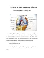
Consistent terminology
for patella position and patellar
tracking are both improving; appre-
ciation of the complexity of the mo-
tion involved is a necessity (Figure
1). Motions that can be measured in-
clude medial and lateral translation
of the patella, axial plane rotation of
the patella (ie, tilt), coronal plane ro-
tation (ie, patellar spin), and sagittal
plane flexion.
3
In vivo and in vitro
studies show that in early flexion,
the patella shifts medially 4 to 9 mm
as it is drawn into the trochlea. The
patellae generally tilt medially in
vitro during early flexion by <4° be-
fore beginning to tilt laterally up to
<4° as flexion progresses to 90°. In
vivo studies of patellar tilt have been
less consistent. Studies of coronal
plane patellar rotation also are not
very consistent, but they generally
demonstrate that the inferior pole of
the patella rotates laterally as knee
flexion progresses. There is much
room for improvement in the clini-
cal evaluation of patellar motion. As
yet, in vivo understanding of patellar
tracking is incomplete.
Dye et al
14
investigated the soft
tissues anterior to the patella and
found differences compared with tra-
ditional anatomic texts. Apparently,
a superficial transverse fascial layer
exists, with a deeper intermediate
oblique aponeurotic layer, both of
which are superficial to the deep rec-
tus femoris fibers, which are directly
applied to the bone of the patella.
Eckhoff et al
15
reported that the sul-
cus of the trochlea in both normal
and osteoarthritic knees is actually
slightly lateral to the midplane be-
tween the medial and lateral femoral
condyles. Their finding is contrary to
the traditional assumption that the
sulcus is in the midline. Radio-
graphic imaging of the patella dem-
onstrated that the geometric center
of the patella was slightly lateral (2.2
± 0.9 mm) to the patellar ridge.
16
Yet
when interpreting imaging studies of
the patellofemoral joint, bony con-
gruence often may not reflect the
real articular congruence. Stäubli
and colleagues
17,18
used magnetic res-
onance arthrograms to demonstrate
that, because of variable thickness of
the articular cartilage on the patella,
images of bone that appear incongru-
ent may actually have excellent car-
tilage congruity.
Clinical Evaluation
It is important to remember that not
all anterior knee pain is associated
with measurable abnormalities of
patellar alignment or individual an-
Anterior Knee Pain
536 Journal of the American Academy of Orthopaedic Surgeons
atomic variations. Patellofemoral
malalignment must not be consid-
ered a synonym for anterior knee
pain. Measurable malalignment of
the patellofemoral joint may or may
not be a key factor in any specific
patient with anterior knee pain.
Studies have failed to be sensitive in
consistently finding radiographic
malalignment in patients with patel-
lofemoral pain.
19
Are radiographic
findings (eg, shallow sulcus, patella
alta, lateral tilt angle) pathologic if
the patient is asymptomatic? Or is
the effect of the preexisting differ-
ence in morphology critical only in
the presence of injury, repetitive
overload, or neuromuscular decom-
pensation? There are no definite an-
swers to these questions.
Misunderstanding of the patho-
genesis and inappropriate treatment
can occur when all pain is assumed
to be associated with some degree of
patellar malalignment. This as-
sumption may result in surgical re-
alignment in patients in whom
alignment may not be the primary
problem. A well-intentioned opera-
tion to realign a normally aligned pa-
tellofemoral joint can lead to a poor
outcome. Imbalances in the extensor
mechanism include dynamic and
static neuromuscular factors. The
patellar position on static imaging is
only part of the pathophysiology . Re-
cent literature has pointed out the
value of recognizing other causes of
patellofemoral pain in patients with
normal anatomic alignment, such as
patellar or quadriceps tendinitis,
20
postoperative neuromas,
21
and sa-
phenous neuritis.
22
The role of the entire leg in the
pathogenesis of anterior knee pain
has come under increased scrutiny.
Witvrouw et al
23
evaluated 282 ado-
lescents (average age, 18.6 years) and
noted that 7% to 10% developed pa-
tellofemoral pain within 2 years. An-
thropometric, physical examination,
psychological, and electromyograph-
ic data were collected prospectively
to discern which factors would pre-
dict the onset of pain. Notable find-
ings were decreased quadriceps and
gastrocnemius flexibility, increased
vastus medialis obliquus (VMO) re-
flex response time and delayed VMO
firing versus the vastus lateralis, de-
creased explosive strength, and in-
creased thumb to forearm mobility.
Factors that did not correlate with
the onset of knee pain included
alignment (ie, Q angle), psychologi-
cal testing, isokinetic strength, and
any of the anthropometric data (eg,
height, weight). Two important
studies found electromyographic dif-
ferences, proving that contraction of
the vastus lateralis came before the
VMO in symptomatic patients com-
pared with control subjects.
24,25
The hip extensor muscles play a
critical role in lower extremity func-
tion. Zhang et al
26
found that the hip
extensors contribute 25% of the
energy absorption during landing.
When the hip musculature does not
absorb its share of the load, other
parts of the extremity must compen-
Figure 1
Clinically relevant patellar position relative to the trochlea. A, Axial view demonstrating medial and lateral translation and patellar
rotation (commonly called tilt). B, Coronal view demonstrating internal and external rotation (commonly called spin). C, Sagittal
view demonstrating flexion. (Adapted with permission from Post WR, Teitge R, Amis A: Patellofemoral malalignment: Looking
beyond the viewbox. Clin Sports Med 2002;21:521-546.)
William R. Post, MD
Volume 13, Number 8, December 2005 537
sate. Deficits in hip strength add to
load on the knee, even independent
of the rotational changes that may
occur in the presence of hip weak-
ness. Providing further evidence of
entire extremity involvement, Baker
et al
27
tested 20 patients with anteri-
or knee pain and found that knee
joint proprioception was abnormal
in both weight-bearing and non–
weight-bearing tests compared with
a control population.
Understanding patellofemoral
disorders does require more than a
thorough understanding of anatomy.
Dye
28
defines the envelope of func-
tion as the “range of load that can be
applied across an individual joint in
a given period without supraphysio-
logic overload or structural failure.”
Essentially, an asymptomatic joint
has adequate tissue homeostasis, so
the amount of load applied to the in-
volved joint is successfully handled.
When the joint is out of homeosta-
sis, pain results. The ability of a j oint
to tolerate loading depends on mul-
tiple factors, not just the radiograph-
ic alignment of the joint. The abso-
lute amount of loading over time is
an important factor in overuse inju-
ries. For example, patients suffering
from anterior knee pain caused by
blunt trauma may have a positive
bone scan (a measure of physiology,
not structure) that resolves over
time as their pain does.
29
The knee is
out of homeostasis on the bone scan
while it is abnormal, but homeosta-
sis is restored over time. Keeping pa-
tients within their pain-free enve-
lope of function, however narrow
that may be, is a key to successful
treatment.
For example, a previously asymp-
tomatic middle-aged, decondition-
ed, sedentary, slightly overweight
woman who rapidly increases her
activity by taking a five-mile hike
up a mountain trail may present 10
days later with anterior knee pain, a
small effusion, peripatellar tender-
ness, and a patellar axial radiograph
suggesting mild patellofemoral ar-
throsis with lateral patellar tilt, and
lateral subluxation. Her increased
activity resulted in loss of joint tis-
sue homeostasis. Relative rest, pain
control, and anti-inflammatory mo-
dalities likely would restore her
daily function, even in the presence
of her preexisting radiographic
“malalignment.” Acute treatment
consists of keeping her within her
new envelope of function (ie, activi-
ties with low enough load that she
is minimally symptomatic), while
working gradually to increase her
envelope of function by weight loss,
strengthening, and flexibility exer-
cises. If such a patient does not seek
care but rather waits out the pain,
she would likely become weaker
from the decreased activity level
and less flexible from the decrease in
activity; also, she might gain weight
because of the inactivity.
Similarly, patients with systemic
illnesses, such as thyroid disorders or
cancer, can develop knee pain as
their muscle weakness decreases
their envelope of function. The next
time such a patient tries to increase
her or his activity level, the envelope
of function is even smaller. The pa-
tient becomes caught in this cycle
and presents much later with a his-
tory of chronic knee pain and radio-
graphic evidence of malalignment.
Rescue from the deconditioned state
is not possible in some patients, and
surgery may be necessary. Theoreti-
cally, a patient who does not respond
to a rehabilitation program has in-
curred such a degree of macrostruc-
tural damage that the joint cannot
return to a homeostatic state. Thus,
surgical intervention to remove the
ongoing focus of inflammation or to
realign the patellofemoral joint to
decrease pathologic loading would be
rational. It is important to remember
that there are no absolute radio-
graphic indications for surgery.
Malalignment can be understood
as a situation “where bony align-
ment, joint geometry, soft tissue re-
straints, neuromuscular control and
functional demands combine to pro-
duce symptoms as a result of abnor-
mally directed loads which exceed
the physiological threshold of the
tissues.”
3
With regard to surger y for
realignment, current clinical stan-
dards for assessing patellofemoral
alignment lack complete informa-
tion, such as patellar spin and sagit-
tal plane flexion. Understanding of
the effect of standard realignment
procedures on all components of
alignment and tracking is currently
limited.
30
Unfortunately, in vivo
understanding of the effect of re-
alignment procedures on three-
dimensional tracking is even more
lacking. With increased appreciation
of the pathophysiology of soft-tissue
pain comes the consideration that
symptomatic relief may occur as a
result of cutting certain soft-tissue
structures, in addition to (or possibly
independent of) any effect that sur-
gery may have on macrostructural
alignment. Even the postoperative
period of relative rest and structured
rehabilitation may contribute to res-
toration of joint homeostasis.
Nonsurgical
Management
Although controversy exists over the
best methods to improve leg strength
in patients with anterior knee pain,
the traditional concept of trying to
achieve isolated VMO exercise is not
supported by extensive and persua-
sive recent literature.
31
One random-
ized study evaluated the effects of
open kinetic chain exercise (non–
weight-bearing) versus closed chain
exercise (weight-bearing) in a group
of patients with anterior knee pain.
32
Although both types of exercise pro-
duced improvements in strength,
pain relief, and return to function,
the closed chain exercises produced
less pain, better triple jump (func-
tional improvement), and less sub-
jective “clicking.” It would be short-
sighted to discard either open or
closed chain exercises entirely.
Several thorough reviews of non-
surgical treatment have been pub-
lished recently;
33,34
many are partic-
Anterior Knee Pain
538 Journal of the American Academy of Orthopaedic Surgeons
ularly notable. Doucette and
Goble
35
reported that 84% of pa-
tients improved after 8 weeks of
quadriceps rehabilitation and
stretching. Patellar axial radiographs
demonstrated some improvement
after treatment, although the values
were within previously published
normal limits at both times, and val-
ues were equivalent between the
symptomatic and asymptomatic
knees. Long-term (7-year) follow-up
of 49 patients treated with quadri-
ceps exercises, rest, and nonsteroidal
anti-inflammatory drugs showed
that nearly 75% of patients main-
tained improvement from 6 months
to 7 years.
36
Many factors were stud-
ied, including radiographs, magnetic
resonance imaging, and other base-
line clinical findings, but none corre-
lated with the treatment result.
37
Unfortunately, no criteria, examina-
tion, or treatment predicted which
patients would respond well. In par-
ticular, patellar taping has generated
much interest, with studies showing
pain relief, alterations in the timing
of VMO contraction, and increased
exercise tolerance.
38,39
Although all of these studies con-
firmed that nonsurgical manage-
ment can be successful and shed
light on the nature of the problem,
only very recently has a double-blind
multicenter placebo-controlled trial
of nonsurgical treatment been re-
ported. Seventy-one subjects aged
<40 years were randomly assigned to
either a placebo or a treatment
group.
40
Subjects were included if
they reported anterior or retropatel-
lar knee pain on at least two of the
following activities: prolonged sit-
ting, stairs, squatting, running,
kneeling, and hopping/jumping. Pa-
tients had symptoms for at least 1
month, an average pain level of 3 on
a 0 to 10 visual analog pain scale,
and insidious onset of symptoms.
The treatment group had six weekly
visits involving patellar taping,
quadriceps training with biofeed-
back, gluteal strengthening, and an-
terior hip and hamstring stretching.
The placebo group had placebo tap-
ing, turned-off ultrasound, and a pla-
cebo “medicated gel.” Thirty-five
percent of patients in the placebo
group believed they were in the ac-
tive treatment group. When mea-
sured by improvement in pain or
function, the treatment group
showed statistically (P ≤ 0.04) better
improvements compared with the
placebo group (which also showed
some improvement).
Therefore, a nonsurgical program
must include activity modification
based on patient history. Athletes
must modify their training, and ad-
justments should be made in work
and daily activities for nonathletes.
Such modifications are important to
get the patient back within his or her
envelope of function. Particular at-
tention also should be paid to flexi-
bility, especially of the quadriceps, a
common deficit in patients with an-
terior knee pain. Strengthening must
be done without causing severe pain.
Strengthening may often be facilitat-
ed by patellar taping. Open or closed
chain exercise programs are individ-
ualized to limit pain, which will fa-
cilitate regular exercise and effective
strengthening. Emphasis on hip
strengthening has also been very
helpful. Nonsurgical management
should be pursued until both the cli-
nician and patient are certain that a
plateau has been reached in the lev-
el of pain and function. This usually
requires at least 3 months of careful
and compliant rehabilitation. Be-
cause very few patients with anteri-
or knee pain do not respond to reha-
bilitation, providers would be well
advised to carefully reconsider the
differential diagnosis when faced
with a patient who has not respond-
ed as expected.
Surgical Management
Because of the success of nonsurgi-
cal management, surgery for anteri-
or knee pain is not necessary in most
patients. Successful surgical treat-
ment requires an accurate diagnosis,
taking particular care to ascertain
whether there are symptoms of pa-
tellar instability or signs of patel-
lofemoral malalignment on physical
examination and imaging studies.
Patients with normal alignment and
no instability may be symptomatic
from tendinosis in the quadriceps or
patellar tendons, pathologic hyper-
trophy and inflammation in the me-
dial plica, or less common causes
(eg, neuromas). Severe damage to the
articular surface of the patella or the
trochlea can at times be the isolated
cause of symptoms.
However, before concluding that
the anterior knee pain is caused by
chondromalacia of the patella, other
causes must be ruled out. Isolated le-
sions of the articular cartilage of the
patellofemoral joint are one of the
less common causes of anterior knee
pain. In such patients, arthroscopic
débridement of Outerbridge grade 2
and 3 chondral lesions can be useful.
In their review of 36 patients with
chondromalacia patellae, Federico
and Reider
41
reported 57.9% good or
excellent results in patients with
traumatic onset; patients with atrau-
matic onset had 41.1% good or ex-
cellent results. All but four patients
thought the surgery was beneficial.
In one recent randomized, non-
blinded study of a similar group of
patients with Outerbridge grade 2
and 3 chondromalacia, bipolar radio-
frequency débridement was com-
pared with mechanical débridement
alone.
42
Both groups improved at fi-
nal 2-year evaluation, but the radio-
frequency group scored significantly
better (P = 0.0006). However, con-
cerns remain about the potentially
damaging long-term effects of radio-
frequency energy on bone and carti-
lage.
43
Although confirmation of the
role of radiofrequency chondroplasty
will depend on future randomized,
blinded studies, these studies
41,42
to-
gether show the positive value of
chondroplasty in carefully selected
patients with grade 2 and 3 lesions.
Lateral release can be effective in
treating a well-defined subset of pa-
William R. Post, MD
Volume 13, Number 8, December 2005 539
tients with anterior knee pain, but it
is seldom needed. Most patients
with pain and a tight lateral retinac-
ulum can be effectively treated non-
surgically. Lateral release may help
by relieving pressure in the lateral
retinaculum, dividing neuromatous
nerves in the lateral retinaculum, or
relieving pressure on the lateral fac-
et of the patella; at present, the exact
mechanism cannot be stated with
certainty. The role of lateral release
in managing anterior knee pain has
been clarified in the past 10 years.
Several studies have shown that the
ideal candidate is a patient with no
history of patellar instability.
44,45
The degree of chondral damage also
seems to be important. Aderinto and
Cobb
46
reported satisfactory results
in only 59% of patients with ad-
vanced patellar arthrosis treated
with lateral release. Conversely,
Shea and Fulkerson
47
reported 92%
good and excellent results after later-
al release when there were no chon-
dral lesions greater than grade 1 and
2 and there was evidence of lateral
tilt on computed tomography.
O’Neill
48
compared the results of
arthroscopic lateral release with
those of open lateral retinacular
lengthening and found slightly bet-
ter results after the lengthening pro-
cedure, although chondral damage
was less severe in this group. This
study raises the question whether a
lengthening procedure is a good al-
ternative to release. The biomechan-
ical effects of lateral release have
been shown to be related to the
length of the release, especially in
the distal direction. Although it is
not known with certainty the clini-
cally necessary amount of release,
extending the release distally to the
level of the tibiofemoral joint line
does result in a measurable increase
in patellar mobility.
49
Inarecentsur-
vey of the International Patellofem-
oral Study Group (a group of clini-
cians with special interest and
expertise in patellofemoral disor-
ders), lateral release was an infre-
quently done procedure. Indications
for the procedure were anterior knee
pain with evidence of a tight lateral
retinaculum on physical examina-
tion.
50
Complications of lateral release
can include persistent or worsening
pain or instability. When present,
these complications can make the
preoperative symptoms seem minor.
Particularly in the setting of a nor-
mally aligned patella that has been
treated with lateral release, medial
subluxation can occur. In this situa-
tion, an excessive lateral release that
included division of the vastus later-
alis tendon also should be suspect-
ed. Medial subluxation must be
suspected clinically in any patient
reporting persistent pain after later-
al release.
51
Symptoms often include
a sense of the patella moving lateral-
ly, a complaint that can mislead cli-
nicians. The cause of this sensation
is the patella’s momentarily sublux-
ating medially out of the trochlea in
early flexion, then snapping back lat-
erally into the trochlea with further
flexion. When the clinician fails to
recognize this diagnosis and instead
interprets the symptoms to be recur-
rent lateral subluxation, further pro-
cedures, such as tibial tuberosity
medial transfer or medial reefing,
may be recommended. However,
such procedures would only worsen
the symptoms.
Medial patellar subluxation must
be confir med by clinical examina-
tion. Two maneuvers have been de-
scribed. Fulkerson
52
recommended
pushing the patella medially with
the knee in extension, then suddenly
flexing the knee. When this repro-
duces the complaint, medial sublux-
ation is likely. Nonweiler and
DeLee
53
suggested examining the in-
volved knee in a lateral position. The
involved knee is placed with the lat-
eral side up, allowing the involved
patella to sag via gravity medially
out of the trochlea. The patient with
medial patellar subluxation will be
unable to flex the knee. Nonsurgical
management can help to confirm
this diagnosis if taping or bracing the
patella into a more lateral position
decreases symptoms. Hughston et
al
54
found that 68% of patients re-
ported improvement in their func-
tional levels and 75% reported sub-
jective improvement by attempts at
repair or reconstruction of the lateral
retinaculum. Surgical management
of this condition involves repair or
reconstruction of the lateral release
defect; although helpful, this is best
considered as a salvage procedure.
Patients with radiographic or ar-
throscopic evidence of lateral patel-
lar tilt and subluxation who have
failed persistent and patient nonsur-
gical management can improve sig-
nificantly after lateral release and
anteromedial tibial tuberosity trans-
fer. Pidoriano et al
55
correlated the
results of anteromedial tibial tuber-
cle transfer with the location of car-
tilage lesions on the patella; they
found that proximal and global pa-
tellar lesions did less well. Their
findings correlate with laboratory
studies showing that anterior tuber-
osity transfer, while decreasing over-
all load, shifts load disproportionate-
ly to the proximal patella. Careful
consideration of the location of car-
tilage lesions is recommended when
contemplating tuberosity transfer,
just as one would do with any other
osteotomy to avoid transferring load
onto articular lesions.
Early weight bearing after antero-
medial tubercle transfer should be
avoided; two series have demon-
strated the potential for fracture dur-
ing full weight-bearing activities be-
tween 4 and 7 weeks.
56,57
Based on
this information, rehabilitation
should include only partial weight
bearing until osteotomy healing is
complete, both radiographically and
clinically. One report indicated that
two athletes sustained tibial frac-
tures while jogging 6 months postop-
eratively; this finding is extremely
uncommon, however.
58
Procedures to restore cartilage in-
tegrity to the patellofemoral joint
have not met with widespread suc-
cess. Efforts are ongoing to evaluate
Anterior Knee Pain
540 Journal of the American Academy of Orthopaedic Surgeons
the usefulness of autologous chon-
drocyte implantation and osteo-
chondral transfers. Only relatively
small numbers of cartilage-restoring
procedures in the patellofemoral
joint have been reported, and overall
results are mixed. Experience has
shown that careful evaluation and
correction of patellofemoral align-
ment must be included.
59-62
Less ag-
gressive procedures, such as chon-
droplasty, microfracture, or abrasion,
may be equally advantageous and
should be considered first-line treat-
ments.
63
Patellofemoral arthroplasty can
be considered in the presence of true
end-stage arthrosis.
64-66
Resurfacing
of the patellofemoral joint should be
done only in low-demand patients
after very careful clinical evaluation
clearly shows that this articulation
is the sole cause of symptoms. A
bone scan may be a helpful adjunc-
tive test in this setting; significant
uptake in the tibiofemoral joint indi-
cates that isolated patellofemoral ar-
throplasty is not appropriate. Mont
et al
67
suggested total knee arthro-
plasty for patients aged >55 years
with primarily patellofemoral arthri-
tis. Special care is needed at the time
of surgery to ensure that the exten-
sor mechanism is well aligned. Sur-
geons undertaking patellofemoral
replacement should be very experi-
enced in patellofemoral realignment
procedures and should be prepared to
combine them with arthroplasty as
needed.
Summary
Despite the prevalence of anterior
knee pain, much is unknown regard-
ing the etiology, pathomechanics,
and management of the many caus-
es of this symptom. To label this set
of disorders as “patellofemoral syn-
drome” is worrisome because it may
deter some clinicians from trying to
reach a more precise diagnosis. Cli-
nicians should strive for the greatest
possible degree of diagnostic accura-
cy and specificity to maximize out-
comes.
A greater understanding of the
natural history of different causes of
anterior knee pain also would be of
great value; learning to predict
which lesions progress over time
would allow the clinician to treat
those lesions more aggressively.
Hypotheses regarding potentially is-
chemic neurologic changes that may
result from excessive soft-tissue ten-
sion may produce insight into new
treatments. Although significant in-
sights have been made in the past 10
years regarding the understanding of
the pathophysiology, diagnosis, and
treatment of anterior knee pain,
there is room for improvement in all
areas. Particularly promising devel-
opments include dynamic magnetic
resonance imaging and advances in
nonsurgical management in treating
the entire extremity, with particular
emphasis on the key role of the hip
muscles in controlling femoral posi-
tion. Improvements in imaging ar-
ticular cartilage may make possible
more precise diagnosis of the loca-
tion and severity of cartilage lesions;
however, clinicians need to be cau-
tious in concluding that the articular
cartilage lesion is the cause of symp-
toms. Clinicians still need to im-
prove their understanding of the role
and boundaries of surgery in anteri-
or knee pain. Currently, nonsurgical
management remains the most pre-
dictable method of treatment.
References
Evidence-based Medicine: Referenc-
es 23, 32, 36, 37, 40, 48, and 63 are
level I or II evidence-based studies.
1. Fulkerson JP: Patellofemoral pain dis-
orders: Evaluation and management.
J Am Acad Orthop Surg 1994;2:124-
132.
2. Post WR: Clinical evaluation of pa-
tients with patellofemoral disorders.
Arthroscopy 1999;15:841-851.
3. Post WR, Teitge R, Amis A: Patel-
lofemoral malalignment: Looking be-
yond the viewbox. Clin Sports Med
2002;21:521-546.
4. Post WR, Fulkerson J: Knee pain dia-
grams: Correlation with physical ex-
amination findings in patients with
anterior knee pain. Arthroscopy
1994;10:618-623.
5. Biedert RM, Stauffer E, Friederich NF:
Occurrence of free nerve endings in
the soft tissue of the knee joint: A his-
tologic investigation. Am J Sports
Med 1992;20:430-433.
6. Dye SF, Vaupel GL, Dye CC: Con-
scious neurosensory mapping of the
internal structures of the human knee
without intraarticular anesthesia.
Am J Sports Med 1998;26:773-777.
7. Sanchis-Alfonso V, Rosello-Sastre E,
Monteagudo-Castro C, Esquerdo J:
Quantitative analysis of nerve chang-
es in the lateral retinaculum in pa-
tients with isolated symptomatic
patellofemoral malalignment: A pre-
liminary study. Am J Sports Med
1998;26:703-709.
8. Biedert RM, Sanchis-Alfonso V:
Sources of anterior knee pain. Clin
Sports Med 2002;21:335-347.
9. Sanchis-Alfonso V, Rosello-Sastre E,
Revert F: Neuralgrowth factor expres-
sion in the lateral retinaculum in
painful patellofemoral malalignment.
Acta Orthop Scand 2001;72:146-149.
10. Witonski D, Wagrowska-Danielewicz
M: Distribution of substance-P nerve
fibers in the knee joint in patients
with anterior knee pain syndrome: A
preliminary report. Knee Surg Sports
Traumatol Arthrosc 1999;7:177-183.
11. Miltner O, Siebert CH, Schneider U,
Niethard FU, Graf J: Patellar hyper-
tension syndrome in adolescence: A
three-year follow up. Arch Orthop
Trauma Surg 2003;123:455-459.
12. Schneider U, Breusch SJ, Thomsen M,
Wenz W, Graf J, Niethard FU: A new
concept in the treatment of anterior
knee pain: Patellar hypertension syn-
drome. Orthopedics 2000;23:581-
586.
13. Katchburian MV, Bull AM, Shih YF,
Heatley FW, Amis AA: Measurement
of patellar tracking: Assessment and
analysis of the literature. Clin
Orthop 2003;412:241-259.
14. Dye SF, Campagna-Pinto D, Dye CC,
Shifflett S, Eiman T: Soft-tissue anat-
omy anterior to the human patella.
J Bone Joint Surg Am 2003;85:1012-
1017.
15. Eckhoff DG, Montgomery WK,
Stamm ER, Kilcoyne RF: Location of
the femoral sulcus in the osteoarthrit-
ic knee. J Arthroplasty 1996;11:163-
165.
16. Asano T, Akagi M, Koike K, Nakamu-
ra T: In vivo three-dimensional patel-
William R. Post, MD
Volume 13, Number 8, December 2005 541
lar tracking on the femur. Clin
Orthop 2003;413:222-232.
17. Staeubli HU, Bosshard C, Porcellini P,
Rauschning W: Magnetic resonance
imaging for articular cartilage:
Cartilage-bone mismatch. Clin
Sports Med 2002;21:417-433.
18. Stäubli HU, Dürrenmatt U, Porcellini
B, Rauschning W: Anatomy and sur-
face geometry of the patellofemoral
joint in the axial plane. J Bone Joint
Surg Br 1999;81:452-458.
19. Laprade J, Culham E: Radiographic
measures in subjects who are asymp-
tomatic and subjects with patellofem-
oral pain syndrome. Clin Orthop
2003;414:172-182.
20. Popp JE, Yu JS, Kaeding CC: Recalci-
trant patellar tendinitis: Magnetic
resonance imaging, histologic evalua-
tion, and surgical treatment. Am J
Sports Med 1997;25:218-222.
21. Kasim N, Fulkerson JP: Resection of
clinically localized segments of pain-
ful retinaculum in the treatment of
selected patients with anterior knee
pain. Am J Sports Med 2000;28:811-
814.
22. Morganti CM, McFarland EG, Cosgar-
ea AJ: Saphenous neuritis: A poorly
understood cause of medial knee pain.
J Am Acad Orthop Surg 2002;10:130-
137.
23. Witvrouw E, Lysens R, Bellemans J,
Cambier D, Vanderstraeten G: Intrin-
sic risk factors for the development of
anterior knee pain in an athletic pop-
ulation: A two-year prospective study.
Am J Sports Med 2000;28:480-489.
24. Cowan SM, Bennell KL, Hodges PW,
Crossley KM, McConnell J: Delayed
onset of electromyographic activity of
vastus medialis obliquus relative to
vastus lateralis in subjects with patel-
lofemoral pain syndrome. Arch Phys
Med Rehabil 2001;82:183-189.
25. Owings TM, Grabiner MD: Motor
control of the vastus medialis oblique
and vastus lateralis muscles is dis-
rupted during eccentric contractions
in subjects with patellofemoral pain.
Am J Sports Med 2002;30:483-487.
26. Zhang SN,Bates BT, Dufek JS: Contri-
butions of lower extremity joints to
energy dissipation during landings.
Med Sci Sports Exerc 2000;32:812-
819.
27. Baker V, Bennell K, Stillman B, Cow-
an S, Crossley K: Abnormal knee joint
position sense in individuals with pa-
tellofemoral pain syndrome.
J Orthop Res 2002;20:208-214.
28. Dye SF: The knee as a biologic trans-
mission with an envelope of function:
A theory. Clin Orthop 1996;325:10-
18.
29. Dye SF, Boll DA: Radionuclide imag-
ing of the patellofemoral joint in
young adults with anterior knee pain.
Orthop Clin North Am 1986;17:249-
262.
30. Mizuno Y, Kumagai M, Mattessich
SM, et al: Q-angle influences tib-
iofemoral and patellofemoral kine-
matics. J Orthop Res 2001;19:834-
840.
31. Malone T, Davies G, Walsh WM:
Muscular control of the patella. Clin
Sports Med 2002;21:349-362.
32. Witvrouw E, Lysens R, Bellemans J,
Peers K, Vanderstraeten G: Open ver-
sus closed kinetic chain exercises for
patellofemoral pain: A prospective,
randomized study. Am J Sports Med
2000;28:687-694.
33. Powers CM: Rehabilitation of patel-
lofemoral joint disorders: A critical re-
view. J Orthop Sports Phys Ther
1998;28:345-354.
34. Crossley K, Bennell K, Green S, Mc-
Connell J: A systematic review of
physical interventions for patellofem-
oral pain syndrome. Clin J Sport Med
2001;11:103-110.
35. Doucette SA, Goble EM: The effect of
exercise on patellar tracking in lateral
patellar compression syndrome. Am
J Sports Med 1992;20:434-440.
36. Kannus P,Natri A, Paakkala T, Jarvin-
en M: An outcome study of chronic
patellofemoral pain syndrome: Seven-
year follow-up of patients in a ran-
domized, controlled trial. J Bone
Joint Surg Am 1999;81:355-363.
37. Natri A, Kannus P, Jarvinen M: Which
factors predictthe long-term outcome
in chronic patellofemoral pain syn-
drome? A 7-yr prospective follow-up
study. Med Sci Sports Exerc 1998;30:
1572-1577.
38. Powers CM, Landel R, Sosnick T, et
al: The effects of patellar taping on
stride characteristics and joint mo-
tion in subjects with patellofemoral
pain. J Orthop Sports Phys Ther
1997;26:286-291.
39. Cowan SM, Bennell KL, Hodges PW:
Therapeutic patellar taping changes
the timing of vasti muscle activation
in people with patellofemoral pain
syndrome. Clin J Sport Med 2002;12:
339-347.
40. Crossley K, Bennell K, Green S,
Cowan S, McConnell J: Physical ther-
apy for patellofemoral pain: A ran-
domized, double-blinded, placebo-
controlled trial. Am J Sports Med
2002;30:857-865.
41. Federico DJ, Reider B: Results of iso-
lated patellar debridement for patel-
lofemoral pain in patients with nor-
mal patellar alignment. Am J Sports
Med 1997;25:663-669.
42. Owens BD, Stickles BJ, Balikian P,
Busconi BD: Prospective analysis of
radiofrequency versus mechanical de-
bridement of isolated patellar chon-
dral lesions. Arthroscopy 2002;18:
151-155.
43. Ryan A, Bertone AL, Kaeding CC,
Backstrom KC, Weisbrode SE: The ef-
fects of radiofrequency energy treat-
ment on chondrocytes and matrix of
fibrillated articular cartilage. Am J
Sports Med 2003;31:386-391.
44. Betz RR, Magill JT III, Lonergan RP:
The percutaneous lateral retinacular
release. Am J Sports Med 1987;15:
477-482.
45. Christensen F, Soballe K, Snerum L:
Treatment of chondromalacia patel-
lae by lateral retinacular release of the
patella. Clin Orthop 1988;234:145-
147.
46. Aderinto J, Cobb AG: Lateral release
for patellofemoral arthritis. Arthros-
copy 2002;18:399-403.
47. Shea KP, Fulkerson JP: Preoperative
computed tomography scanning and
arthroscopy in predicting outcome af-
ter lateral retinacular release.
Arthroscopy 1992;8:327-334.
48. O’Neill DB: Open lateral retinacular
lengthening compared with arthro-
scopic release: A prospective, ran-
domized outcome study. J Bone Joint
Surg Am 1997;79:1759-1769.
49. Marumoto JM, Jordan C, Akins R: A
biomechanical comparison of lateral
retinacular releases. Am J Sports
Med 1995;23:151-155.
50. Fithian DC, Paxton E, Post W, Panni
AS: Lateral retinacular release: A sur-
vey of the International Patellofemo-
ral Study Group. Arthroscopy 2004;
20:463-468.
51. Fulkerson JP: Diagnosis and treat-
ment of patients with patellofemoral
pain. Am J Sports Med 2002;30:447-
456.
52. Fulkerson JP: A clinical test for medi-
al patella tracking (medial sublux-
ation). Tech Orthop 1997;12:144.
53. Nonweiler DE, DeLee JC: The diagno-
sis and treatment of medial sublux-
ation of the patella after lateral reti-
nacular release. Am J Sports Med
1994;22:680-686.
54. Hughston JC, Flandry F, Brinker MR,
Terry GC, Mills JC III: Surgical correc-
tion of medial subluxation of the pa-
tella. Am J Sports Med 1996;24:486-
491.
55. Pidoriano AJ, Weinstein RN, Buuck
Anterior Knee Pain
542 Journal of the American Academy of Orthopaedic Surgeons
DA, Fulkerson JP: Correlation of pa-
tellar articular lesions with results
from anteromedial tibial tubercle
transfer. Am J Sports Med 1997;25:
533-537.
56. Stetson WB, Friedman MJ, Fulkerson
JP, Cheng M, Buuck D: Fracture of the
proximal tibia with immediate
weightbearing after a Fulkerson os-
teotomy. Am J Sports Med 1997;25:
570-574.
57. Bellemans J, Cauwenberghs F, Brys P,
Victor J, Fabry G: Fracture of the prox-
imal tibia after Fulkerson anterome-
dial tibial tubercle transfer: A report
of four cases. Am J Sports Med 1998;
26:300-302.
58. Godde S, Rupp S, Dienst M, Seil R,
Kohn D: Fracture of the proximal tibia
six months after Fulkerson osteoto-
my: A repor t of two cases. J Bone
Joint Surg Br 2001;83:832-833.
59. Brittberg M, Lindahl A, Nilsson A,
Ohlsson C, Isaksson O, Peterson L:
Treatment of deep cartilage defects in
the knee with autologous chondro-
cyte transplantation. N Engl J Med
1994;331:889-895.
60. Peterson L, Minas T, Brittberg M,
Nilsson A, Sjogren-Jansson E, Lindahl
A: Two- to 9-year outcome after autol-
ogous chondrocyte transplantation of
the knee. Clin Orthop 2000;374:212-
234.
61. Minas T: Autologous chondrocyte
implantation for focal chondral de-
fects of the knee. Clin Orthop 2001;
391:S349-S361.
62. Peterson L, Brittberg M, Kiviranta I,
Akerlund EL, Lindahl A: Autologous
chondrocyte transplantation: Biome-
chanics and long-term durability.
Am J Sports Med 2002;30:2-12.
63. Knutsen G, Engebretsen L, Ludvigsen
TC, et al: Autologous chondrocyte
implantation compared with micro-
fracture in the knee: A randomized
trial. J Bone Joint Surg Am 2004;86:
455-464.
64. Argenson JN, Guillaume JM, Aubani-
ac JM: Is there a place for patellofem-
oral arthroplasty? Clin Orthop 1995;
321:162-167.
65. Kooijman HJ, Driessen AP, van Horn
JR: Long-term results of patellofem-
oral arthroplasty: A report of 56 ar-
throplasties with 17 years of follow-
up. J Bone Joint Surg Br 2003;85:836-
840.
66. Krajca-Radcliffe JB, Coker TP: Patel-
lofemoral arthroplasty: A 2- to 18-year
followup study. Clin Orthop 1996;
330:143-151.
67. Mont MA, Haas S, Mullick T, Hun-
gerford DS: Total knee arthroplasty
for patellofemoral arthritis. J Bone
Joint Surg Am 2002;84:1977-1981.
William R. Post, MD
Volume 13, Number 8, December 2005 543