Báo cáo hóa học: " Investigating the complexity of respiratory patterns during the laryngeal chemoreflex" pot
Bạn đang xem bản rút gọn của tài liệu. Xem và tải ngay bản đầy đủ của tài liệu tại đây (299.31 KB, 8 trang )
BioMed Central
Page 1 of 8
(page number not for citation purposes)
Journal of NeuroEngineering and
Rehabilitation
Open Access
Research
Investigating the complexity of respiratory patterns during the
laryngeal chemoreflex
Andrei Dragomir
1
, Yasemin Akay
1
, Aidan K Curran
2
and Metin Akay*
1
Address:
1
Harrington Department of Bioengineering, Ira A. Fulton School of Engineering Arizona State University, Tempe, AZ 85287, USA and
2
Department of Physiology, Dartmouth Medical School, NH 03756, USA
Email: Andrei Dragomir - ; Yasemin Akay - ; Aidan K Curran - ;
Metin Akay* -
* Corresponding author
Abstract
Background: The laryngeal chemoreflex exists in infants as a primary sensory mechanism for
defending the airway from the aspiration of liquids. Previous studies have hypothesized that
prolonged apnea associated with this reflex may be life threatening and might be a cause of sudden
infant death syndrome.
Methods: In this study we quantified the output of the respiratory neural network, the diaphragm
EMG signal, during the laryngeal chemoreflex and eupnea in early postnatal (3–10 days) piglets. We
tested the hypothesis that diaphragm EMG activity corresponding to reflex-related events involved
in clearance (restorative) mechanisms such as cough and swallow exhibit lower complexity,
suggesting that a synchronized homogeneous group of neurons in the central respiratory network
are active during these events. Nonlinear dynamic analysis was performed using the approximate
entropy to asses the complexity of respiratory patterns.
Results: Diaphragm EMG, genioglossal activity EMG, as well as other physiological signals (tracheal
pressure, blood pressure and respiratory volume) were recorded from 5 unanesthetized
chronically instrumented intact piglets. Approximate entropy values of the EMG during cough and
swallow were found significantly (p < 0.05 and p < 0.01 respectively) lower than those of eupneic
EMG.
Conclusion: Reduced complexity values of the respiratory neural network output corresponding
to coughs and swallows suggest synchronous neural activity of a homogeneous group of neurons.
The higher complexity values exhibited by eupneic respiratory activity are the result of a more
random behaviour, which is the outcome of the integrated action of several groups of neurons
involved in the respiratory neural network.
Background
The laryngeal chemoreflex (LCR) has been investigated in
many epidemiological and physiological studies as a
putative exogenous stressor that may contribute to the
pathogenesis of sudden infant death syndrome (SIDS) [1-
3]. The triple-risk model proposed for SIDS states that
death occurs at the confluence of three factors – a inher-
ently vulnerable infant, exposed to an exogenous stressor
during a critical period of postnatal development [4]. The
LCR is elicited when liquid reaches the laryngeal mucosal
Published: 20 June 2008
Journal of NeuroEngineering and Rehabilitation 2008, 5:17 doi:10.1186/1743-0003-5-17
Received: 20 December 2007
Accepted: 20 June 2008
This article is available from: />© 2008 Dragomir et al; licensee BioMed Central Ltd.
This is an Open Access article distributed under the terms of the Creative Commons Attribution License ( />),
which permits unrestricted use, distribution, and reproduction in any medium, provided the original work is properly cited.
Journal of NeuroEngineering and Rehabilitation 2008, 5:17 />Page 2 of 8
(page number not for citation purposes)
receptors. Commonly, the LCR response consists of a
series of events that may be categorized as conservative (in
terms that they try to preserve the limited oxygen reserves
without removing the reflex causing stimulus) such as
apnea, bradycardia and redistribution of blood flow or
restorative (events that try to clear the stimulus and restore
the normal functioning of the airway): swallowing and
coughing [1]. Previous studies have suggested that while
swallowing and apnea are predominant in the postnatal
period, cough emerges as a stronger response as the ani-
mals develop into adulthood [5].
The manifestations of LCR consist of swallowing and
coughing, which occur frequently, apnea (usually associ-
ated with bradycardia), startle, laryngeal constriction and
arousal from sleep. Swallowing and coughing are the pri-
mary manifestations, while the others may or may not
appear depending on the type and strength of the stimu-
lus. Apneas usually follow a period of swallowing and
coughing, while coughing is usually associated with prior
arousal. Swallowing and coughing remove fluids from the
pharyngeal airway, while apnea combined with the laryn-
geal constriction prevent aspiration. Generally the con-
servative and restorative aspects of the reflex are mutually
exclusive [1]. Prolonged apneas pose paradoxically a great
danger: even if together with the resultant hypoxia and
bradycardia they are part of a preventive mechanism, they
might become lethal if the system is not restored in a
timely manner [5]. Previous studies indicated apnea dura-
tion to be strongly influenced by the stimulus type (water
being much more effective than saline solutions) but even
more by a central neural mechanism that perpetuates res-
piratory depression, altered central neural processing of
receptor input being a highly relevant factor [6]. The
whole LCR duration was found to be prolonged by vul-
nerabilities of the neurons in the rostral ventral medulla
(RVM) and to enhance the disruption of stable respiratory
patterns within this context, thus strengthening its rele-
vance in SIDS [1].
In recent studies we have investigated the complexity of
respiratory patterns during eupnea and hypoxia using
nonlinear dynamic analysis and time-frequency analysis
of the phrenic neurogram during early maturation [7,8].
Our results suggested that during severe hypoxia (gasp-
ing) the complexity of the respiratory neural networks is
reduced and this might be due to the silencing of neurons
responsible for activities in the early phase of the phrenic
neurogram.
In the current study, we aim at gaining insight into the
output of the respiratory network in piglets during the
LCR and assess the changes in respiratory patterns com-
plexity during cough, swallow and early recovery after
apnea, when compared to eupnea. We aim at proving that
during the LCR the activity of the respiratory neural net-
works is taken over by a homogeneous group of neurons;
hence we should observe reduced complexity in the respi-
ratory patterns during the key restorative events. Obvi-
ously, vulnerability within some of these neurons might
be fatal.
Quantitative changes in the complexity of biomedical sig-
nals have been traditionally assessed using nonlinear
dynamics analysis methods [9-11]. Generally, physiologi-
cal signals are complex and thought to originate from
complex nonlinear systems [12-14]. Since respiratory
motor output depends on the integrated properties of the
central respiratory neural network, and such a system has
complex dynamic behaviour, the respiratory patterns
present irregular (complex) features that reflect the
dynamics of the underlying neural network. Therefore,
nonlinear dynamics methods have been preferred to spec-
tral analysis and time domain or time-frequency analysis
methods in the cases when information about the system
generating the output is needed [11].
The approximate entropy (ApEn), which is a method
commonly used to asses irregularity (complexity) of bio-
logical signals [7,15], was chosen for our analysis. Since
many biological signals have short data length (100–5000
points) and traditional nonlinear dynamics analysis
methods are largely dependent on the length of the data
sequence [15,16], the approximate entropy method has
been proposed as an ideal tool for these cases [17]. ApEn
is computationally efficient and produces accurate esti-
mates in the case of short data segments.
Methods
Experiments
Experiments were performed on 5 unanesthetized chron-
ically instrumented intact piglets ranging in age from 3 to
10 days. All experimental protocols and surgeries were
approved by The Institutional Animal Care and Use Com-
mittee of Dartmouth College. Animals were anesthetized
using isoflurane in O
2
. Two-wire electro-myographic
(EMG) electrodes were sewn into the diaphragm through
a subcostal incision in the right upper quadrant of the
abdomen to monitor the diaphragm EMG (EMGdia)
activity. Another set of wires was inserted into the gen-
ioglossus through a submental incision to monitor gen-
ioglossal EMG (EMGgg) activity. A 2.7 mm-diameter
catheter was placed in the trachea just below the cricoid
cartilage to record endotracheal pressure and exteriorized
between the shoulder blades on the animals back. The
wires were tunneled subcutaneously and exited the skin at
the top of the skull. Respiration was measured by using a
barometric plethysmograph modified to allow continu-
ous gas flow [1]. A dual-lumen umbilical catheter was
inserted into the femoral artery, with one lumen con-
Journal of NeuroEngineering and Rehabilitation 2008, 5:17 />Page 3 of 8
(page number not for citation purposes)
nected to a transducer to measure arterial BP, while the
second lumen was used to withdraw blood-gas samples.
To stimulate the LCR, a pharyngeal catheter was placed
through the nose at the time of experiments. EEG elec-
trodes were screwed into the skull over the left frontal,
right occipital and right parietal regions, while EOG elec-
trodes were placed lateral to and just above each eye. A
pair of EMG wires was placed in the neck muscles posteri-
orly. EEG, EOG and neck EMG were used to determine
animals arousals and sleep states. These wires were also
exteriorized at the top of the skull and, along with dia-
phragm anf GG EMG wires, were attached to brass con-
nectors and placed in a plastic connector. The connector
was sealed and attached to the skull with acrylic adhesive.
The connector could be attached to recording leads to
acquire data from conscious animals.
The animals were studied ~24 h after the surgery. The
EMGdia and EMGgg were amplified and band- pass fil-
tered from 10–300 Hz. Respiration, endotracheal pres-
sure, blood pressure and animal temperatures were
recorded continuously. All signals were sampled at 1000
Hz and recorded using a data acquisition system (Power-
Lab, ADInstruments).
LCR characterization
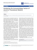
Figure 1 displays some typical signal tracings during the
LCR. Coughing was detected by a massive increase in
EMGdia activity that preceded forceful expiratory activity,
easily identifiable by an increase on the tracheal pressure
tracing [1]. Swallow was associated with a negative deflec-
tion on the tracheal pressure tracing and a burst visible on
the EMGgg. Apneas were defined as periods of silence on
the EMGdia and EMGgg (reflecting no breathing activity)
that lasted longer than the last 2 normal breaths before
the moment of stimulus application. Early recovery
breaths are considered the first bursts visible on the EMG-
dia following the apnea and they are the outcome of the
systems' efforts to restore normal activity. The end of the
LCR was considered when 5 regular (eupneic) consecutive
breath bursts were observed. Generally, apnea occurs after
coughing and swallowing activities, which appear at the
Typical tracings during the LCR in a 10 days old pigletFigure 1
Typical tracings during the LCR in a 10 days old piglet. Example of the events undergoing during the LCR. After the
stimulus, swallowing is visible on the EMGgg tracing and coughing is visible on the EMGdia. Apnea results in the cessation of
respiratory activity and this is visible on all channel.
Journal of NeuroEngineering and Rehabilitation 2008, 5:17 />Page 4 of 8
(page number not for citation purposes)
onset of LCR. These manifestations of the reflex are mutu-
ally exclusive. There was no coughing observed without
prior arousal. Arousal was identified by characteristic
small amplitude, high frequency EEG tracings, as well as
large amplitude bursts on the EOG and increased activity
on the neck EMG.
Approximate entropy
The approximate entropy is a statistical measure that
smooths transient interference and can suppress the influ-
ence of noise by properly setting of the algorithms param-
eters. It can be employed in the analysis of both stochastic
and deterministic signals [17,18]. This is crucial in the
case of biological signals, which are outputs of complex
biological networks and may be deterministic or stochas-
tic, or both. ApEn provides a model-independent measure
of the irregularity of the signals. The algorithm summa-
rizes a time series into a non-negative number, with
higher values representing more irregular systems [17,18].
The approximate entropy estimates are calculated using
segments X(i) through X(N - m + 1) defined by X(i) = [x(i),
, x(i + m - 1)]. The difference between X(i) and X(j), d
[X(i), X(j)] as the maximum absolute difference between
their related scalar elements can be estimated as:
d [X(i), X(j)] = max
k = 0,m-1
[|x(i + k) - x(j + k)] ≤ r
(1)
assuming that all the differences between the correspond-
ing elements will be less than the threshold r.
For any given X(i), the ratio of the difference between X(i)
and X(j) smaller than the threshold r to the total number
of vectors (N - m + 1) is obtained as:
The approximate entropy, ApEn(m,r), can be estimated as
a function of the parameters m and r as follows:
where
In practice, the approximate entropy values can be esti-
mated for a signal with N samples as:
ApEn(m, r, N) = [
Φ
m
(r) -
Φ
m+1
(r)] (5)
The parameter m is the embedding dimension of the ana-
lyzed signals and the parameter r is the threshold to sup-
press the noise in the signal. Throughout this study we
have chosen m = 2 as described in previous works
[11,17,18]. The parameter r can be chosen as 0.1SD(x(i)),
where SD(x(i)) represents the standard deviation of the
original signal x(i).
Results
Our objective in this study was the investigation of
changes in the complexity of the central respiratory net-
work of the piglets during the LCR. EMGdia, EMGgg as
well as other physiological signals needed to completely
characterize the manifestations of the reflex were
recorded. 5 piglets, aged 3–10 days, were used for the
experiments. The LCR was elicited by injecting 0.05 ml
water into the larynx via a nasal catheter. The recorded sig-
nals were detrended by removing their mean before anal-
ysis using ApEn was performed.
The respiratory volume, EMGgg, EMGdia and tracheal
pressure recordings corresponding to a reflex elicited in a
10 days old piglet are shown in Figure 1. Totally, the reflex
lasts ~30 sec; the water stimulus first triggers the swallow,
which is immediately followed by a cough and afterwards
apnea. Apnea duration is ~6 sec, with the system subse-
quently attempting to recover. There are several early
recovery breaths which show a characteristic pattern. They
have shorter duration than regular breaths and their early
phase (first half) activity seems decreased, resembling pat-
terns in gasping following hypoxia [7]. Regular respiratory
activity is restored after ~30 sec, counted when 5 consecu-
tive regular breaths appear [1]. In the presented case swal-
lowing precedes the cough but during the experiments we
observed swallows also succeeding the cough as well as
after apnea.
To investigate how the complexity of respiratory patterns
change during the LCR, we split the respiratory patterns
into 5 characteristic groups: regular (eupneic) breaths
(breaths occurring before the stimulus was given), swal-
lows, coughs, early recovery breaths (first breath burst vis-
ible on the EMGdia after apnea) and recovered breaths (at
least 5 consecutive breaths similar to the regular breaths
before stimulus but occurring not earlier than 25 sec after
the stimulus). The latter condition was imposed based on
observations from previous studies which determined an
average duration of 20–25 sec for the water-elicited LCR
in early postnatal piglets [1].
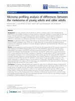
Figure 2 displays the average approximate entropy (com-
plexity) values measured for the 5 piglets under study. The
values represent means ± standard error of 3 separate
measurements for each subject, corresponding to 3 elic-
ited reflexes. It is easily observable that the complexity val-
Ci Ni Nm fori Nm
r
m
r
m
( ) ( ) /( ) , ,=−+=−+11 1
(2)
ApEn( ,) lim () ()mr r r
N
mm
=−
⎡
⎣
⎤
⎦
→∞
+
ΦΦ
1
(3)
Φ
m
r
m
i
Nm
rCiNm() ln ()/( )=−+
=
−+
∑
1
1
1
(4)
Journal of NeuroEngineering and Rehabilitation 2008, 5:17 />Page 5 of 8
(page number not for citation purposes)
ues are highest in the case of regular (eupneic) breaths; the
recovery breaths have values similar to the regular ones,
indicating that the system restored its normal functioning
after the reflex. Swallow and cough bursts, despite their
longer time duration exhibit very low complexity values,
indicating that respiratory networks' output during these
events are the result of the activity of a homogenous group
of neurons. Early recovery breaths show relatively low val-
ues too, which might be an indication that the preceding
apnea silences some groups of neurons within the central
respiratory network. The generally observed trend
throughout our experiments was that the first breath fol-
lowing apnea had the lowest entropy value, while subse-
quent breaths exhibited continuously increasing values.
Generally, after ~30 sec the entropy of the breaths return
to values comparable to those before the stimulus appli-
cation.
We used the analysis of variance (ANOVA) to compare the
significance of the differences in the means of the result-
ing approximate entropy values. Thus, swallows had sig-
nificantly lower values than regular breaths (p < 0.01),
and also than recovered breaths (p < 0.05). Coughs had
significantly lower values (p < 0.05) than regular and
recovered breaths. Early recovery breaths were signifi-
cantly different when compared to the regular breaths (p
< 0.01) and fully recovered breaths (p < 0.05), but fully
recovered breaths complexity values were not significantly
different when compared to the regular breaths (p > 0.1).
Approximate entropy values during LCR: Approximate entropy values ± standard error for the 5 characteristic groups of res-piratory patterns during the LCR: regular (eupneic) breaths, swallowing, coughing, early recovery breaths following apnea and fully recovered breathsFigure 2
Approximate entropy values during LCR: Approximate entropy values ± standard error for the 5 characteristic groups
of respiratory patterns during the LCR: regular (eupneic) breaths, swallowing, coughing, early recovery breaths following apnea
and fully recovered breaths. Results represent averages of entropy values of 3 measurements for each of the 5 animals under
study.
Eupnea Swallowing Coughing Early Recovery Recovered
0.7
0.8
0.9
1
1.1
1.2
1.3
1.4
ApEn
LCR Events
Journal of NeuroEngineering and Rehabilitation 2008, 5:17 />Page 6 of 8
(page number not for citation purposes)
To further characterize the changes undergone by the res-
piratory patterns during the LCR, we have also studied the
breathing temporal patterns. Figure 3 presents compara-
tively the EMGdia tracings of a 10 days old piglet. The top
plot corresponds to a regular eupneic breath occurring
before the application of the LCR stimulus (0.05 ml water
solution). The middle plot shows an early recovery breath,
occurring immediately after an apnea period. The bottom
plot presents a breath occurring ~30 sec after the stimulus
application. All three plots correspond to the same
induced LCR.
The shorter duration of the burst and the signal shape in
the middle plot, resembles those of hypoxic bursts (gasp-
ing) studied in our previous works [7,8]. The resemblance
to gasping extends to the fact that early recovery breaths
following apnea are characterized by brief, intense inspir-
atory efforts of the diaphragm and other respiratory mus-
cles. Previous studies agreed that gasping is the result of a
Typical EMGdia tracings for eupneic, early recovery and recovered breathsFigure 3
Typical EMGdia tracings for eupneic, early recovery and recovered breaths. EMGdia tracings corresponding to reg-
ular breathing activity (top plot), early recovery breath, following apnea (middle plot) and a fully recovered breath (bottom
plot) of a 10 days old piglet.
0 100 200 300 400 500 600 700 800 900 1000
−50
0
50
Amplitude, uV
Regular (Eupneic) Breath
0 200 400 600 800 1000
−50
0
50
Amplitude, uV
Early Recovery Breath
0 200 400 600 800 1000
−50
0
50
Time, msec
Amplitude, uV
Recovered Breath
Journal of NeuroEngineering and Rehabilitation 2008, 5:17 />Page 7 of 8
(page number not for citation purposes)
unique medullary pattern generator which does not con-
tribute to eupneic breathing [19]. We hypothesize (and
intend to test this hypothesis in future studies) that the
mechanism responsible for the respiratory activity during
early recovery might be similar to the one involved in
gasping, where all inspiratory neurons fire simultaneously
at the beginning of the inspiratory period [19]. On the
other hand, the pattern exhibited in the bottom plot
highly resembles the one on the top plot, indicating sys-
tems' full recovery after the critical respiratory disruption
associated to the LCR.
Furthermore, Table 1 displays average durations (means ±
standard errors) of LCR related events and of eupneic
breaths for the 5 piglets under study. The events are the
same ones that were considered for the approximate
entropy estimations presented above. The results reinforce
the previous observations, suggesting that the early recov-
ery breaths occurring after apnea exhibit shorter duration
possibly due to an apnea-influenced mechanism that
silences part of the neural activity. We interpret these res-
piratory efforts as a last resort attempt of the system to
restore normal activity. As expected, breaths occurring
after recovery from LCR have similar durations with regu-
lar eupneic breaths. Coughing and swallowing have sig-
nificantly longer durations. Another interesting
observation is that older animals (8 and 10 days) exhibit
longer duration of respiratory activities, when compared
to younger ones (3 days), results that agree with previous
studies that investigated changes in the respiratory system
in the context of early maturation [5,9]. This is due to the
fact that respiratory premotor and motor neurons
undergo rapid changes in biochemical and bioelectric
properties during the first month of postnatal life. Thus,
there is an increase in the complexity of the dendritic tree
of respiratory neurons as it changes from a bipolar to a
multipolar morphology [20,21].
Discussion and conclusion
Coughing and swallowing are part of a defense mecha-
nism that develops in fetus and continues in postnatal life
aiming to protect the airway from fluid ingestion. Failure
of these mechanisms might result in life threatening con-
ditions. Apnea plays also an important role in preserving
the limited oxygen resources, without, however, removing
the offending stimulus. Paradoxically, prolonged apnea
resulting from vulnerabilities within groups of neurons in
the central respiratory network might be fatal [5]. Our
results support this supposition, the early recovery breaths
after apnea presenting significantly reduced complexity
values and shorter duration than regular breaths, suggest-
ing that apnea silences part of the neural activity via a
mechanism that might be similar with that involved in
gasping [7,8].
Reduced complexity values of the respiratory neural net-
work output corresponding to coughs and swallows sug-
gest synchronous neural activity of a homogeneous group
of neurons that might be taking over respiratory activity
under emergency conditions. The higher complexity val-
ues exhibited by eupneic respiratory activity are the result
of a more random behavior, which is the outcome of the
integrated action of several groups of neurons involved in
the respiratory neural network.
The whole succession of events aiming at protecting the
laryngeal airway is commonly known as the laryngeal
chemoreflex (LCR). It involves coughing, swallowing,
apnea, laryngeal constriction, startle and bradycardia. Our
findings suggest that respiratory patterns show signifi-
cantly reduced complexity throughout the duration of
LCR. This poses the organism under great threat when
combined with an underlying neural vulnerability and in
conjunction with failed cardiorespiratory and arousal
responses to physiological stimuli often encountered dur-
ing early maturation. This supports the results of previous
studies indicating LCR as part of the risks associated to
sudden infant death syndrome (SIDS) [1,5].
Competing interests
The authors declare that they have no competing interests.
Table 1: Average durations (in msec) of respiratory activity during the laryngeal chemoreflex 5 piglets, 3–10 days old, for each piglet
the reflex was elicited 3 times.
LCR EVENTS
Piglet Regular breath Swallowing Coughing Early recovery Recovered breath
1 (3 days) 490.66 ± 20.34 728.33 ± 40.03 891.66 ± 27.79 358.00 ± 16.06 474.66 ± 14.37
2 (3 days) 472.33 ± 22.16 775.00 ± 23.43 862.33 ± 33.67 381.33 ± 19.81 462.33 ± 17.23
3 (3 days) 563.00 ± 16.18 801.66 ± 18.40 899.00 ± 21.62 392.66 ± 23.16 570.33 ± 20.04
4 (8 days) 641.66 ± 31.26 910.33 ± 32.84 991.33 ± 41.28 401.33 ± 20.55 621.00 ± 28.43
5 (10 days) 766.00 ± 24.78 926.33 ± 34.15 1004.66 ± 38.44 441.66 ± 18.22 767.33 ± 23.81
Mean 586.73 ± 29.34 828.33 ± 23.71 929.79 ± 34.82 394.99 ± 21.63 576.13 ± 26.25
Publish with BioMed Central and every
scientist can read your work free of charge
"BioMed Central will be the most significant development for
disseminating the results of biomedical research in our lifetime."
Sir Paul Nurse, Cancer Research UK
Your research papers will be:
available free of charge to the entire biomedical community
peer reviewed and published immediately upon acceptance
cited in PubMed and archived on PubMed Central
yours — you keep the copyright
Submit your manuscript here:
/>BioMedcentral
Journal of NeuroEngineering and Rehabilitation 2008, 5:17 />Page 8 of 8
(page number not for citation purposes)
Authors' contributions
AD performed the data processing and analysis and
drafted the manuscript, YA participated in the study
design and results interpretation, AKC performed the
experiments, MA guided the data procesisng and analysis,
interpreted the results and contributed to writing the
manuscript.
Acknowledgements
This work was supported in part by NIH grant (HL 65732) made to Dr
Akay and Parker Francis Foundation and the Charles H. Hood Foundation
made to Dr. Curran.
References
1. Velde L Van der, Curran AK, Filiano JJ, Darnall RA, Bartlett D Jr,
Leiter JC: Prolongation of the laryngeal chemoreflex after
inhibition of the rostral ventral medulla in piglets: a role in
SIDS? J Appl Physiol 2003, 94:1883-1895.
2. Page M, Jeffery H, Post R, Woods J: Stimulated pharyngeal reflux
can lead to life-threatening apnea if swallowing and arousal
are depressed. J SIDS Infant Mortal 1996, 2:281-293.
3. Harding R, Johnson P, McClelland ME: Liquid-sensitive laryngeal
receptors in the developoing sheep, cat and monkey. J Physiol
1977, 277:409-422.
4. Filiano JJ, Kinney HC: A perspective on neuropathological find-
ings in victims of the Sudden Infant Death Syndrome. Biol
Neonate 1994, 65:194-197.
5. Thach BT: Maturation and transformation of reflexes that
protect the laryngeal airway from liquid aspiration from fetal
to adult life. Am J Med 2001, 111:69S-77S.
6. Lawson EE: Prolonged central respiratory inhibition following
reflex-induced apnea. J Appl Physiol 1981, 50(5):874-879.
7. Akay M, Sekine N: Investigating the complexity of respiratory
patterns during recovery from severe hypoxia. J Neural Eng
2004, 1:16-20.
8. Akay M: Hypoxia silences the neural activities in the early
phase of the phrenic neurogram of eupnea in piglet. J Neuro-
eng Rehab 2005, 2:1-9.
9. Akay M, Sekine M, Moodie KL: Nonlinear dynamics of respira-
tory patterns during maturation. Methods Inf Med 2004,
43:99-101.
10. Kuusela TA, Jartti TT, Tahvanainen KUO, Kaila TJ: Nonlinear meth-
ods of biosignal analysis in assessing terbutaline-induced
heart rate and blood pressure changes. Am J Physiol Heart Circ
Phyisiol 2002, 282(2):H773-H781.
11. Akay M: Biomedical Signal Processing New York: Academic Press; 1994.
12. Freeman WJ: Tutorial on neurobiology: from single neuron to
brain chaos. Int J Bifurc Chaos 1992, 2:451-482.
13. Garde S, Regalado MG, Schechtman VL, Khoo MCK: Nonlinear
dynamics of heart rate variability in cocaine-exposed
neonates during sleep. Am J Physiol Heart Circ Physiol 2001,
280:H2920-2928.
14. Glass L, Mackey MC: From clocks to chaos: the rhythms of life Princeton:
University Press; 1988.
15. Pincus SM: Greater signal regularity may indicate increased
system isolation. Math Biosci 1994, 122:161-181.
16. Lipsitz LA, Pincus SM, Morin RJ, Tong S, Eberle LP, Gootman PM:
Preliminary evidence for the evolution in complexity of
heart rate dynamics during autonomic maturation in neona-
tal swine. J Auton Nerv Syst 1997, 65:1-9.
17. Pincus SM, Huang WM: Approximate entropy, statistical prop-
erties and applications. Commun Stat Theory Methods 1992,
21:3061-3077.
18. Pincus SM: Approximate entropy as a measure of system
complexity. Proc Natl Acad Science 1991, 88:2297-2301.
19. St John WM, Zhou D, Fregosi RF: Expiratory neural activities in
gasping. J Appl Physiol 1989, 66:223-231.
20. Cameron WE, Jodkovski JS, Fang K, Guthrie RD: Electrophysiolog-
ical properties of developing phrenic motoneurons in the
cat. J Neurophysiol 1991, 65:671-679.
21. Hilaire G, Duron B: Maturation of the mammalian respiratory
system. Physiological Reviews 1999, 79:325-360.