Human Musculoskeletal Biomechanics Part 13 doc
Bạn đang xem bản rút gọn của tài liệu. Xem và tải ngay bản đầy đủ của tài liệu tại đây (387.72 KB, 14 trang )
Elements of Vascular Mechanics
231
vascular wall (see also at viscosity). Modern cellular physiology has proven, that separate
from contraction control molecular mechanisms will ensure the dephosphorylation of
myosin light chains, terminating the actomyosin crossbridge cycle, which means that
contraction and relaxation can be controlled somewhat separately in vascular smooth
muscle (Schubert 2008). An other feature, we have to mention is the myogenic contraction.
Passive stress on vascular muscle, especially from small arteries, will induce its active
contraction. Such processes can be observed in vivo and form an important mechanism for
tissue perfusion autoregulation.
While large arteries will not change their lumen to affect volume blood flow in a sensible
manner, smaller arteries and veins can contract until their lumen fully disappears. The
extent of contraction, the vascular “tone” is delicately set at different points of the
circulation and in different times. Several ten types of cytoplasma membrane and some
cytoplasmic receptors have been identified in vascular muscle affecting vascular
contractility. Their amount and the extent of contraction or relaxation induced varies in
different vascular territories. Also, thousands of drug molecules have been isolated or
synthetized that affect vascular contractility, some of the most frequently used
cardiovascular drugs are among them. While earlier it was thought that the amount of
receptors is specific for the tissue, now we now that even receptor molecule expression is
under physiological control, altered receptor expression and altered receptor sensitivity will
form important part of vascular remodeling processes.
8. Viscosity of the vessel wall
For methodical reasons, because it is very difficult to study them under reproducible
conditions, vessel wall viscosity is an unduly neglected area. Most authors agree that vessels
are not only elastic, but viscoelastic (Apter 1966, Azuma 1971, T Bauer 1982, Bergel 1964,
Craiem 2008, Fung 1984, Goto 1966, Greven 1976, Hasegawa 1983, Nadasy 1988,
Orosz 1999a, 1999b, Steiger 1989, Toth 1998, Zatzman 1954). Vessels show all the three
typical viscotic phenomena, the creep (viscotic elongation at continuous stress, Fig 5a.),
the stress relaxation (decreasing stresses after unit-step elongation, Fig 5b.) and hysteresis
loops (difference between upward and downward routes of the stress-strain curves, Fig.
5c.).
Viscosity might be essential in distributing the force among parallelly connected
components of the wall, dampening sudden force elevations on them, preventing their
rupture or overwear. There is an agreement that at least part of vascular viscosity will go on
in the smooth muscle cells themselves. Our explanation was that passive slide between actin
and myosin filaments, with breaking and reestablishment of latching cross-bridges could
explain vascular viscosity. Viscous elongation this way could be restored by ATP dependent
slow contraction and being reversible (Fig. 5a). In pathologic tissue, devoid of functionable
smooth muscle cells, a slow but inherent viscous dilation of extracellular connective tissue fibers
goes toward the fatal rupture of the wall (Fig 5b). Viscoelasticity of the wall can be modeled
with Maxwell or Kelvin models, containing one viscous, one parallelly connected and one
serially connected elastic units (Fung 1984, Orosz 1999a 1999b). In case of the simple
acellular aneurysmal tissue we have identified a fairly continuous stoichiometric ratio
between the three viscoelastic components which, first gives some insight into the molecular
organizational principles of vascular viscoelasticity (Toth 1998).
Human Musculoskeletal Biomechanics
232
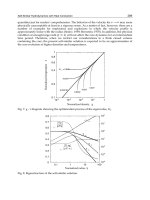
Fig. 5. Blood vessel wall viscosity. a. Viscous creep of contracted human umbilical arterial
segments. Slow creep in oxygenized nKR (●), sped up by doubling distending pressure (□),
or by applying smooth muscle relaxant, sodium nitrite (○) or with calcium-free solution (▲).
Viscosity is also decreased by inhibiting the energy metabolism of smooth muscle cells by 2-
deoxy-glucose (∆). (From Nadasy 1988, with permission of Akadémiai Kiadó) b. Stress
relaxation and tensile strength of human aneurysmic tissue. Strip from brain aneurysm sac.
Stepwise elevation of length, force recorded as a function of time (with permission of
Karger). c. In vivo pressure-diameter pulsatile hysteresis loops recorded in the rabbit
thoracic aorta. Each loop corresponds to one cardiac cycle. Taken at different levels of
bleeding hypotension. (Nadasy, Csaki, Porkolab and Monos, unpublished).
9. Biomechanics of different vascular segments
9.1 Windkessel artery and distributing artery biomechanics
Elasticity is the very essence of Windkessel artery function (Milnor 1982, Zieman 2005). With
each ventricular contraction at rest about 70 ml of blood is pushed into the large arteries,
close to the heart. These vessels are containing a fairly large number of concentric elastic
sheets intertwined with layers of smooth muscle cells in a fishbone pattern, visibly
connecting neighboring elastic sheets. (Clark 1985). At physiological stresses and above
them these vessels are more elastic than more peripheral vessels with less elastic tissue
(Stemper 2007, Fig. 4b.). With aging and hypertension, rigidity of these vessels increases
with a concomitant increase of diameter (Farasat 2008, Giumelly 1999, Safar 2005). In vivo
Elements of Vascular Mechanics
233
elasticity is frequently measured in form of pulse wave velocity (Huotari 2010, Westerhof
2007), aortic compliance (Long 2004, Mersich 2005), input impedance (Mazzaro 2005) or
augmentation index (Safar 2005). Exercise training can stimulate elastin production and
reduce high-stress stiffness (DeAndrade 2010). Elastin production is stimulated by periodic
stress, that is, by pulse-pressure. The produced elastin will form parallelly connected sheets,
that are fairly stretched even at physiological diastolic pressures and thus take part of the
force from smooth muscle and collagen. Diameter to wall thickness ratios can thus be
relatively large in elastic vessels. Too large periodicity in stretch, however, will speed up the
disintegration of elastic lamellae, a typical feature in aged and chronically hypertensive
large arteries (Greenwald 2007). An unsettled question is pulsatile viscosity. We have found
a profound hysteresis of the pressure-diameter curves in vivo (Nadasy 2007 and
unpublished, Fig. 5c.).
9.2 Resistance artery biomechanics
Resistance arteries have limited amount of elastic tissue, the real arterioles none at all. Their
most important function is to offer a relative large but controllable resistance which makes
controlled in space and time) flow distribution toward the tissues. They are characterized by
relatively thick walls and a large diapason between most relaxed and most contracted
diameters (Szekeres 1998, Fig. 3b.) and by massive myogenic response (Fig 4d.). Pulse
pressure is dampened usually en route in large arteries, remaining undulations will support
only a limited elastica production of medial cells. In hypertension, however pressure
undulations can increase in resistance sized arteries with biomechanically and histologically
observable elevation in elastin production. In later phases of the disease, however, these
elastic lamellae will be disrupted. Similar alterations can happen with aging (Arribas 1999,
Briones 2003, Gonzales 2005,2006, Intengan 1998, 2001, Laurant 1997, Nadasy 2010a,
Takeuchi 2005). Even more important are the segmental geometry alterations. The great
circulatory physiologist Folkow realized first that morphological wall thickening might
reduce lumen and stabilize elevated resistance and hypertension. He supposed to happen it
with an elevation of wall mass (hypertophic wall remodeling, Folkow 1971, 1990, 1995).
Later, Mulvany has proven that morphological restriction of the lumen with increased wall
thickness can happen without alteration in wall mass (eutrophic remodeling, Mulvany 1990,
1992). The idea emerged that what essentially happens first is a morphological stabilization
of a contracted diameter (Mathiasen 2007, Nadasy 2010a). Now we have a picture that both
in hypertension and aging there is a morphological lumen restriction of resistance vessels
(Dickhout 2000, Frisbee 1999, James 2006, Jeppesen 2004, Kvist 2003, Matrai 2010, McGuffy
1996 Moreau 1998, Muller-Delp 2002, Mulvany 1996, Nadasy 2010a, 2010b, Najjar 2005,
Orlandi 2006, Pose-Reino 2006, Riddle 2003, Rizzoni 2006, Rodriguez-Porce 2006, Stacy 1989,
Varbiro 2000). We believe that the fact, that substances inducing immediate blood pressure
rise have independent from biomechanical effects trophic action on the resistance artery
walls is not contradictory to the biomechanical control theory. With their additional effects
on vascular smooth muscle protein expression, in the real situation, they promote existing
biomechanical control processes (Nadasy 2010a, Safar 1997, Simon 1994, Toyuz 2005). Even
more important than changes in segmental geometry, can be the network alterations.
Rarefaction and course deviations in hypertension also increase local resistance (Greene
1989, Harper 1978, Nadasy 2000, 2010b, Prasad 1995).
Human Musculoskeletal Biomechanics
234
9.3 Biomechanics of veins
Veins are frequently referred to as being distensible. However, similarly to all vessels, veins
also turn rigid when sufficiently stretched (Fig. 4e). Most in vitro and in vivo studies show
that the transition between the distensible and rigid sections of the pressure-diameter
characteristic curve – similarly to arteries and all other vessels – lies around typical
physiological pressures (Berczi 2005, Molnar 2006, Molnar 2010, Monos 1983, 1995, 2003,
Raffai 2008, Stooker 2003, Zamboni 1996,1998 ). That makes it possible to insert venous
grafts into the arterial system (Monos 1983).
10. Conclusion
Geometry and viscoelasticity controlled both in the short and long runs. Viscoelastic
units, the evidence of mechanically driven continuous vessel wall remodeling. The
vascular mechanical failure: A biomechanical explanation for the thick vessel syndrome.
The possibility to produce mechanical work at the expense of chemical energy, the ability to
restructure the active and passive force-bearing components, even degrade or synthesize
them (vascular remodeling) makes the vascular wall an unusually complicated viscoelastic
material.
Short term control of segmental geometry is most effective in resistance arteries. Contraction
of the outer circumferential smooth muscle layer – because of the incompressibility of the
wall – presses the inner layers into the lumen, inducing substantial decrease in lumen
diameter and elevation in wall thickness. The hemodynamic effect will be much increased
local vascular resistance. Short term control of elasticity will be an important physiological
function of the smooth muscle of large arteries. When contracting, they stress upon the
elastic membranes reducing high-stress isobaric elastic modulus of the wall. This improves
adjustment of vascular impedance to altered ventricular function. Long term control of
vascular lumen will be driven by endothelial shear (to keep it constant, Murray-Rodbard
law). Normally, several mechanisms point toward such a balanced situation. Endothelial
shear can alter several proteins’ expression in the wall, the induced acute vasodilation can
morphologically stabilize, agonists released in response to shear might contribute to
alteration in the morphological lumen. Even substances with primary tissue effects might
have additional direct or indirect vascular effects that help adjust vascular lumen to altered
tissue function and blood flow needs (feed-forward control). In a phylogenetically unusual
situation, however, such adaptation processes can “derail” and work against formation of
an optimal morphological vascular lumen. Vessel wall thickness - on the long term - will be
controlled to stabilize tangential stress – if there is no change in tissue composition
(Folkow-Rodbard-Mulvany’s law). In case of periodic stress, smooth muscle cells will be
stimulated to produce elastin (Burton-Roach-Kadar’s law), which reduces high-stress
modulus. Elastic lamellae produced will bear part of the force, leaving less stress on
parallelly connected smooth muscle and collagen, allowing thus lesser wall thicknesses.
While the viscoelastic properties of the contributing molecules are poorly described, studies
on blood vessels with extreme histological composition suggest that intracellular contractile
fibers, elastic tissue and collagen are organized in viscoelastic units. The number of serially
and parallely connected such units plastically adapts to lengths and forces applied. There
seems to be a stoichiometrically determined connection between series and parallel elasticity
and viscosity of such viscoelastic units. Viscosity – together with elasticity – helps even
distribution of the forces among the parallelly connected elements of the vascular wall.
Elements of Vascular Mechanics
235
Restoration of elongated viscous units will be possible at the expense of ATP energy by
smooth muscle contraction, if this viscous elongation happened by breaking up, passive
sliding and reformation of “latching” actomyosin cross-bridges (intracellular viscosity). If
viscous elongation happens between extracellular fibers, migration, adhesion and
contraction of smooth muscle elements, with subsequent connective tissue production fixing
the restored length might restore the original situation. Study of aneurysmic tissue, where
no contractile elements are present to prevent slow but fatal viscous dilation, make it
probable, that such restoring processes are continuously going on in healthy vascular
tissues. Based on biomechanical experience, we can suppose that if common mechanisms to
distribute the force to smooth muscle and elastic components fail, there is a possibility for
the vascular wall to prevent fatal rupture to develop, by increasing the amount of collagen
in the wall. By this, however, the adaptation to periodic stresses (large vessels), the ability to
control resistance (small arteries) and the ability to reduce stress by contraction (veins) will
be lost. With loss of smooth muscle, the “ropes” of collagenous tissue cannot be pulled and
fixed together, new and new collagenous masses should be produced to prevent slow
passive viscotic creep and fatal rupture. In case of large vessels that will alter the pressure
distribution in the radial direction of the wall and will interfere with vasa vasorum blood
supply of the vessel wall itself. The “blood vessel wall failure” will have a common course,
independently of the original pathology that has induced it. That yields a simple
biomechanical explanation for the “thick vessel syndrome” and for its amazing analogies
with the aging process.
11. Acknowledgement
This work and studies leading to this work have been supported by Hungarian National
Grants OTKA TO 32019 and 42670, the Health Science Council of Hungary (ETT 128/2006)
by the Hungarian Space Agency (BO 00080/03) as well as by the Hungarian Hypertension
Society and the Hungarian Kidney Foundation.
12. References
Abramson DI Ed. Blood Vessels and Lymphatics Academic Press New York and London,
1962.
Albinsson S. Nordstrom I. Hellstrand P. Stretch of the vascular wall induces smooth muscle
differentiation by promoting actin polymerization. J Biol Chem 279:34849-55,2004.
Altman PL, Dittmer DS eds. Biology Data Book 2nd edn. Vol III. Federation of American
Societes for Experimental Biology, Bethesda, Maryland 1974.
Apter JT, Rabinowitz M, Cunnings DH. Correlation of viscoelastic properties of large
arteries with microscopic structure. Circ Res 19:104-121,1966.
Arribas SM, Daly CJ, McGrath IC. Measurements of vascular remodeling by confocal
microscopy. Methods Enzymol 307:246-273,1999.
Azuma T, Hasegawa M. A rheological approach to the architecture of arterial walls. Japn J
Physiol 21:27-47,1971.
Bauer RD, Busse R, Schabert A. Mechanical properties of arteries. Biorheology 19:409-
424,1982.
Human Musculoskeletal Biomechanics
236
Bayliss WM. On the local reactions of the arterial wall to changes of internal pressure. J
Physiol (London) 28:200-223,1902.
Bérczi V, Molnár A, Apor A, Kovács V, Ruzics Cs, Várallyay Cs, Hüttl K, Monos E,
Nádasy GL. Non-invasive assessment of human large vein diameter, capacity,
distensibility and ellipticity in situ: dependence on anatomical location, age, body
position and pressure Eur J Appl Physiol 95:283-289,2005.
Bergel DH. The static elastic properties of the elastic wall. J Physiol 156:445-457, 1961.
Bergel DH. Arterial viscoelasticity. In: Pulsatile Blood Flow, Attinger ED ed. McGraw Hill,
New York, 1964. pp. 275-292.
Briones AM, Gonzalez JM, Somoza B, Giraldo J, Daly CJ, Vila E, Gonzalez MC, McGrath
JC, Arribas SM. Role of elastin in spontaneously hypertensive rat small mesenteric
artery remodeling. J Physiol 552:185-195,2003.
Burton AC. Relation of structure to function of the tissue of the wall of blood vessels.
Physiol Rev 34:619-642,1954.
Busse R Bauer RD, Sattler T, Schabert A. Dependence of elastic and viscous proerties on
circumferential wall stress at two different muscle tones. Pflügers Arch 390:113-
119,1981.
Clark JM, Glagov S Transmural organization of the arterial media. The lamellar unit
revisited. Arteriosclerosis 5:19-34,1985.
Cliff WJ. Blood Vessels, Cambridge University Press, Cambridge, 1976.
Clyman RI, McDonald KA, Kramer RH. Integrin receptors on aortic smooth muscle cells
mediate adhesion to fibronectin, laminin and collagen Circ Res 67:175-186,1990.
Cox RH. Three-dimensional mechanics of arterial segments in vitro: Methods. J Appl
Physiol 36:381-384,1974.
Cox RH. Arterial wall mechanics and composition and the effects of smooth muscle
activation. Am J Physiol 229:807-812,1975a.
Cox RH. Pressure dependence of the mechanical properties of arteries in vivo. Am J Physiol
229:1371-1375,1975b.
Cox RH. Passive mechanics and connective tissue composition of canine arteries. Am J
Physiol 234:H533-H541,1978.
Cox RH, Bagshaw RJ. Effects of hypertension and its reversal on canine arterial wall
properties. Hypertension 12:301-309,1988.
Craiem D, Rojo FJ, Atienza JM, Armentano RL, Guinea GV. Frctional-order viscoelasticity
applied to describe uniaxial stress relaxation of human arteries. Physics Med Biol
53:4543-4554,2008.
DeAndrade Moraes-Teixeira J, Felix A, Fernandes-Santos C, Moura AS, Mandarim-de-
Lacerda CA, deCarvalho JJ. Exercise training enhances elastin, fibrillin and nitric
oxide in the aorta wall of spontaneously hypertensive rats. Exp Mol Pathol 89:351-
357,2010.
Dickhout JG, Lee RM. Increased medial smooth muscle cell length is responsible for
vascular hypertrophy in young hypertensive rats. Am J Physiol Heart Circ Physiol
279:H2085-H2094,2000.
Elements of Vascular Mechanics
237
Discher D, Dong C, Fredberg JJ, Guilak F, Ingber D, Janmey P, Kamm RD, Schmid-
Schonbein GW, Weinbaum S. Biomechanics: Cell research and applications for the
next decade. Ann Biomed Eng 37:847-859,2009.
Dobrin PB, Rovick AA. Influence of vascular smooth muscle on contractile mechanics and
elasticity of arteries. Am J Physiol 217:1644-1651, 1969.
Dobrin PB. Mechanical properties of arteries Physiol Rev 58:397-460,1978.
Duling BR, Gore RW, Dacey RG Jr, Damon DR. Methods for isolation, cannulation and in
vitro study of single microvessels. Am J Physiol 241:H108-H116, 1981.
Farasat SM, Morrell CH, Scuteri A, Ting CT Yin FCP, Spurgeon HA, Chen CH, Lakatta EG,
Najjar SS Pulse pressure is inversely related to aortic root diameter. Implications
for the pathogenesis of systolic hypertension Hypertension 51:196-202,2008.
Folkow B. The hemodynamic consequences of adaptive structural changes of the resistance
vessels in hypertension. Clin. Sci 41:1-12,1971.
Folkow B. “Structural factor” in primary and secondary hypertension. Hypertension 16:89-
101,1990.
Folkow B. Hypertensive structural changes in systemic precapillary resistance vessels: how
important are they for in vivo haemodynamics? J Hypertens 13:1546-1559,1995.
Frisbee JC, Lombard JH. Development and reversibility of altered skeletal muscle arteriolar
structure and reactivity with high salt diet and reduced renal mass hypertension.
Microcirculation 6:215-22,1999.
Fung YC. Biodynamics. Circulation. Springer Verlag, New York, 1984.
Fung YC, Liu SQ. Determination of the mechanical properties of the different layers of blood
vessels in vivo. Proc Natl Acad Sci US 92:2169-2173,1995.
Gabella G. Structural apparatus of force transmission in smooth muscles. Physiol Rev
64:455-477,1984.
Giummelly P, Lartaud-Idjouadiene I, Marque V, Niederhoffer N, Chillon JM, Capdeville-
Atkinson C, Atkinson J. Effects of aging and antihypertensive treatment on aortic
internal diameter in spontaneously hypertensive rats. Hypertension 34:207-
211,1999.
Gonzalez JM, Briones AM, Starcher B, Conde MV, Somoza B, Daly C, Vila E, McGrath I,
Gonzalez MC, Arribas SM. Influence of elastin on rat small artery mechanical
properties. Exp Physiol 90:463-468,2005.
Gonzalez JM, Briones AM, Somoza B, Daly CJ, Vila E, Starcher B, McGrath JC, Gonzalez
MC, Arribas SM. Postnatal alterations in elastic fiber organization precede
resistance artery narrowing in SHR. Am J Physiol Heart Circ Physiol 291:H804-
812,2006.
Goto M, Kimoto Y. Hysteresis and stress relaxation of the blood vessels studied by a
universal tensile-testing instrument. Jap J Physiol 16:169-184,1966.
Gow BS. The influence of vascular smooth muscle on the viscoelastic properties of blood
vessels. In: Cardiovascular Fluid Dynamics, Bergel DH ed. Academic, New York,
1972. pp. 66-110.
Greene AS, Tonellato PJ, Lombard LJ, Cowley AW Jr. Microvascular rarefaction and tissue
vascular resistance in hypertension. Am J Physiol 256:H126-H131,1989.
Human Musculoskeletal Biomechanics
238
Greenwald SE, Newman DL, Denyer HT. Effect of smooth muscle activity on the static and
dynamic elastic properties of rabbit carotid artery. Cardiovasc Res 16:86-94,1982.
Greenwald SE. Ageing of the conduit arteries. J Pathol 211:157-172,2007.
Greven K. The time course of creep and stress relaxation in the relaxed and contracted
smooth muscle. Bulbring E, Shuba MF eds, Raven Press, New York, 1976. pp. 223-
228.
Harper RN, Moore MA, Marr MC, Watts LE, Hutchins PM Arteriolar rarefaction in the
conjunctiva of human essential hypertensives Microvascular Research 16:369-
372,1978.
Hasegawa M. Rheological properties and wall structures of large veins. Biorheology 20:531-
545,1983.
Hayashi K, Naiki T. Adaptation and remodeling of vascular wall; biomechanical response to
hypertension. J Mech Behav Biomed Materials 2:3-19,2009.
Hegedus K Some observations on reticular fibers in the media of the major cerebral arteries.
A comparative study of patients without vascular disease and those with ruptured
berry aneurysms. Surg Neurol 22:301-307,1984.
Heistad DD, Armstrong ML, Baumbach GL, Faraci FM. Sick vessel syndrome. Recovery of
atherosclerotic and hypertensive vessels. Hypertension. 26:509-513,1995.
Herlihy JT, Murphy RA. Length-tension relationship of smooth muscle of the hog carotid
artery. Circul Res 33:275-283,1973.
Herman P, Kocsis L, Eke A. Fractal branching pattern in the pial vasculature in the cat. J
Cerebr Blood Flow Metabol 21:741-753,2001.
Hudetz AG, Mark G, Kovach AGB, Monos E. The effect of smooth muscle activation on the
mechanical properties of pig carotid arteries. Acta Physiol Acad Sci Hung 56:263-
273, 1980.
Huotari MJ, Maatta K, Nadasy GL, Kostamovaare J. A photoplethysmographic pulse wave
analysis for arterial stiffness in extremities. Artery Research 4: 155,2010. (A)
Intengan HD, Schiffrin EL. Mechanical properties of mesenteric resistance arteries from
Dahl salt-resistant and salt-sensitive rats: role of endothelin-1. J Hypertens
;16:1907-1912,1998.
Intengan HD, Schiffrin EL. Vascular remodeling in hypertension: roles of apoptosis,
inflammation, and fibrosis. Hypertension 38:581-587,2001.
Jackson PA, Duling BR. Myogenic response and wall mechanics of arterioles. Am J Physiol
257:H1147-H1155,1989.
James MA, Tullett J, Hemsley AG, Shore AC. Effects of aging and hypertension on the
microcirculation. Hypertension 47:968-974,2006.
Jeppesen P, Gregersen PA, Bek T. The age-dependent decrease in the myogenic response of
retinal arterioles with the Retinal Vessel Analyzer. Grafes Arch Clin Exp Ophthalm
242:914-919,2004.
Kadar A, Veress B, Jellinek H. Relationship of elastic fibre production with smooth muscle
cells and pulsation effect in large vessels. Acta Morphol Acad Sci Hung 17:187-
200,1969.
Kamiya A Togawa T. Adaptive regulation of wall shear stress to flow change in the canine
carotid artery. Am J Physiol 239:H14-H21,1980.
Elements of Vascular Mechanics
239
Koens MJW, Faraj KA, Wismans RG, van der Vliet JA, Krasznai AG, Cuijpers VMJI, Jansen
JA, Daamen WF, van Kuppevelt TH Controlled fabrication of triple layered and
molecularly defined collagen/elastin vascular grafts resembling the native blood
vessel. Acta Biomaterialia 6:4666-4674,2010.
Kuo L, davis MJ, Chilian WM. Myogenic activity in isolated subepicardial and
subendocardial coronary arterioles. Am J Physiol 255:H1558-H1562,1988.
Kvist S, Mulvany MJ. Contrasting regression of blood pressure and cardiovascular structure
in declipped renovascular hypertensive rats. Hypertension 41:540-545,2003.
Laurant P, Touyz RM, Schiffrin EL. Effect of pressurization on mechanical properties of
mesenteric small arteries from spontaneously hypertensive rats. J Vasc Res 34:117-
125, 1997.
Lee RT, Huang H. Mechanotransduction and arterial smooth muscle cell:new insight into
hypertension and atherosclerosis. Ann Med 32:233-235,2000.
Liu SQ, Fung YC. Zero-stress state of arteries. J Biomech Eng 110:82-84,1988.
Long A, Rouet L, Bissery A, Goeau-Brissoniere O, Sapoval M. Aortic compliance in healthy
subjects: Evaluation of tissue Doppler imaging. Ultrasound Med Biol 30:753-
759,2004.
Lorant M, Nadasy GL, Monos E: Changes in network characteristics of saphenous vein after
long-term head-up tilt position of the rat. Physiol Res 52:525-531,2003
Lundholm L, Mohme-Lundholm E. Length at inactivated contractile elements, length-
tension diagram, active state and tone of vascular smooth muscle. Acta Physiol
Scand 68:347-359,1966.
Mathiasen ON, Buus N, Larsen ML, Mulvany JM. Small artery structure adapts to
vasodilation rather than to blood pressure during antihypertensive treatment. J
Hypert 25:1027-1034,2007.
Matrai M, Mericli M, Nadasy GL, Varbiro Sz, Szekeres M, Banhidy F, Acs N, Monos E,
Szekacs B: Gender differences in biomechanical properties of intramural coronary
resistance arteries of rats, an in vitro microarteriographic study J Biomech 40:1024-
1030,2007.
Matrai M, Szekacs B, Mericli M, Nadasy GL, Szekeres M, Banhidy F, Bekesi G, Monos E, Sz
Varbiro: Biomechanics and vasoreactivity of female intramural coronaries in
angiotensin II induced hypertension. Acta Physiol Hung 97:31-40,2010.
Mazzaro L, Almasi SJ, Shandas R, Gates PE. Aortic imput impedance increases with age in
healthy men and women. Hypertension 45:1101-1106,2005.
McGuffee LJ, Little SA. Tunica media remodeling in mesenteric arteries of hypertensive rats.
Anat Rec 246:279-292,1996.
Mersich B, Rigo J Jr, Besenyei C, Lenard Z, Studinger P, Kollai M. Opposite changes in
carotid versus aortic stiffness during healthy human pregnancy. Clin Sci 209:103-
107,2005.
Milnor WM. Hemodynamics Wiulliams and Wilkins, Baltimore/London 1982.
Molnár AA, Apor A, Kristóf V, Nádasy GL, Preda I,
Hüttl K, Acsády G, Monos E, Bérczi
V: Generalized changes in venous distensibility in postthrombotic patients Thromb
Res 117:639-45, 2006.
Human Musculoskeletal Biomechanics
240
Molnar G, Nemes A, Kekesi V, Monos E, Nadasy GL. Maintained geometry, elasticity and
contractility of human saphenous vein segments stored in a complex tissue culture
medium Eur J Vasc Endovasc Surg 40:88-93,2010.
Monos E, Hudetz AG, Cox RH. Effect of smooth muscle activation on incremental elastic
properties of major arteries. Acta Physiol Hung 53:31-39,1979.
Monos E. Csengôdy J Does haemodynamic adaptation take place in the vein grafted into an
artery? Pfluegers Archiv 384:177-182,1983.
Monos E. Biomechanics of the Vascular Wall, Medicina, Budapest, 1986 (In Hungarian)
Monos E, Berczi V, Nadasy GL. Local control of veins: Biomechanical, metabolic, and
humoral aspects Physiol. Rev. 75:611-666, 1995.
Monos E, Lóránt M, Dörnyei G, Bérczi V, Nádasy Gy: Long-term adaptátion mechanisms in
extremity veins supporting orthostatic tolerance. (Review) News Physiol Sci 18:210-
214,2003
Moreau P, d'Uscio LV, Luscher TF. Structure and reactivity of small arteries in aging.
Cardiovasc Res 37:247-253,1998.
Muller-Delp J, Spier SA, Ramsey MW, Lesniewski LA, Papadopoulos A, Humphrey
JD, Delp MD. Effects of aging on vasoconstrictor and mechanical properties of
rat skeletal muscle arterioles. Am J Physiol Heart Circ Physiol 282:H1843-1854,
2002.
Mulvany HJ, Warshaw DM. The anatomical location of the series elastic component in rat
vascular smooth muscle. J Physiol (London) 314:321-330,1981.
Mulvany MJ, Aalkjer C. Structure and function of small arteries. Physiol Rev 70:921-
961,1990.
Mulvany MJ. The development and regression of vascular hypertrophy. J Cardiovasc
Pharmacol 19 (Suppl 2):S22-S27,1992.
Mulvany MJ. Effects of angiotensin converting enzyme inhibition on vascular remodelling
of resistance vessels in hypertensive patients. J Hypertens 14(Suppl.6.):S21-
S24,1996
Nádasy G.L., E. Monos, E. Mohácsi, J. Csépli, A. G. B. Kovách: Effect of increased luminal
blood flow on the development of the human arterial wall. Comparison of
mechanical properties of double and single umbilical arteries in vitro. Blood
Vessels 18:139–143, 1981.
Nádasy G.L., E. Mohácsi, E. Monos, J. L. Lear, A. G. B. Kovách: A simple model describing
the elastic properties of human umbilical arterial smooth muscle. Acta Physiol
Hung 70:75–85, 1987.
Nádasy G. L., Monos E., Mohácsi E., Kovách A.G.B.: The background of
hysteretic properties of the human umbilical arterial wall. Smooth muscle
contraction and hysteresis of the pressure–radius curves. Acta Physiol Hung
71:347–361, 1988.
Nadasy GL, Varbiro S, Acs N, Szekacs B, Lorant M, Jackel M, Kerenyi T, Monos E:
Intramural coronary resistance artery network remodelling in chronically
angiotensin II-infused female rats J Physiol (London) 526(Suppl. S):133P, 2000.
Nadasy GL, Szekeres M, Dezsi L, Varbiro Sz, Szekacs B, Monos E: Brief communication
Preparation of intramural small coronary artery and arteriole segments and
Elements of Vascular Mechanics
241
resistance artery networks from the rat heart for microarteriography and for in situ
perfusion video mapping Microvasc Res 61:282-286,2001.
Nádasy G, Mericli M, Mátrai M, Várbíró Sz, Szekeres M, Ács N, Monos E, Székács B:
Gender differences in biomechanics of intramural coronary resistance arteries of
the rat. Acta Physiol Hung 92:287-288,2005.
Nádasy Gy: Arterial blood supply and tissue needs. In: Physiology and Maintenance., OOP
Hanninen and M Atalay eds., Chapter 6.54.7.2. UNESCO Encyclopedia of Life
Support Systems, EOLSS, www.eolss.net (2007a).
Nádasy GL, Monos E. Biomechanical principles of vascular wall design in health and in
disease: some mathematics of angiogenesis. Acta Phsiol Hung 94:377-379,2007b.
Nadasy GL, Varbiro Sz, Szekeres M, Kocsis A, Szekacs B, Monos E, Kollai M: Biomechanics
of resistance artery wall remodeling in angiotensin-II hypertension and after its
recovery Kidney Blood Press Res 33:37-47,2010a.
Nadasy GL, Szekacs B, Varbiro Sz, Sekeres M, Wappler E, Szalai E, Simon A, Monos E.
Analog biomechanical effects of aging and hypertension in resistance blood vessels
Acta Physiol. Hung. 97: 462, 2010b.
Najjar SS, Scuter A, Lakatta EG. Arterial aging: is it an immutable cardiovascular risk factor?
(Review) Hypertension 454-462,2005.
Orlandi A, Bochaton-Piallat ML, Gabbiani G, Spagnoli LG. Aging, smooth muscle cells and
vascular pathobiology: Implications for atherosclerosis. Atherosclerosis 221-230,
2006.
Osol G, Halpern W. Myogenic properties of cerebral blood vessels from normotensive and
hypertensive rats. Am J Physiol 249:H914-H921,1985.
Orosz M, Molnarka G, Toth M, Nadasy GL, Monos E: Viscoelastic behavior of vascular wall
simulated by generalized Maxwell models - a comparative study Med Sci Mon
5:549-555, 1999.
Orosz M, Molnárka Gy, Nádasy Gy, Raffai G, Kozmann Gy, Monos E: Validity of
viscoelastic models of blood vessel wall Acta Physiol Hung 86:265-271, 1999.
Oxlund H. Relationship between the biomechanical properties, composition and molecular
structure of connective tissues. Connect Tissue Res 15:65-72,1986.
Pose-Reino A, Rodriguez-Fernandez M, Hayik B, Gomez-Ulla F, Carrera-Nouche MJ,
Gude-Sampedro F, Estevez-Nunez JC, Mendez-Naya I. Regression of alterations
in retinal microcirculation following treatment for arterial hypertension. J Clin
Hypertens 8:590-595,2006.
Prasad A, Dunhill GS, Mortimer PS, MacGregor GA. Capillary rarefaction in the forearm
skin in essential hypertension. J Hypertens 13:265–268,1995.
Pries AR, Reglin B, Secomb TW. Remodeling of blood vessels: responses of diameter and
wall thickness to hemodynamic and metabolic stimuli. Hypertension 46:725-
731,2005.
Raffai G, Lódi C, Illyés G, Nádasy G, Monos E: Increased diameter and enhanced myogenic
response of saphenous vein induced by two-week experimental orthostatsis are
reversible Physiol Res 57:175-183,2008.
Rhee AY, Brozovich FV. Force maintenance in smooth muscle: analysis using sinusoidal
perturbations. Archives of Biochemistry & Biophysics. 410:25-38,2003.
Human Musculoskeletal Biomechanics
242
Riddle DR, Sonntag WE, Lichtenwalner RJ. Microvascular plasticity in aging. (Review)
Ageing Research Reviews 2:149-168,2003.
Rizzoni D, Agabiti-Rosei E. Small artery remodeling in hypertension and diabetes. Curr
Hypert Rep 8:90-95,2006.
Roach MR , Burton AC. The reason for the shape of the distensibility curves of arteries. Can
J Biochem Physiol 35:681-690,1957.
Rodbard S. Negative feedback mechanisms in the architecture and function of the
connective tissue and cardiovascular tissues. Perspect Biol Med 13:507-527, 1970.
Rodbard S. Vascular caliber Cardiology 60:4-49,1975.
Rodriguez-Porcel M, Zhu XY, Chade AR, Amores-Arriaga B, Caplice NM, Ritman EL,
Lerman A, Lerman LO. Functional and structural remodeling of the myocardial
microvasculature in early experimental hypertension. Am J Physiol Heart Circ
Physiol. 290:H978-H984,2006.
Rossitti S, Lofgren J. Vascular dimensions of the cerebral arteries follow the principle of
minimum work. Surgery 54:347-350,1963.
Safar ME, van Bortel LM, Struijker-Boudier HA. Resistance and conduit arteries following
converting enzyme inhibition in hypertension. J Vasc Res 34:67-81, 1997.
Safar ME. Systolic hypertension in the elderly: arterial wall mechanical properties and the
renin-angiotensin-aldosterone system. J Hypertension 23:673-681,2005.
Schmidt-Nielsen K. Animal Physiology. Adaptation and Environment, Cambridge
University Press, Cambridge 1979.
Schubert R, Lidington D, S-S Bolz. The emerging role of calcium sensitivity regulation in
promoting myogenic vasoconstriction. Cardiovasc Res 77:8-18,2008.
Schwartz CJ, Werthessen NT, Wolf A. Eds. Structure and Function of the Circulation.
Volumes 1-2-3. Plenum Press, New York and London, 1980.
Shimazu T, Hori M, Mishima M, KIbatake A, Kodama K, Nanto S, Inoue M. Clinical
assessment of elastic properties of large coronary arteries: Pressure-diameterv
relationship and dynamic incremental elastic modulus. Int J Cardiol 13:27-
45,1986.
Siegman MJ, Butler TM, Mooers SV, Davies RE. Crossbridge attachment, resistance to
stretch and viscoelasticity in resting mammalian smooth muscle. Science 191:383-
385,1976.
Simon G, Abraham G, Altman S. Stimulation of vascular glycosaminoglycan synthesis by
subpressor angiotensin II in rats. Hypertension 23(1 Suppl):I148-51,1994.
Somlyo AP, Somlyo AV. Vascular smooth muscle I. Normal structure, pathology,
biochemistry and biophysics. Pharmacol Rev 20:197-272, 1968.
Stacy DL, Prewitt RL. Effects of chronic hypertension and its reversal on arteries and
arterioles. Circul Res 65:869-879,1989.
Steiger HJ, Aaslid R, Reule HJ. Growth of aneurysms can be understood as passive yield to
blood pressure. Acta Neurochir (Wien) 100:74-78,1989.
Stemper BD, Yoganandan N, Pintar FA. Mechanics of arterial subfailure with increasing
loading rate J Biomech 40:1806-1812,2007.
Elements of Vascular Mechanics
243
Stooker W, Gok M, Sipkema P, Niessen HWM, BaidoshviliA, Westerhof N, Jansen AK,
Wildevuur CRH, Eijsman L Pressure-Diameter relationship in the human greater
saphenous vein. Ann Thorac Surg 76:1533-1538,2003.
Szekeres M, Nadasy GL, Dezsi L, Orosz M, Tôkés A, Monos E: Segmental differences in
geometric, elastic and contractile characteristics of small intramural coronary
arteries J Vasc Res 35:332-344,1998.
Szekeres M, Nádasy GY L, Kaley G, Koller A: Nitric oxide and prostaglandins modulate
pressure-induced myogenic responses of intramural coronary arterioles J.
Cardiovasc Pharmacol 43:242-249,2004.
Szentiványi M, Nádasy GL, Tóth M, Kopcsányi V, Jedrákovits A, Monos E: Biomechanics of
the saphenous artery and vein in spontaneous hypertension Pathophysiology
4:295-302,1998.
Takeuchi K, Ideishi M, Tashiro T, Morishige N Yamada T, Saku K, Urata H. Higher
small arterial elasticity in hypertensive patients treated with angiotensin II receptor
blockers. Hypertens Res. 2005,28:639-644,2005.
Toth M, Nadasy GL, Nyary I, Kerenyi T, Orosz M, Molnarka G, Monos E: Sterically
inhomogenous viscoelastic behavior of human saccular aneurysms J Vasc Res
35:345-355,1998.
Touyz RM. Intracellular mechanisms involved in vascular remodelling of resistance arteries
in hypertension: role of angiotensin II. Exp Physiol. 90:449-455,2005.
VanDijk AM, Wieringa PA, Van der Meer , Laird DJ. Mechanics of resting isolated single
vascular smooth muscle cells from bovine coronary artery Am J Physiol 246:C277-
C287,1984.
Várbiro S, Nádasy GL, Monos E, Vajo Z, Ács N, Miklós Z, Tőkés A, Székács B Effect
of ovariectomy and hormone replacement therapy on small artery biomechanics
in angiotensin-induced hypertension in rats. J Hypertension 2000; 18:1587-
1595,2000.
Vidik A, Danielsen CC, Oxlund H. On fundamental and phenomenological models,
structure and mechanical properties of collagen, elastin and glycosaminoglycan
complexes. Biorheology 19:437-451,1982.
Westerhof BE, Guelen I, Stok WJ, Wesseling KH, Spaan JAE, Bos JB, Stergiopulos N
Arterial pressure transfer characteristics:effects of travel time. Am J Physiol
292:H800-H807,2007.
Wolinsky H, Glagov S. Structural basis for the static mechanical properties of the aortic
media Circ Res 20:99-111,1967.
Zamboni, P. Marcellino, MG. Portaluppi, F. Manfredini, R. , Feo, CV. Quaglio, D. Liboni, A.
The relationship between in vitro and in vivo venous compliance measurement.
International Angiology 15, 149-152,1996.
Zamboni, P. Portaluppi, F. Marcellino, MG. Quaglio, D. Manfredini, R. Feo, CV. Stoney,
RJ. In vitro versus in vivo assessment of vein wall properties. Annals of Vascular
Surgery 12:324-329,1998.
Zamir M. Shear forces and blood vessel radii in the cardiovascular system. J Gen Physiol
69:449-461,1977.
Human Musculoskeletal Biomechanics
244
Zatzman M, Stacy RW, Randall J, Eberstein A. Time course of stress relaxation in isolated
arterial segments. Am J Physiol 177:299-307,1954.
Zieman SJ, Melenovsky V, Kass DA . Mechanisms, pathophysiology, and therapy of arterial
stiffness Arterioscler Thromb Vasc Biol 25:932-943,2005.