Biosensors Emerging Materials and Applications Part 7 pot
Bạn đang xem bản rút gọn của tài liệu. Xem và tải ngay bản đầy đủ của tài liệu tại đây (1.65 MB, 40 trang )
Aptamer Sensors Combined with Enzymes for Highly Sensitive Detection
231
is amenable to hybridization inhibition upon binding to the aptamer target. We modified the
aptamer with an avidin-conjugated enzyme and we succeeded in detecting thrombin, IgE
(Fukasawa et al., 2009), and vascular endothelial growth factor (VEGF) (in preparation) via
enzymatic activity measurement.
The second system makes use of the structural changes that aptamers undergo upon
binding to their target molecules (Fig. 3(b)). We created a "capturable" aptamer by adding a
sequence to it that gave it a new structure. Capturable aptamers cannot hybridize with
CaDNA unless their target molecules are present. In this case, the structure of a capturable
aptamer in the presence of its target molecule changes to a different structure from that
which was present in the absence of the target molecule. We succeeded in the design of a
capturable aptamer for thrombin (Abe et al., 2011) and a mouse prion protein (Ogasawara et
al., 2009). In these studies, although fluorescent labeling was used for detection, enzyme
labeling enabled a 10-fold lower detection of mouse prion protein than fluorescent labeling
(unpublished data).
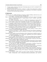
Fig. 3. The scheme of a single aptamer-based B/F separation system. (a) In the absence of a
target molecule, the aptamers are trapped by the immobilized beads containing CaDNA,
whereas in the presence of the target protein, aptamers that bind to the target are not
trapped. The target protein can therefore be detected by means of simple B/F separations,
and by measuring the fluorescence or enzymatic activity of the labeled aptamer in the
supernatant. (b) The aptamer, which is able to be captured, undergoes a conformational
change upon binding to the target molecule. This change induces the exposure of a partial
single-strand that hybridizes with the CaDNA. Otherwise, any unbound capturable aptamer
does not hybridize with the CaDNA and is removed by the bound/free separation.
Biosensors – Emerging Materials and Applications
232
Of these two types of single aptamer-based B/F separation systems, the first can be easily
designed, because it does not require any additional sequences, whereas the second system
requires careful design of the additional sequence of the aptamer with structural prediction.
However, the benefit of the second system is that it can eliminate many interfering
compounds. The first system can eliminate enzyme-modified aptamers that do not bind to
the target molecule, but it is difficult to eliminate interfering compounds because aptamers
that bind to the target molecule are present in the supernatant. It is therefore necessary to
select a particular system to suit the needs of each particular target molecule.
Wei et al. reported a different type of single aptamer based B/F separation system without
complementary DNA being present (Wei & Ho, 2009). They utilized steric hindrance
between enzyme-modified antibodies and antigen-modified target-binding aptamers. They
used fluorescein-modified aptamers and anti-fluorescein horseradish peroxidase (HRP)-
conjugated antibody. The antibody cannot bind to the fluorescein-modified aptamer due to
steric hindrance without its target molecule. The aptamers change conformation upon
binding to the target molecule, and then the antibodies can bind to them. Since the aptamers
were immobilized on the solid support, this sensing system enabled B/F separation to occur
using an aptamer.
2.2 Homogeneous sensing
To measure the target molecules without B/F separation, regulation of signal output is
required. Jhaveri et al. reported aptamers that changed their structure upon binding to the
target molecule, which resulted in the regulation of fluorescent signals (Jhaveri et al., 2000).
If we can introduce enzyme signal amplification into a signaling aptamer, a highly sensitive
detection can be performed without the need for B/F separation. Reported homogeneous
detection systems using enzymes are based on two strategies: enzyme activity regulation by
the target molecule, and DNA amplification accompanied by the target molecule binding to
aptamers.
2.2.1 Enzyme activity regulation by the target molecule
If we can find an enzyme that catalyzes a reaction with a target molecule, we can construct
an effective sensing system such as the glucose sensor, which is already on the market and is
being used daily. However, it is difficult to screen an enzyme that reacts with a given target
molecule. Protein engineering allows us to improve the enzyme substrate specificity, and we
have reported such examples (Igarashi et al., 2004), but it is still difficult to change the
substrate specificity dramatically. Then we constructed an enzyme that has a novel subunit
that can regulate enzymatic activity allosterically based on the aptamer. If the target
molecule activates enzymatic activity, we can quantify the target molecule via an enzyme
activity measurement. We named this sensing system the Aptameric Enzyme Subunit (AES)
(Ikebukuro et al., 2008; Yoshida et al., 2009; Yoshida et al., 2006a, b, 2008).
An AES consists of two aptamers: an enzyme-inhibiting aptamer and a target molecule-
binding aptamer. The enzyme does not generate signals because the AES inhibits enzymatic
activity when it is not bound to the target molecules. However, upon binding of the target
molecules to the AES, the AES changes its conformation, which results in a loss of enzyme
inhibitory activity. Then we can measure the target molecule concentration via enzyme
activity measurements without the need for B/F separation. Therefore, an AES acts as an
enzyme subunit that can regulate its activity via the target molecule binding allosterically.
Aptamer Sensors Combined with Enzymes for Highly Sensitive Detection
233
Figure 4 shows a design strategy for an AES. To act as an AES, the binding ability of an
enzyme-inhibiting moiety against an enzyme should decrease upon binding of the target
molecule to the target molecule-binding moiety. We used a 31-mer thrombin-binding
aptamer (TBA) that we optimized as the enzyme-inhibiting aptamer (Fig. 4(a)) (Ikebukuro et
al. 2005b). The TBA forms a G-quadruplex structure that plays an important role in its
inhibitory activity. Then we inserted the target molecule-binding moiety into a loop region
of the G-quadruplex that does not critically affect its binding ability against thrombin. This
was done by inserting the DNA-binding domain into the TBA (Yoshida et al., 2006b) (Fig.
4(b)). DNA binding would disrupt the TBA's structure, resulting in an increase of thrombin
activity. Next, we inserted an adenosine-binding aptamer into the TBA (Yoshida et al.,
2006a) (Fig. 4(c)). We expected that adenosine binding would stabilize the TBA structure
rather than disrupt it. As expected, we observed a decrease in thrombin activity that was
dependent on the adenosine concentration. However, it was not obvious whether most
aptamer stabilization occurred because of the aptamer's structure, or whether there was also
influence from the TBA's structure upon binding to the target molecule. Then, we designed
different types of AESs for the purpose of universal molecule sensing (Fig. 4(d)).
Fig. 4. Aptameric enzyme subunits using a thrombin-inhibiting aptamer. The target-binding
aptamer was inserted into a loop of thrombin-inhibiting aptamer that was not a critical
region for thrombin recognition. a) The structure of 31-mer thrombin-inhibiting aptamer b)
The AES inhibits thrombin activity without a target DNA. Target DNA hybridization
induces a destruction of the structure of thrombin-inhibiting aptamer, resulting in an
increase of thrombin activity. c) There is more inhibition of thrombin activity when the AES
binds to the target molecule as compared to when there is no target binding. d) The AES
inhibits thrombin activity without a target molecule. Target molecule binding induces a
break in hybridization between the target molecule binding aptamer and additional
complementary DNA, resulting in an increase of thrombin activity.
We split the TBA into two parts in the same region where a target-binding aptamer was
inserted. One strand is connected with the target-binding aptamer and another strand is
Biosensors – Emerging Materials and Applications
234
connected with its complementary strand (Fig. 4(d)). Without the target molecule, the target-
binding aptamer moiety hybridizes with its complementary strand, which results in the
stabilization of the TBA conformation. Then the TBA moiety inhibits thrombin enzymatic
activity. Target molecule binding disrupts complementary base pairing and results in a
single-stranded nucleic acid structure, which would destabilize the structure of TBA and
increase thrombin enzymatic activity. Compared with former AESs, we would be able to
design a type of AES that is easily split. We succeeded in designing a type of split AES for
sensing adenosine (Yoshida et al., 2006a), IgE (Yoshida et al., 2008) and insulin (Yoshida et
al., 2009).
Chelyapov and Fletcher et al. reported similar sensing systems for AESs (Chelyapov, 2006;
Fletcher et al., 2010). Chelyapov used an aptamer that inhibited Russell’s viper venom factor
X activator (RVV-X), and Fletcher et al. used an aptamer that inhibited EcoRI.
AESs are advantageous because they sense rapidly and easily. Target molecule binding
transduces enzymatic activity immediately. In addition, an AES does not require the
modification of an enzyme with an aptamer. Therefore, enzymatic activity can be fully
utilized. To design AESs for highly sensitive detection, it is most important that the aptamer
has powerful enzyme inhibitory activity. When we used an aptamer with weak inhibitory
activity, we had to add a large quantity of it in order to completely inhibit thrombin activity.
Then most of the aptamer in solution will not bind to enzyme It is difficult to detect low
concentrations of target molecules because target molecules bind to AESs that do not bind to
enzyme. Therefore, we should use enzyme-inhibiting aptamers that have a high inhibitory
activity.
2.2.2 Real-time PCR or RCA assay
Fredriksson et al. reported a proximity ligation assay (PLA) (Fredriksson et al., 2002). The PLA
depends on the simultaneous and proximate recognition of target molecules by pairs of
affinity probes modified with oligonucleotides. Each modified oligonucleotide can be
hybridized with connector DNA, resulting in the formation of amplifiable DNA through
ligation between modified oligonucleotides. Then we can detect target molecules through PCR
amplification without B/F separation. Fredriksson et al. reported a PLA using an aptamer (Fig.
5(a)). Although PLA and immuno-PCR require oligonucleotide modification with affinity
probes, oligonucleotide modification with an antibody is a cumbersome process. On the other
hand, the aptamer can be easily connected to oligonucleotides by DNA synthesis. Therefore,
the aptamer is more suitable for immuno-PCR and the PLA than the antibody.
Di Giusto et al. reported protein detection by rolling cycle amplification (RCA) based on
proximity extension (Di Giusto, 2005) (Fig. 5(b)). This method used a circular aptamer and
an aptamer that had a complimentary sequence with a part of a circular aptamer that could
bind to the target molecule simultaneously. They reported circularization of the aptamer,
enabling it to stabilize without loss of function. When both aptamers bind to the target
molecule, complementary DNA hybridizes with a part of the circular DNA, and the rolling
cycle amplification reaction starts. This method can detect protein, without the need for
carrying out B/F separation or ligation.
Although proximity ligation or an extension assay will achieve highly sensitive detection of
proteins without B/F separation, they require two aptamers that can bind to the target
molecule simultaneously. There are some reports of protein detection by PCR or RCA that
employs the conformational change of an aptamer. For PCR, binding to the target molecule
Aptamer Sensors Combined with Enzymes for Highly Sensitive Detection
235
should induce a conformational change of the aptamer, and when the aptamer hybridizes to
its complementary DNA, this will serve as a primer binding site (Yang & Ellington, 2008)
(Fig. 5(c)). Then we can detect the target molecule by ligation of the aptamer to
complementary DNA followed by PCR amplification. On the other hand, for RCA, Yang et
al. designed an aptamer sequence for proximity ligation within the internal aptamer (Yang
et al., 2007) (Fig. 5(d)). Upon binding of the target molecule, both the 5’ end and 3’ end form
a stem and join with each other. Then an aptamer is formed by ligation of circular DNA, and
it is amplified by RCA
Fig. 5. Biosensing based on different methods of DNA amplification, accompanied by target
molecule binding. a) Proximity ligation assay. Two aptamers are ligated after binding to the
proximate site of target molecules, resulting in the detection of the target through PCR
amplification. b) Proximity extension assay. An aptamer is circularized and a primer
sequence that is complementary to a part of the circularized aptamer is added to the other
aptamer. Proximate binding of both aptamers to the target molecules induce a RCA reaction.
c) Target molecule binding induces a conformational change in the aptamers. Then, the
aptamer hybridizes and ligates with probe DNA, resulting in the formation of amplifiable
DNA, which enables detection of the target through PCR amplification. d) Target molecule
binding induces a conformational change of the aptamer, resulting in the formation of
circular DNA by intramolecular ligation. Circular DNA is amplified by RCA.
Biosensors – Emerging Materials and Applications
236
Conformational change of an aptamer is an attractive strategy for biosensing because only
one aptamer is required. However, to design drastic conformational changes of the aptamer
would be time-consuming. Although there are many reports of biosensing using
conformational changes of aptamers, only a few target protein-binding aptamers are used
because their conformational changes have been thoroughly studied. Wu et al. reported a
universal aptamer sensing system using RCA (Wu et al., 2010). As previously mentioned,
the structure of aptamers is stabilized upon binding to a target molecule, resulting in
inhibition of hybridization with the captured DNA that is a part of the complimentary DNA
of the aptamer. Wu et al. utilized free capture DNA that was not hybridized with an
aptamer for formation of circular DNA by ligation, followed by RCA. This sensing system
does not require careful design of the aptamer's desired conformational change. However,
the addition of DNA to an aptamer or hybridization with an aptamer before target molecule
binding results in decreasing binding affinity of the aptamer.
3. Transduction of binding events into measurable signals by enzymes
Enzymes can transduce binding events to various measurable signals and amplify them. As
mentioned above, enzymes are combined with aptamer sensors using various sensing
schemes. Table 1 shows a list of enzymes combined with aptamer sensors. There are many
reports that aptamer sensors have been combined with ribozyme or deoxyribozyme (Breaker,
2002; Kuwabara et al., 2000). (Deoxy)ribozyme is attractive for use as a labelling tool of
aptamer sensors because it can easily be connected to an aptamer by synthesis, whereas
enzyme connections often require chemical crosslinking that sometimes causes a decrease in
enzymatic activity. However, compared with enzymes, there is limited use for
(deoxy)ribozyme combinations in detection schemes because their activities are much less than
that of enzymes and they catalyze fewer types of reactions than enzymes. In the following
subsection, we describe the features of enzymes and detection schemes. We have focused on
electrochemical biosensors because they can be constructed with low cost and high sensitivity.
3.1 Oxidoreductase
Electrochemical sensing applications using aptamers are rapidly increasing (Cho et al.,
2009). Electrochemical sensing systems enable highly sensitive detection of target molecules,
and these systems can be readily miniaturized at a low cost. Therefore, an electrochemical
sensing system is suitable for POCT. In fact, the most frequently used biosensor is a glucose
biosensor, based on electrochemical sensing using glucose dehydrogenase. Since glucose-
sensing systems are well-established and used commercially, they are attractive tools for
sensing systems of various biomarkers that use aptamers.
We first reported thrombin sensing using an aptamer conjugated with pyrroloquinoline
quinone-dependent glucose dehydrogenase (PQQGDH) (Ikebukuro et al., 2004; Ikebukuro
et al., 2005a). PQQGDH has a high catalytic activity (about 5000 U/mg protein). We used
glutaraldehyde to crosslink PQQGDH with avidin. Biotin-modified aptamers were labeled
by PQQGDH through avidin-biotin interaction. Thiol-modified aptamers were immobilized
on an Au electrode. A sandwich structure was formed on the Au electrode, and we observed
a current that was dependent on the target molecule concentration via PQQGDH activity
mediated by 1-methoxy-5-methylphenazinium methyl sulfate with a low detection limit of
10 nM. However, cross-linking between PQQGDH and avidin resulted in a decrease in
enzymatic activity. Then we reported the accomplishment of PQQGDH labeling without
Aptamer Sensors Combined with Enzymes for Highly Sensitive Detection
237
Name Detection type
Polymerase Fluorescence
Phi29 polymerase Fluorescence or electrochemical
Dehydrogenase Electrochemical
Peroxidase (HRP) Electrochemical, Chemiluminescence or Fluorescence
Alkaliphosphatase Electrochemical, Chemiluminescence or Fluorescence
Nuclease Fluorescence
Protease Fluorescence or others
Table 1. Enzyme list for signal amplification in aptamer sensors
crosslinking using a PQQGDH-binding aptamer (Abe et al., 2010; Osawa et al., 2009). The
PQQGDH-binding aptamer that we screened was bound to PQQGDH with high affinity
(K
d
: c.a. 40 nM) and specificity, and it did not affect PQQGDH activity. Enzyme labeling of
target-binding aptamer via noncovalent bonding with enzyme-binding aptamer would help
us to make a construct for highly sensitive detection.
3.2 Polymerase
Since the development of Immuno-PCR in 1992 (Sano et al., 1992), polymerases have been
used as biosensor signal amplification tools. As contrasted with the cumbersome step of
antibody modification using oligonucleotides, aptamers are easily applicable to similar
assays that use immuno-PCR. If the aptamer has sufficient length for primer binding, it can
be amplified directly (Fischer et al., 2008). Since a PCR reaction can amplify DNA
exponentially, signal amplification by polymerase enables more highly sensitive detection
than by ELISA. The limit of detection of a given ELISA is, in general, enhanced 100 to 10000-
fold by the use of PCR as a signal amplification system. The disadvantage of PCR is the
requirement of a longer reaction time than for other enzyme reactions. Many researchers
have attempted time reduction of PCR, and they succeeded in a PCR that took 20 minutes
using Lab-on-a-chip technology (Kim et al., 2009; Kopp et al., 1998).
Phi29 polymerase has been used to catalyze RCA, and it is also used for signal amplification.
As contrasted with a typical DNA polymerase, Phi29 polymerase can amplify hundreds of
copies of a circular DNA template isothermally. This unique amplification was utilized for
biosensing that could not be performed by a typical DNA polymerase. Isothermal
amplification has a great advantage for use with biosensing because there is no requirement
for specific devices.
The reaction products are ordinarily measured by fluorescence using Sybr® Green I or a
related molecule that can generate a fluorescent signal upon specific recognition of double-
stranded DNA. In addition, since RCA can isothermally produce a long strand of DNA that is
connected to the aptamer, the aptamer can be labelled by fluorescence or enzymatic methods
via DNA probe hybridization. A molecular beacon that can recognize DNA with more
specificity than Sybr® Green I and can generate a fluorescent signal upon DNA binding will
enable real-time detection with high specificity. Since RCA products have many probe binding
Biosensors – Emerging Materials and Applications
238
sites, multiple enzyme labelling in a RCA product will enable a 10 to 100-fold signal
amplification compared with modification of an aptamer with an enzyme (Zhou et al., 2007).
3.3 Alkaline phosphatase and horseradish peroxidase
Alkaline phosphatase (ALP) and HRP are mainly used as biosensors when combined with
an antibody and an aptamer. The most important advantage of these enzymes is that we can
use commercial avidin conjugates, as well as commercial antibody conjugates. Then we can
easily apply them to various sensing systems.
ALP catalyzes the dephosphorylation of various substrates, and is used in various sensing
systems such as chemiluminescent detection, fluorescence detection and electrochemical
detection. ALP allows a nonreductive substrate, ascorbic acid 2-phosphate, to be converted
into reducing agent ascorbic acid at an electrode's surface. Finally, silver ions were reduced
and deposited on the electrode surface as metallic silver, which was determined by linear
sweep voltammetry. Zhou et al. combined RCA, to be used for the detection of Platelet-
Derived Growth Factor (PDGF), with ALP by using an electrochemical assay based on silver
deposition (Zhou et al., 2007). They succeeded in the detection of PDGF
with a low detection
limit of 10 fM. Xiang et al. combined diaphorase with ALP for further signal amplification
(Xiang et al., 2010). They used p-aminophenylphosphate (p-APP) as a substrate for ALP.
ALP catalyzes the dephosphorylation of p-APP to p-aminophenol (p-AP), and the p-AP was
then subjected to an electrochemical oxidation process that caused it to change to p-
quinonimine (p-QI) on the electrode. Diaphorase catalyzes the reduction of p-QI to p-AP,
coupled with NADH oxidation. Successful thrombin detection occurred with a low
detection limit of 8.3 fM. The dual amplified detection strategy substantially lowered the
detection limit by four orders of magnitude compared to common single enzyme-based
schemes.
HRP catalyzes reduction of various substrates that is accompanied by hydrogen peroxide
oxidation. Using a specific mediator such as 3,3',5,5'-tetramethylbenzidine (TMB), HRP has
been applied to electrochemical detection. TMB was also used for enhancement of surface
plasmon resonance imaging (SPRI) (Li et al., 2007).
3.4 Nuclease
Specific nucleases are used for fluorescence signal amplification using a molecular beacon as
the substrate. The molecular beacon is a stem-loop type of DNA that is labeled with a
fluorescent molecule and has a quencher at each termini (Tyagi & Kramer, 1996). Although
fluorescence is quenched with stem-loop structure formation, fluorescence is observed upon
binding to the target DNA or the target molecule when structural disruption of the
molecular beacon is induced. Although most molecular beacons bind to DNA, we can
design the transduction of any molecule by controlling the binding event of the molecule to
an aptamer so that specific DNA signals are transmitted, which are then detected by a
molecular beacon. A simple example is the modification of complementary DNA of a
molecular beacon with an aptamer in a sandwich assay. Xue et al. used Nb.BbvC I, which is
one of the nick-end labeling nucleases used for fluorescence signal amplification (Xue et al.,
2010). The molecular beacon recognizes the modified DNA of the aptamer, and then
Nb.BbvC I cleaves the hybrid of the molecular beacon with the aptamer. Since Nb.BbvC I
introduces a nick to the strands of the molecular beacon, the molecular beacon then
dissociates from the aptamer. The released target strand could then hybridize to another
Aptamer Sensors Combined with Enzymes for Highly Sensitive Detection
239
molecular beacon and initiate a second cycle of cleavage. Each DNA strand modified by an
aptamer has the capability to go through many such cycles.
Fletcher et al. also used a molecular beacon inserted into the EcoRI recognition sequence
(Fletcher et al., 2010). They used EcoRI to inhibit the aptamer and DNA, which consisted of
target-binding of the DNA and the complementary DNA of EcoRI that would inhibit the
aptamer. The binding of the target DNA induces hybridization of the complementary DNA
to the EcoRI-inhibiting aptamer, resulting in an increase of fluorescence via cleaving of the
molecular beacon by active EcoRI.
3.4 Protease
Since TBA is well characterized, some researchers, including ourselves, have used thrombin
as a detection enzyme, utilizing ability of TBA inhibiting thrombin activity (Pavlov et al.,
2005; Yoshida et al., 2006a). Protease activity was measured using a synthetic peptide
labeled with a fluorescent molecule as the substrate. In the case of a protease such as
thrombin and RVV-X factor X activator, we can measure protease activity via observation of
the coagulation that results from enzymatic activity. Chelyapov constructed a biosensor that
can evaluate RVV-X activity with the naked eye, using microspheres for signal amplification
(Chelyapov, 2006). Chelyapov succeeded in the detection of VEGF with a low detection limit
of 5 fmol. Despite semi-quantitative or qualitative assays, visible detection is suitable for
POCT because it does not require specific devices.
4. Conclusion
Many aptamer sensors have been reported for the past two decades. However, antibodies
are still commonly used for diagnostics because unlike aptamers, many kinds of antibodies
can be utilized. Although different kinds of aptamers have been increasing every year, it is
difficult to replace aptamer sensors with existing antibody-based devices. Therefore, we
should not use aptamers as alternatives for antibodies, but instead, we should utilize their
unique properties accompanied with their molecular structure for constructing sensors.
There is a strong need for aptamer sensors to be developed for theranostics and POCT, since
there is substantial growth in the demand for biomarkers that will be used in drug
development and in vitro diagnosis.
As mentioned above, certain properties of aptamers enable us to construct biosensors that
are suitable for POCT. They can easily measure target molecules with high sensitivity and
rapidity. Aptamers enable us to construct homogeneous biosensors that can use any
enzyme. Most homogeneous sensing systems that use antibodies require specific devices or
are based on the aggregation of beads, resulting in a sandwich formation. However, we can
construct various homogeneous biosensors, including those based on electrochemical
systems, utilizing various enzyme activities. The AES is a most ideal sensing system because
it can amplify signals without any cumbersome processes, although optimization would
require rigorous control of the structural change of the aptamer in order to enable highly
sensitive detection. If we can obtain the aptamer that inhibits glucose dehydrogenase, we
would be able to construct attractive biosensors.
One of advantages of aptamers for theranostics is that they can measure target molecules by
binding to them. Homogeneous detection with capturable aptamers enable the detection of
a target molecule using a single aptamer. We can detect any molecules, from cells to small
molecules, based on the same sensing strategies, and we do not have to select and optimize
Biosensors – Emerging Materials and Applications
240
two affinity probes. As a short-term goal, we should develop biosensors for novel
biomarkers, since aptamers would be excellent candidates for affinity probes that facilitate
the construction of a biosensing system for any biomarker.
5. Acknowledgment
This work was supported by the 2009 Industrial Technology Research Grant Program of the
New Energy and Industrial Technology Development Organization (NEDO) of Japan.
6. References
Abe, K.; Ogasawara, D.; Yoshida, W.; Sode, K. & Ikebukuro, K., (2011). Aptameric sensors
based on structural change for diagnosis. Faraday Discuss. Vol. 149, pp. 93-106.
Abe, K.; Sode, K. & Ikebukuro, K., (2010). Constructing an improved pyrroloquinoline
quinone glucose dehydrogenase binding aptamer for enzyme labeling. Biotechnol.
Lett. Vol. 32, No. 9, pp. 1293-1298.
Berezovski, M.V.; Lechmann, M.; Musheev, M.U.; Mak, T.W. & Krylov, S.N., (2008).
Aptamer-facilitated biomarker discovery (AptaBiD). J. Am. Chem. Soc. Vol. 130,
No. 28, pp. 9137-9143.
Breaker, R.R., (2002). Engineered allosteric ribozymes as biosensor components. Curr. Opin.
Biotechnol. Vol. 13, No. 1, pp. 31-39.
Chelyapov, N., (2006). Allosteric Aptamers Controlling a Signal Amplification Cascade
Allow Visual Detection of Molecules at Picomolar Concentrations†. Biochemistry
Vol. 45, No. 7, pp. 2461-2466.
Cho, E.J.; Lee, J.W. & Ellington, A.D., (2009). Applications of aptamers as sensors. Annu.
Rev. Anal. Chem. Vol. 2, pp. 241-264.
Cox, J.C. & Ellington, A.D., (2001). Automated selection of anti-protein aptamers. Bioorg.
Med. Chem. Vol. 9, No. 10, pp. 2525-2531.
Di Giusto, D.A., (2005). Proximity extension of circular DNA aptamers with real-time
protein detection. Nucleic. Acids. Res. Vol. 33, No. 6, pp. e64.
Ellington, A.D. & Szostak, J.W., (1990). In vitro selection of RNA molecules that bind specific
ligands. Nature Vol. 346, No. 6287, pp. 818-822.
Fischer, N.; Tarasow, T. & Tok, J., (2008). Protein detection via direct enzymatic
amplification of short DNA aptamers. Anal. Biochem. Vol. 373, No. 1, pp. 121-128.
Fletcher, S.J.; Phillips, L.W.; Milligan, A.S. & Rodda, S.J., (2010). Toward specific detection of
Dengue virus serotypes using a novel modular biosensor. Biosen. Bioelectron. Vol.
26, No. 4, pp. 1696-1700.
Fredriksson, S.; Gullberg, M.; Jarvius, J.; Olsson, C.; Pietras, K.; Gústafsdóttir, S.; Östman, A.
& Landegren, U., (2002). Protein detection using proximity-dependent DNA
ligation assays. Nat. Biotechnol. Vol. 20, No. 5, pp. 473-477.
Fukasawa, M.; Yoshida, W.; Yamazaki, H.; Sode, K. & Ikebukuro, K., (2009). An Aptamer-
Based Bound/Free Separation System for Protein Detection. Electroanalysis Vol. 21,
No. 11, pp. 1297-1302.
Han, K.; Liang, Z.Q. & Zhou, N.D., (2010). Design Strategies for Aptamer-Based Biosensors.
Sensors Vol. 10, No. 5, pp. 4541-4557.
Igarashi, S.; Okuda, J.; Ikebukuro, K. & Sode, K., (2004). Molecular engineering of PQQGDH
and its applications. Arch. Biochem. Biophys. Vol. 428, No. 1, pp. 52-63.
Aptamer Sensors Combined with Enzymes for Highly Sensitive Detection
241
Ikebukuro, K.; Kiyohara, C. & Sode, K., (2004). Electrochemical detection of protein using a
double aptamer sandwich. Anal. Lett. Vol. 37, No. 14, pp. 2901-2909.
Ikebukuro, K.; Kiyohara, C. & Sode, K., (2005a). Novel electrochemical sensor system for
protein using the aptamers in sandwich manner. Biosens. Bioelectron. Vol. 20, No.
10, pp. 2168-2172.
Ikebukuro, K.; Okumura, Y.; Sumikura, K. & Karube, I., (2005b). A novel method of
screening thrombin-inhibiting DNA aptamers using an evolution-mimicking
algorithm. Nucleic. Acids. Res. Vol. 33, No. 12, pp. e108.
Ikebukuro, K.; Yoshida, W.; Noma, T. & Sode, K., (2006). Analysis of the evolution of the
thrombin-inhibiting DNA aptamers using a genetic algorithm. Biotechnol. Lett.
Vol. 28, No. 23, pp. 1933-1937.
Ikebukuro, K.; Yoshida, W. & Sode, K., (2008). Aptameric enzyme subunit for homogeneous
DNA sensing. Biotechnol. Lett. Vol. 30, No. 2, pp. 243-252.
Jhaveri, S.; Kirby, R.; Conrad, R.; Maglott, E.; Bowser, M.; Kennedy, R.; Glick, G. & Ellington,
A., (2000). Designed signaling aptamers that transduce molecular recognition to
changes in fluorescence intensity. J. Am. Chem. Soc Vol. 122, No. 11, pp. 2469-2473.
Kim, H.; Dixit, S.; Green, C.J & Faris, G.W., (2009). Nanodroplet real-time PCR system with
laser assisted heating. Optics Express Vol. 17, No. 1, pp. 218-227
Knight, C.G.; Platt, M.; Rowe, W.; Wedge, D.C.; Khan, F.; Day, P.J.; McShea, A.; Knowles, J.
& Kell, D.B., (2009). Array-based evolution of DNA aptamers allows modelling of
an explicit sequence-fitness landscape. Nucleic. Acids. Res. Vol. 37, No. 1, pp. e6.
Kopp, M.U.; Mello, A.J. & Manz, A., (1998). Chemical amplification: continuous-flow PCR
on a chip. Science Vol. 280, No. 5366, pp. 1046-1048.
Kuwabara, T.; Warashina, M. & Taira, K., (2000). Allosterically controllable ribozymes with
biosensor functions. Curr. Opin. Chem. Biol. Vol. 4, No. 6, pp. 669-677.
Li, D.; Song, S. & Fan, C., (2010). Target-responsive structural switching for nucleic acid-
based sensors. Acc. Chem. Res. Vol. 43, No. 5, pp. 631-641.
Li, Y.; Lee, H.J. & Corn, R.M., (2007). Detection of protein biomarkers using RNA aptamer
microarrays and enzymatically amplified surface plasmon resonance imaging.
Anal. Chem. Vol. 79, No. 3, pp. 1082-1088.
Noma, T. & Ikebukuro, K., (2006). Aptamer selection based on inhibitory activity using an
evolution-mimicking algorithm. Biochem. Biophys. Res. Commun. Vol. 347, No. 1,
pp. 226-231.
Noma, T.; Ikebukuro, K.; Sode, K.; Ohkubo, T.; Sakasegawa, Y.; Hachiya, N. & Kaneko, K.,
(2006a). A screening method for DNA aptamers that bind to specific, unidentified
protein in tissue samples. Biotechnol. Lett. Vol. 28, No. 17, pp. 1377-1381.
Noma, T.; Sode, K. & Ikebukuro, K., (2006b). Characterization and application of aptamers
for Taq DNA polymerase selected using an evolution-mimicking algorithm.
Biotechnol. Lett. Vol. 28, No. 23, pp. 1939-1944.
Ogasawara, D.; Hachiya, N.S.; Kaneko, K.; Sode, K. & Ikebukuro, K., (2009). Detection
system based on the conformational change in an aptamer and its application to
simple bound/free separation. Biosens. Bioelectron. Vol. 24, No. 5, pp. 1372-1376.
Osawa, Y.; Takase, M.; Sode, K. & Ikebukuro, K., (2009). DNA Aptamers that Bind to
PQQGDH as an Electrochemical Labeling Tool. Electroanalysis Vol. 21, No. 11, pp.
1303-1308.
Pavlov, V.; Shlyahovsky, B. & Willner, I., (2005). Fluorescence detection of DNA by the
catalytic activation of an aptamer/thrombin complex. J. Am. Chem. Soc. Vol. 127,
No. 18, pp. 6522-6523.
Biosensors – Emerging Materials and Applications
242
Pestourie, C.; Cerchia, L.; Gombert, K.; Aissouni, Y.; Boulay, J.; De Franciscis, V.; Libri, D.;
Tavitian, B. & Duconge, F., (2006). Comparison of different strategies to select
aptamers against a transmembrane protein target. Oligonucleotides Vol. 16, No. 4,
pp. 323-335.
Sano, T.; Smith, C.L. & Cantor, C.R., (1992). Immuno-PCR: very sensitive antigen detection
by means of specific antibody-DNA conjugates. Science Vol. 258, No. 5079, pp. 120-
122.
Savory, N.; Abe, K.; Sode, K. & Ikebukuro, K., (2010). Selection of DNA aptamer against
prostate specific antigen using a genetic algorithm and application to sensing.
Biosens. Bioelectron. Vol. 26, No. 4, pp. 1386-1391.
Tuerk, C. & Gold, L., (1990). Systematic evolution of ligands by exponential enrichment:
RNA ligands to bacteriophage T4 DNA polymerase. Science Vol. 249, No. 4968, pp.
505-510.
Tyagi, S. & Kramer, F.R., (1996). Molecular beacons: probes that fluoresce upon
hybridization. Nat. Biotechnol. Vol. 14, No. 3, pp. 303-308.
Wei, F. & Ho, C M., (2009). Aptamer-based electrochemical biosensor for Botulinum
neurotoxin. Anal. Bioanal. Chem. Vol. 393, No. 8, pp. 1943-1948.
Wu, Z S.; Zhang, S.; Zhou, H.; Shen, G L. & Yu, R., (2010). Universal aptameric system for
highly sensitive detection of protein based on structure-switching-triggered rolling
circle amplification. Anal. Chem. Vol. 82, No. 6, pp. 2221-2227.
Xiang, Y.; Zhang, Y.; Qian, X.; Chai, Y.; Wang, J. & Yuan, R., (2010). Ultrasensitive aptamer-
based protein detection via a dual amplified biocatalytic strategy. Biosens.
Bioelectron. Vol. 25, No. 11, pp. 2539-2542.
Xue, L.; Zhou, X. & Xing, D., (2010). Highly sensitive protein detection based on aptamer
probe and isothermal nicking enzyme assisted fluorescence signal amplification.
Chem. Commun. Vol. 46, No. 39, pp. 7373.
Yang, L. & Ellington, A., (2008). Real-time PCR detection of protein analytes with
conformation-switching aptamers. Anal. Biochem. Vol. 380, No. 2, pp. 164-173.
Yang, L.; Fung, C.W.; Cho, E.J. & Ellington, A.D., (2007). Real-time rolling circle
amplification for protein detection. Anal Chem Vol. 79, No. 9, pp. 3320-3329.
Yoshida, W.; Mochizuki, E.; Takase, M.; Hasegawa, H.; Morita, Y.; Yamazaki, H.; Sode, K. &
Ikebukuro, K., (2009). Selection of DNA aptamers against insulin and construction
of an aptameric enzyme subunit for insulin sensing. Biosens. Bioelectron. Vol. 24,
No. 5, pp. 1116-1120.
Yoshida, W.; Sode, K. & Ikebukuro, K., (2006a). Aptameric enzyme subunit for biosensing
based on enzymatic activity measurement. Anal. Chem. Vol. 78, No. 10, pp. 3296-
3303.
Yoshida, W.; Sode, K. & Ikebukuro, K., (2006b). Homogeneous DNA sensing using enzyme-
inhibiting DNA aptamers. Biochem. Biophys. Res. Commun. Vol. 348, No. 1, pp.
245-252.
Yoshida, W.; Sode, K. & Ikebukuro, K., (2008). Label-free homogeneous detection of
immunoglobulin E by an aptameric enzyme subunit. Biotechnol. Lett. Vol. 30, No.
3, pp. 421-425.
Zhou, L.; Ou, L J.; Chu, X.; Shen, G L. & Yu, R Q., (2007). Aptamer-based rolling circle
amplification: a platform for electrochemical detection of protein. Anal. Chem. Vol.
79, No. 19, pp. 7492-7500.
13
Enhancing the Performance of Surface-based
Biosensors by AC Electrokinetic Effects
- a Review
Protiva Rani Roy, Matthew R. Tomkins and Aristides Docoslis
Department of Chemical Engineering, Queen’s University, Kingston, ON
Canada
1. Introduction
Miniaturized surface based biosensors are a cost effective and portable means for the
sensing of biologically active compounds. With advents in micro- and nanotechnology, the
design of surface based biosensors can be adapted for various detection goals and for
integration with multiple detection techniques. In particular, the issue of pathogen detection
is an important challenge with applications in defence, health care, food safety, diagnostics
and clinical research. The research of micro-fluidic analytical systems, such as surface based
biosensors or “lab-on-a-chip” designs, have gained increasing popularity, not only due to
the enhancement of the analytical performance, but also due to their reduced size, decreased
consumption of reagents and the ability to integrate multiple technologies within a single
device. Although conventional pathogen detection methods are well established, they are
greatly restricted by the assay time. For pathogens that typically occur at low
concentrations, the mass transfer required for detection is diffusion limited and incubation
is often needed in order to enhance the concentration of the target analyte. AC electrokinetic
effects provide a means for biosensors to detect pathogens quickly and at lower
concentrations, thus overcoming these bottlenecks.
2. Overview of AC electrokinetic phenomena
AC electrokinetics deals with the movement of a particle and/or the fluid by means of an
AC electric field and has received considerable attention for improving the capture of
analytes. An example of an AC electrokinetic force is dielectrophoresis (DEP) where a non-
uniform electric field acts on an uncharged particle. When acting on a fluid, AC
electrokinetic forces can induce AC electroosmosis and AC electrothermal effects. These
forces can create non-uniform streamlines to convex and mix (Li, 2004), or even to separate a
mixture of particle sizes (Green & Morgan, 1998)
.
Most bioparticles, such as cells and
viruses, behave as dielectrically polarized particles in the presence of an external field.
Using AC electric fields for particle manipulation offers several advantages, such as
allowing operation at low voltages, which is important for portable devices and minimizing
electrolysis and chemical reactions. The following will provide a brief overview of AC
electrokinetic forces with applications for use in biosensors, as comprehensive reviews of
AC electrokinetic forces in general are available elsewhere (Ramos et al., 1998).
Biosensors – Emerging Materials and Applications
244
DEP is a force acting on the induced dipole of a polarizable particle in a suspending fluid in
the presence of a non-uniform electric field (Pohl, 1951). It was first defined by Pohl in 1951,
and was used to remove suspended particles from a polymer solution. Pethig & Markx
(1997) provides a review of applied DEP in the field of biotechnology. In brief, if a particle,
such as a bacterium or virus, is more polarizable than the surrounding medium, the particle
undergoes positive DEP (pDEP) and tends towards areas of high electric field strength (Fig
1a–Left). If a particle is less polarizable than the surrounding medium, it undergoes negative
DEP (nDEP) and tends towards areas of electric field minima (Fig 1a–Right).
Fig. 1. AC electrokinetic effects generated by a pair of electrodes (horizontal gold or black
bars) located on the surface of a non-conductive substrate. a) A schematic representation of
a particle undergoing pDEP (left) and nDEP (right) in a non-uniform electric field. b) The
reported mechanism for AC-electroosmosis where the arrows indicate fluid flow driven
down towards the electrode gap and out along the surface of the electrode due to the force
of the tangential component of the electric field on the ions in solution. Adapted from
Morgan & Green (2003). c) Circulation pattern of fluid near the electrode edge created by
electrothermal effects where the arrows indicate the net force on a suspended particle with
an r
p
of 200 nm. The colour intensity indicates the magnitude of the fluid velocity with an
scale bar in log
10
m/s. The circulation zones appear to be similar to a microfluidic system
subjected to AC electroosmotic flow. (Tomkins et al., 2008).
The time averaged dielectrophoretic force for a spherical particle in an electric field with a
constant phase is presented in equation 1.
Enhancing the Performance
of Surface-based Biosensors by AC Electrokinetic Effects - a Review
245
**
2
32
**
2Re
2
pm
DEP
p
RMS
pm
Fr E
(1)
The equation shows that the DEP force (F) is a function of a particle’s size (r
P
), both the
particle and the medium’s complex permittivities (
*
p
&
*
m
) as well as the gradient of the
applied electric field (E). Since the force of DEP varies with particle size and the electric field
gradient, it allows for the separation between different sized cells. Alternatively, by
measuring the velocities of single cells as a function of distance and voltage, DEP can be
used to characterize their electrical properties (Pohl & Pethig, 1977; Burt et al., 1990;
Humberto et al., 2008). However, the most attractive application of DEP is that it can be
integrated within a biosensor with a pair of electrodes in order to amplify a pathogen’s
concentration at a sensor surface. The use of either pDEP or nDEP causes the deterministic
motion of particles towards the desired location; yet, it is a short range force. The same
electric field for applied DEP can have an effect on the medium as well by causing fluid
flows and thereby overcoming limitations due to diffusion by enhancing the movement of
particles from the bulk to the local area of the sensor (Sigurdson et al., 2005).
AC electroosmosis and AC electrothermal effects produce similar flow patterns in some
cases, but they are of different origin. AC electroosmotic flow is typically produced from the
interaction of the nonuniform electric field and the diffuse electrical double layer formed by
the polarization of the electrode by the counter ions in an electrolyte solution (Fig 1b). The
tangential component of the electric field (E
t
) at the electrode surface applies a force (F) on
the ions present, pushing them out across the surface of the electrode and thus dragging
fluid down into the center of the gap. The time averaged fluid velocity due to AC
electroosmosis is presented in equation 2.
*
1
Re
2
q
ot
x
E
u
(2)
AC electroosmosis is a function of the surface charge density (σ
qo
), fluid viscosity (η) and the
reciprocal debye length (κ). At low frequencies, the majority of the potential drop occurs at the
double layer near the electrodes. Therefore, the remaining voltage drop across the electrodes is
small in comparison and since the tangential component of the electric field must be
continuous the resulting velocity due to AC electroosmosis is negligible. At high frequencies,
the potential across the double layer is very small and results in virtually no induced charge,
again causing negligible AC electroosmosis effects. AC electroosmosis dominates at
frequencies between 100 and 100,000 Hz while above 100,000 Hz, AC electrothermal flow is
predominant. AC electrothermal flow arises by uneven Joule heating of the fluid, which gives
rise to nonuniformities in conductivity and permittivity. These nonuniformities interact with
the electric field to generate flow, often in circulating patterns (Fig. 1c) (Feng et al., 2007). The
time averaged body force on the medium responsible for the generation of AC electrothermal
fluid flow for a constant phase electric field is presented in equation 3.
2
22
()
11
()
24
()
m
em
mCR
f
TEE E T
(3)
Biosensors – Emerging Materials and Applications
246
AC electrothermal fluid flow is a function of:
and
the effects of temperature on the
gradients of permittivity and conductivity respectively; and
CR
, the charge relaxation time
of the medium defined as the ratio of a medium’s permittivity to its conductivity. The first
term on the right hand side of equation 3 is the Coloumbic contribution while the second
term is the dielectric contribution to the total force. The Columbic term dominates at
frequencies below the charge relaxation time.
Due to the range of effective frequencies, voltages and ease of application, a number of
researchers have proposed techniques to enhance the activity of microfluidic sensors by
using AC electrohydrodynamic flows (Sigurdson et al., 2005; Hoettges et al., 2003; Gagnon &
Chang, 2005; Wu et al., 2005a; Sauli et al., 2005; Hou et al., 2007; Wu et al., 2005b). This
chapter will review the use of AC electrokinetics to develop biosensors for pathogens as
well as the different detection techniques employed.
3. Manipulation of bioparticles by AC Electrokinetics
Before surface based biosensors can identify a target bioparticle, that bioparticle must first
move from the bulk sample towards the sensing element and then become captured or
detected. As demonstrated in the previous section, AC electrokinetics effects can be used to
affect both the movement of bioparticles from the bulk. Through AC electroosmosis or AC
electrothermal flows bioparticles are continuously brought towards the sensing element
overcoming any diffusion limitations. With DEP, the bioparticles are retained in proximity
to the sensing element allowing for more time for capturing or detection to take place.
Without these driving forces, biosensors can suffer from poor detection limits because of the
low number distribution of molecules in the detection region and limited physical
sensitivity of the transducer. The literature presented will demonstrate how AC
electrokinetics has been employed to manipulate cells, viruses and DNA for the
performance enhancement of surface based biosensors.
3.1 Biological cells
Cells, including bacteria and yeast, represent the largest sized bioparticles in the category of
pathogens and are generally the most easily influenced by AC electrokinetic effects. One of
the first reports dealing with the manipulation of cells was presented by Dimitrov & Zhelev
(1987) where the manipulation, dielectrophoretic mobility, and dielectrophoretic coefficients
of individual cells were examined under different conditions. The capability to move cells
based on their dielectric properties allowed for DEP to be useful in the separation of
mammalian cells (Gascoyne et al., 1992), viable and nonviable cells (Markx et al., 1994; Oblak
et al., 2007; Li & Bashir, 2002; Talary et al., 1996; Jen & Chen, 2009), microorganisms (Markx
et al., 1995) and human breast cancer cells from blood cells (Becker et al., 1995). This cell
sorting allows for the screening of cells prior to exposure to a biosensor’s surface thus
providing a means of rapid sample sorting.
Depending on the sensing location and the dielectric properties of the pathogen of interest,
the electrode design can be important consideration. Interdigitated castellated
microelectrodes have been widely used for cell manipulation and separation (Betts, 1995;
Oblak et al., 20007; Pethig et al., 1992; Pethig, 1996) as this design allows for the differential
focusing and collection of cells at distinct electrodes areas under the influence of both
positive and negative dielectrophoretic forces (Gascoyne et al., 1992). In 1991 the first
Enhancing the Performance
of Surface-based Biosensors by AC Electrokinetic Effects - a Review
247
polynomial electrode design was reported to produce a well defined non-uniform electric
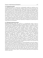
field for the study and application of nDEP (Huang & Pethig, 1991). An example of this is
presented in Fig. 2 where E. coli and M. Lysodeikticus are separated using a polynomial
electrode setup. Recently, a simple and novel curved electrode design has been used for the
separation of airborne microbes from beads or dust that are present in airborne
environmental samples, an important task prior to the real-time detection of airborne
microbes (Sungmoon et al., 2009).
Fig. 2. Separation of E. coli (experiencing nDEP) and M. lysodeikticus (experiencing pDEP) in
a polynomial electrode after application of a 4 V
PP,
100 kHz signal in a suspending medium
of 280 mM mannitol with a conductivity of 550 µS cm-
1
(Markx et al., 1994). Reused with
permission.
In order for quantitative and qualitative studies to take place on a single cell or a small
population of cells, the isolation and accurate positioning of the target must first be
accomplished. Negative dielectrophoresis in particular has emerged as a powerful tool for
this role. Under the influence of nDEP bioparticles are typically driven to regions away from
the electrodes. The E. Coli in fig. 2 are collected in a nDEP “trap” or “cage” at the center
because the electric field at that point is a localized minimum. This concept can be expanded
to arrays of microelectrodes, thus enabling the precise placement and retention of multiple
pathogenic samples (Frenea et al., 2003).
3.2 Viruses
Representing some of the smallest size pathogenic bioparticles, the manipulation of virus
particles is made difficult due to the presence of Brownian motion. To overcome the random
stochastic motion, the manipulation of submicron sized particles requires large deterministic
forces. Since DEP scales with a particle’s volume, an electric field gradient of sufficient
magnitude must be generated to provide a powerful enough force and necessitates the use
of electrodes separated by only a few microns (Mullery et al., 1996; Green & Morgan, 1997).
Reducing the dimensions of the electrodes in a biosensor will decrease the voltage required
to produce a given electrical field strength and, as a result, reduce both the power dissipated
in the system and the temperature increment (Castellanos et al., 2003). This is particularly
beneficial for portable systems that run on low power.
A number of reports currently exist on the subject of AC electrokinetic manipulation of
viruses (Park et al., 2007; Akin et al., 2004; Wu et al., 2005a; de la Rica et al., 2008; Müller et al.,
1996; Schnelle et al., 1996).
In many of these cases, successful virus collection results from
Biosensors – Emerging Materials and Applications
248
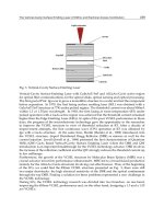
Fig. 3. Fluorescence image of nDEP collected vesicular stomatitis virus in TSE after
fluorescent staining. The microelectrodes have a central gap measuring 2 µm across.
a combination of DEP and electrohydrodynamic flows (Ramos et al., 1999). In 1998, Green &
Morgan reported the manipulation of a mammalian virus, herpes simplex virus type 1, both
by positive and negative DEP over a frequency range of 10 kHz-20 MHz using a polynomial
microelectrode array with a gap of 2
m. More recently, Docoslis et al. (2007) demonstrated
the collection of vesicular stomatitis virus in buffered solutions of physiologically relevant
conductivity using microelectrodes with a gap measuring 2 µm across (Fig. 3).
3.3 DNA
DNA offers a potential tool for the selective detection of pathogens by means of detecting
the presence or absence of genetic sequences found in specific pathogens. A DNA molecule
consists of two strands of deoxyribonucleotides held together by hydrogen bonding and
takes a random conformation in water. Under slightly basic conditions the DNA molecule
becomes negatively charged and a counter ion cloud surrounds the molecule. This counter
ion cloud can be displaced in the presence of an electric field, increasing the ionic
polarizability of the molecule (Hölzel & Bier, 2003). When an electrostatic field is applied,
DNA polarizes, and every part of the DNA orients along the field lines, stretching it into an
approximately straight shape. Due to the field non-uniformity, stretched DNA
dielectrophoretically moves towards the electrode edge until one end comes into contact. On
the basis of this behaviour many researchers have used AC electrokinetics to manipulate
DNA (Walti et al., 2007; Lapizco-Encinas & Palomares, 2007; Washizu et al., 1995 & 2004;
Dewarrat et al., 2002; Asbury et al., 2002; Washizu, 2005; Tuukkanen et al., 2006; Chou et al.,
2002; Kawabata & Washizu, 2001; Yamamoto et al., 2000; Wang et al., 2005). For example, a
modified interdigitated microelectrode array, termed “zipper electrode” by the authors, has
been reported to concentrate a wide range of nanoparticles of biological interest, such as the
influenza virus and DNA (Hübner et al., 2007). Fig. 4 shows the fluorescence microscopy
recorded for the trapping of stained λ-phage DNA in a floating electrode device. The figure
shown here is recorded 10 sec after the application of an electric field with a voltage of 200
V
pp
and a frequency of 30 Hz.
The manipulation of DNA by AC electrokinetic effects has been applied in the biological
field and reviewed recently by Washizu (2005). The versatility of DNA allows for it to be
used as a sensing, or analytical device and AC electrokinetic effects play an important role
in the manipulation of this biological tool. AC Electrokinetics has been used to perform
molecular surgery for the reproducible cutting of DNA at any desired position along the
Enhancing the Performance
of Surface-based Biosensors by AC Electrokinetic Effects - a Review
249
Fig. 4. Dielectrophoretic trapping of λ-phage DNA molecule when 30 Hz, 200 Vpp signal
was applied in a floating electrode device (Asbury et al., 2002). Reused with permission.
DNA molecule (Yamamoto et al., 2000). Gene mapping has also found AC electrokinetics
useful as a means for manipulating DNA to bring it into contact with enzymes in order to
search for binding locations, and thus mapping the gene (Kurosawa et al., 2000). Similarly
manipulating and stretching DNA is useful for determining the order of the nucleotide
bases for gene sequencing (Washizu et al., 2005), and for measuring molecular sizes by
counting base pairs (Washizu & Kurosawa, 1990). AC electrokinetically manipulated
DNA can still undergo molecular interactions and has been used to achieve the selective
binding of foreign single stranded DNA (Kawabata & Washizu, 2001). As a detection and
sensing tool, once the DNA is brought close enough to touch an electrode, if the electrode
edge consists of an electrochemically active metal, such as aluminum, then the DNA
becomes permanently anchored there (Washizu et al., 2004). Alternatively, the DNA can
be trapped dielectrophoretically and it has been demonstrated by a number of researchers
that trapped DNA can be used as a selective bioreceptor towards the development of
pathogen biosensors (Gagnon et al., 2008; Lagally et al., 2005; Cheng et al., 1998a; Cheng et
al., 1998b).
4. Detection of AC-electrokinetically trapped particles
Research over the last decade has shown that there is no shortage of analytical methods that
can be successfully interfaced with AC electrokinetically enhanced sampling in a surface-
based biosensor. The most promising candidates include methods that rely on optical
(absorbance measurement, Raman, confocal microscopy, fluorescent intensity, etc.), mass
based (quartz crystal microbalance, surface acoustic wave, etc.), electrical, or electrochemical
(potentiometric, amperometric, conductometric, coulometric, impedimetric) (Velusamy et
al., 2010) detection. Optical and electrochemical sensors tend to be the most popular for
pathogen analysis due to their selectivity and sensitivity. In general it is convenient to
incorporate conventional optical or electrochemical devices with microfluidic detection
systems. Successful implementation of these methods requires that the concentration
amplification effect achieved by AC electrokinetics be combined with a selective target
retention method. The latter can be accomplished with the immobilization of a target-
specific molecule, such as a strand of DNA, an antibody, a protein, or an enzyme, or a more
complex biological system such as a membrane, cell or tissue (Velusamy et al., 2010). This
type of molecular recognition ensures that the captured bioparticle will remain on the sensor
surface even after the electric field is turned off. The sensitivity of a surface based biosensor
Biosensors – Emerging Materials and Applications
250
is thus directly affected by the packing density of the sensing element bound to the surface.
Methods for surface functionalization have included the use of thiol interactions (Park &
Kim, 1998; Radke & Alocilja, 2005; Bhatia et al., 1989), avidin-biotin interactions (Costanzo et
al., 2005), self-assembled monolayer coated electrodes (Wana et al., 2009), polymer coated
electrodes (Livache et al., 1998) and size specific capillary flow trapping (Hamblin et al.,
2010). A number of proof-of-principle studies have demonstrated that a combination of AC
electrokinetics with a molecular recognition method can substantially improve the
sensitivity of a biosensor (Yang, 2009; Yang et al., 2006; Yang et al., 2008). In principle,
decorating the surface of the biosensor with antibodies allows for easy substitution when
targeting a multitude of pathogens. The ability to replace specific bioreceptors on demand
for the particular screening of a target pathogen gives this method high flexibility.
4.1 Optical detection
Optical based detections vary in their type and application. This section will focus on the
most commonly used, namely: absorbance measurement, surface enhanced Raman
scattering, and fluorescence.
4.1.1 Absorbance based measurements
An optical system was first described by Price et al., (1988) to detect dielectrophoretically
trapped bacterial cells by monitoring the changes in light absorbance through the
suspension as bacteria collected at an electrode array by pDEP. Later on, Pethig et al. (1992)
reported a dual beam optical spectrometer with improved sensitivity for the detection of
yeast cells collected by both nDEP and pDEP (Talary & Pethig, 1994). The mechanism of
pathogen detection by absorbance measurements based on dielectrophoretic immuno-
capture is illustrated in Fig. 5. The immuno-capture of the bacterial cells under DEP after
15 and 30 min of sampling was found to be 82% and 74% more efficient than that achieved
without DEP. The immuno-captured bacterial cells were detected by sandwich format
ELISA on the chips. The absorbance signals by DEP assisted immuno-capture were
reported to be enhanced by 64.7–105.2% for samples containing 10
3
–10
6
cells/20 L (Yang,
2009).
Fig. 5. Mechanism of nDEP immuno-capture: The area of collection (inter-electrode gap) is
functionalized with a target-specific reactive component, an antibody in this case. Application
of a spatially non-uniform electric field (dashed lines) causes nearby antigens to undergo
nDEP and collect midway between the electrodes. Once collected, the immobilized antigens
can be reacted with an optically active component.
Enhancing the Performance
of Surface-based Biosensors by AC Electrokinetic Effects - a Review
251
4.1.2 Fluorescence-based detection
Fluorescence is by far the most frequently used optical signalling method for the monitoring
and detection of AC-electrokinetically trapped bioparticles due to its high level of sensitivity
and low background noise (Hübner et al., 2007; Wong et al., 2004b; Cui et al., 2002; Yang et
al., 2008). Using fluorescent imaging, Docoslis et al. (2007) detected captured virus (vesicular
stomatitis virus) and later explored numerical simulations of the system to better
understand the processes involved (Wood et al., 2007). The virus was captured from
physiologically relevant ionic strength media (880 mS m
-1
) at low concentrations (<10
6
PFU
mL
-1
). The numerical simulations revealed that with a quadrupolar microelectrode the
capturing of the virus was achieved by both DEP for the short range capture and
electrothermal fluid flow to overcome diffusion limitations. Others were also able to achieve
virus capture at low ionic strengths (1-100 mS m
-1
) and higher particle concentrations (>10
6
particles mL
-1
) (Hughes et al., 1998; Hughes et al., 2001; Pethig et al., 1992; Grom et al., 2006;
Morgan & Green, 1997). The dielectrophoretic capture and detection of a food borne
pathogen, Listeria monocytogenes, was accomplished with the aid of the heat shock protein 60
(Hsp60) immobilized on a sensor’s surface (Koo et al., 2009). Hsp60 is a receptor for the
Listeria adhesion protein (LAP), a house keeping enzyme of Listeria monocytogenes during
the intestinal phase infection. Both fluorescent microscopy and ELISA were used to detect
the binding of target cells with the receptor. The enhancement of binding with the aid of
DEP was found to be 60% higher than without. As discussed in section 3.3, single stranded
DNA can be used as a receptor to detect the specific sequence of a pathogen’s genetics.
Lagally et al. (2005) described an integrated system where bacterial cells where
electrokinetically concentrated from a continuous-flow and detected via DNA-rRNA
hybridization. After pDEP trapping the bacterial cells, the cells were lysed by chaotropic salt
and the released DNA was denatured by endonuclease. The E. Coli cells were detected by
fluorescent detection via the sequence specific hybridization of an rRNA-directed optical
molecular beacon with the denatured DNA. This integrated microsystem is capable of the
sequence specific genetic detection of 25 cells within 30 min. After hybridization, the
percentage of the fluorescence was observed to increase with time and a linear relationship
was found between the number of trapped cells and the percentage of maximum
fluorescence. Others have reported the optical detection of cells (e.g., carcinoma cells,
malarially-parasitized cells) where DEP was used to separate infected cells from healthy
cells. Once lysed, the infected cells were identified with fluorescent probes on a bioelectronic
chip (Gascoyne et al., 2004; Cheng et al., 1998a, 1998b).
4.1.3 Raman spectroscopy
Raman spectroscopy allows for analyte identification through the inspection of its
“chemical fingerprint” on the basis of the vibrational, rotational and other low-frequency
modes. Typically, for Raman detection, the signal provided by a low concentration surface
based biosensor is not strong enough for detection. The use of surface enhanced Raman
scattering (SERS) is often needed and can be achieved through the use of metal
nanoparticles. The metal nanoparticles must be either chemically bonded to the bacteria
or settle in the proximity of the bacteria in order to increase the scattering (Hou et al.,
2007; Cheng et al., 2007). An on-chip detection of pathogens using surface enhanced
raman spectroscopy (SERS) has been reported recently by Hou et al. (2007), where the
Raman signals of the pathogens were enhanced by the presence of ~80–100 nm silver
Biosensors – Emerging Materials and Applications
252
nanoparticles. Combined with a discharge driven vortex for target concentration, SERS
was successfully used in the detection of bioparticles at a concentration of 10
4
CFU/mL in
the presence of silver nanoparticles (Hou et al., 2007). A continuous flow system for
bioparticle sorting was presented by Cheng et al. (2007) where, once sorted, the detection
of the pathogen was accomplished via SERS. This integrated chip used DEP for a
combination of filtering, focusing, sorting and trapping with a throughput of 500
particles/s (Cheng et al., 2007).
4.2 Mass based detection
Pathogenic particles with length scales on the order of nanometers can individually weight
as little as tens of picograms. In order for mass based detection to succeed, either very
sensitive detection methods or significant pathogen amplification is necessary. The
following sections will examine how AC electrokinetics has been used to improve the mass
based detection sensitivity and sampling for quartz crystal microbalances and cantilever
based detection methods.
4.2.1 Quartz crystal microbalance detection
A quartz crystal microbalance (QCM) utilizes a piezoelectric quartz crystal that has a
fundamental resonance frequency which changes in accordance to the amount of mass
attached to the crystal surface. Fatoyinbo et al. (2007) developed for the first time an
integrated system where yeast cells were concentrated on an electrode surface by DEP and
then quantified by a QCM system. The steady-state response predicted from the frequency
shift analysis of nanoparticle-loaded DEP-QCM has shown significant improvements in
rates of particle detection. The work was done at a concentration of 10
8
nano-spheres/mL
and detection was achieved five times faster than other QCM surface loading techniques
described in the literature.
4.2.2 Cantilever detection
Similar in concept to the QCM, a cantilever acts as a free-standing platform whose resonant
frequency decreases with the addition of mass. As more bioparticles become deposited on
the surface, the shift becomes more pronounced. The combination of AC electrokinetics with
a cantilever beam was recently achieved and allowed for the rapid collection of human
cancer cells (Park et al., 2008). Using two conductive cantilevers situated across from one
another over a well, Park et al. used pDEP to direct the human cancer cells onto the
cantilever surface. Fig. 6 demonstrates the setup of a series of cantilevers where the change
in resonant frequency is measured using a laser Doppler vibrometer. However, sensitivity
remained an issue as culturing of up to 7 days was required in order for the cell mass to be
detected. nDEP collection of E. Coli was achieved by Tomkins et al. (submitted, 2011)
through the use of polynomial electrodes on a cantilever surface. By using a poly-L-lysine
layer on the cantilever to act as a non-specific layer for the electrostatic retention of bacteria,
a shift in frequency was detected after 30 minutes of collection from a concentration of 10
8
particles /mL. In order to maximize the sensitivity of a cantilever beam, the most desirable
location for collection is at the cantilever tip, furthest away from the anchor. However, Islam
et al. (2007) successfully applied AC electroosmotic flow to drive polystyrene particles to a
point near the anchor and detected a mass change after drying.
Enhancing the Performance
of Surface-based Biosensors by AC Electrokinetic Effects - a Review
253
Fig. 6. (a) A schematic diagram of pairs of cantilevers. Each opposing cantilever acted as one
half of an electrode pair when inducing pDEP. The arrow indicates the direction of flow.
(b) A ‘living’ cantilever with human cervical cancer cells (Park et al., 2008). Reused with
permission.
4.3 Electrical or electrochemical detection
When biosensors employ an electrical or electrochemical sensing element, many of the
features needed for AC electrokinetics are already present. These methods are easier to
interface with miniaturized devices than optical methods because they employ electrical
signals and do not need an often bulky optical measuring system. Microelectrodes for
applied AC electrokinetics can be easily added into a microfluidic channel using standard
photolithographic techniques and their integration with an electrical diagnostic chip allows
for the sharing of features or power sources. Moreover, some electrical sensing methods do
not require a labelling step for sensing target pathogens which makes the on-chip enhanced
sampling provided by AC electrokinetics an attractive asset. Electrical sensing methods can
be separated into 4 subclasses depending on the type of signal being measured:
amperometric (changes in current), conductometric (changes in conductance or resistance),
impedimetric (changes in resistance to an AC current), and coulometric (changes in
capacitance). This section will focus on recent electrical or electrochemical sensing methods
that have used AC electrokinetics.
4.3.1 Amperometric detection
By measuring the change in current as pathogens pass between a pair of sensing electrodes,
it is possible to detect single cells in solution. AC elecktrokinetics can be used to position or
manipulate these single cells into the proper location to achieve sensing. Utilizing the
Coulter-counter principle Pandey & White (2004) used dielectrophoresis to detect a single
cell (Chinese hamster ovary, CHO) as it was driven to pass through a micro-aperture (10-
25μm in diameter, comparable to the size of the cells being tested) in a silicon nitride
membrane. Detection of a cell was achieved by recording the decrease in the ionic current
caused from the passage of a single cell as it passed through the micro-aperture. Live
bacteria were also detected amperometrically by first using pDEP to trap the bacteria and
then using AC induced fluid flow to move the cells until they formed a bridge across
micron-sized electrode gaps (Beck et al., 2005). The cells were first captured at the electrode
edges by applying an electric field (1.5 V
pp
, 1MHz). The cells were then transported along
Biosensors – Emerging Materials and Applications
254
the length of the electrode into the gap by exploiting an electric field induced flow at a lower
voltage (0.5 V). The two electrodes tapered to a point small enough that a single bacterium
would completely bridge the electrodes and detection could be achieved.
4.3.2 Conductometric detection
Direct measurement of the conductance between two electrodes with a nano-sized gap can
be a highly sensitive technique for detecting bioparticles. A series of reports have been
published by Suehiro et al. to detect dielectrophoretically trapped bacteria by measuring
changes in conductance (1999; 2003a; 2003b; 2003c; 2005; 2006). The bacteria were collected
within a small gap (5
m) between the microelectrode arrays by trapping the cells at the
electrode edge with pDEP. After collection, an improved detection method was described
by this group using electropermeabilization (Suehiro et al., 2003b; 2005). While cells can be
destroyed using AC electric fields within a specific frequency window (Menachery &
Pethig, 2005), electropermeabilization causes the cell membrane to become permeable in
order to increase the apparent conductivity of the trapped bacteria. Once applied, the
bacterial cell wall leaks intracellular ions into the surrounding medium and transiently
increases the conductance (Fig. 7). Using this method, the detection time of yeast cells and E.
Coli cells was observed to shorten by two orders of magnitude to 15 min and 3 hr,
respectively and the sensitivity was improved to 10
2
CFU/mL.
Fig. 7. A schematic diagram of electropermeabilization. The cells are first trapped, and then
ruptured via an increased electric field followed by the subsequent release of intercellular
ions to the surrounding area (Suehiro et al., 2003b). Reused with permission.
Selectivity for these detection methods was demonstrated by exploiting the different
dielectric properties of cell mixtures. Selective detection of viable cells from a mixture of
viable and non-viable cells was achieved using DEP collection at two different electric field
frequencies. At 100 kHz the viable and nonviable bacteria were trapped near an electrode
corner due to positive DEP and their conductances changed proportionally with time. At 1
Enhancing the Performance
of Surface-based Biosensors by AC Electrokinetic Effects - a Review
255
MHz only viable bacterial cells were trapped by positive DEP as the conductance change
over time was less remarkable (Suehiro et al., 2003c). The increase in conductance indicated
that certain areas of the electrode gap had been bridged by trapped bacteria.
To enhance the detection of dielectrophoretically collected particles, metal nanoparticles
have been used to transform nonconductive trapped particles into conductive interparticle-
connected entities through metal deposition. For example, silver particles attached to DEP
trapped bioparticles bridged the gap between two microelectrodes by silver nucleation
(Velev & Kaler, 1999). Latex particles coated with protein A were dielectrophoretically
trapped between micron-sized gold electrodes and stabilized by a non-ionic surfactant.
Adsorption of protein A onto the latex surface yielded a sensing interface for the specific
association of the human immunoglobulin (IgG) antigen. The association of the human
immunoglobulin on the surface was probed by the binding of secondary gold labelled anti-
human IgG antibodies, followed by the catalytic deposition of a silver layer on the gold
nanoparticles. The silver layer bridged the gap between the two microelectrodes, resulting
in a resistance of 50-70
, whereas the negative control gave a resistance of 10
3
. The lower
detection limit for this model sensor was calculated at 2
10
-13
- 210
-14
M.
4.3.3 Impedimetric detection
Impedimetric detection is one of the most promising techniques for developing label-free,
real time, and non-invasive methods for bioparticle detection. Milner et al. (1998) first
proposed a differential impedance method for the quantitative detection of DEP captured
bacteria and opened the door for biosensors where non-visible sub-micrometer bioparticles,
such as viruses and DNA fragments, could be quantitatively investigated. This is not to
suggest that impedimetric detection can only be used in isolation. In conjunction with
optical monitoring, impedance has been used for the characterization of prohibitively small
bioparticles (Guan et al., 2004).
Dielectrophoretic impedence measurement (DEPIM), a new method reported by Suehiro et
al., occurs when there is an impedance change as interdigitated microelectrodes are
connected due to the trapping and pearl-chain formation of cells by DEP (Suehiro et al.,
1999). A ‘pearl-chain’ occurs during capture when bioparticles become dielectrophoretically
attracted towards one another and form strings of particles resembling a chain of pearls.
This pearl chain can enhance sensing by being electrically connected in parallel within the
electrode gap, thus increasing the conductance and capacitance between the electrodes. The
conductance, G
T
and capacitances, C
T
between the electrodes are found to increase
proportionally with the increase in cell concentration. By fitting the measured G
T
and C
T
values, a linear calibration chart was derived that enables the absolute measurements of cell
concentration. This method accurately assayed E. Coli cells suspended in solution at a
concentration of 10
5
CFU/mL within 10 min. DEPIM has been used to concentrate bacterial
cells (L. monocytogenes) from dilute solutions (10
5
cfu/mL) in order to detect the metabolic
activity of the bacteria and provide enhanced sensitivity for the biosensor. (Sjöberg et al.,
2005). After trapping the cell on the sensor surface, impedance sensing arose from the
differences in the physical properties, i.e., differences in conductivity and permittivity,
between the particles and the suspending medium as well as the changes in the geometric
form of the collected particle on the electrode array. Other studies found that the changes in
the permittivity of the dielectric between the electrodes are proportional to the total volume
of the suspending medium replaced by the DEP collected particles (Allsopp et al., 1999).
Thus, a linear relationship between the capacitance change and cell concentration was found.