Biosensors Emerging Materials and Applications Part 9 pptx
Bạn đang xem bản rút gọn của tài liệu. Xem và tải ngay bản đầy đủ của tài liệu tại đây (2.6 MB, 40 trang )
16
Organic-inorganic Interfaces for a New
Generation of Hybrid Biosensors
Luca De Stefano
1
, Ilaria Rea
1
, Ivo Rendina
1
, Michele Giocondo
3
,
Said Houmadi
3
, Sara Longobardi
2
and Paola Giardina
2
1
CNR-IMM Institute for Microelectronics and Microsystems, National Research Council
2
Department of Organic Chemistry and Biochemistry, University of Naples “Federico II”
3
CNR-IPCF Institute for Chemical and Physical Processes, National Research Council
Italy
1. Introduction
Biosensors have by far moved from laboratories benches to the point of use, and, in some
cases, their represent technical standards and commercial successes in applications of social
interest, such as medical diagnostic or environmental monitoring. Based on biological
molecules, but also on their bio-inspired synthetic counterparts, biosensors employ different
transducers (optical, potentiometric, volt-amperometric, colorimetric, and so on) converting
the molecular interaction information into a measurable electric signal. As the result of a real
multi-disciplinary field of science and technology, biosensors can take advantage from each
improvement and progress coming from other disciplines: new features and better
performances have been reached in the last year due to simplified fabrication
methodologies, deep integration of optical or electrical transducers, and, last but not least,
microfluidic circuits. More recently, nanostructured components dramatically increased
biosensors reliability especially in public health and environmental monitoring.
Nevertheless, there is still a pressing demand of innovations which could lead to smaller,
faster, and cheaper biosensors systems with ability to provide not only accurate information
but also feedback actions to the real world. The fabrication of a new generation of hybrid
biodevices, where biological, or bio-inspired, molecules are fully integrated with a micro or
a nano technological platform, strongly depends on the bio-compatibilization treatments of
the devices surfaces. The design and the realization of bio/non-bio interfaces with specific
properties, such as chemical stability, wettability, and biomolecules immobilization ability,
are key features in the miniaturization and optimization processes of biosensors. In
particular, protein immobilization is a hot topic in biotechnology since commercial
solutions, as in the case of DNA microarrays, are not still available. Proteins are, due to their
composition, a class of very heterogeneous macromolecules with variable properties. For
these reasons, it is extremely difficult to find a common surface suitable for different
proteins with a broad range in molecular weight and physical–chemical properties such as
charge and hydrophobicity. A further aspect is the orientation of the bound proteins, that
could be of crucial relevance for quantitative analysis, interaction studies, and enzymatic
reactions. Many different surfaces, and chemical treatments of these surfaces, have been
Biosensors – Emerging Materials and Applications
312
investigated in the last years, but an universal solution for all the applications
aforementioned could not be identified.
Following this very actual theme, our main focus in this chapter is to discuss different
applications in biosensing of a special class of amphiphilic proteins: the hydrophobins.
These proteins self-assemble in a nanometric biofilm at the interfaces between water and air,
or on the surfaces covered by water solution. New functionalities can be added to the
biosensors surfaces without using any chemical or physical treatment, just covering them by
a self-assembled protein biofilm.
The main topics covered in the following paragraphs are: origin and properties of the
hydrophobins; deposition methods of the hydrophobins biofilm on different surfaces and
the characterizations techniques we use to determine the physical properties of these bio-
interfaces; the features exhibited by the hydrophobins covered surfaces, and finally, the
biosensors systems based on hydrophobins biofilms.
We outline in this chapter how the peculiarities of these proteins can be of interest in the
technological field, beyond their large utilization in biotechnology, nowadays at industrial
level. Moreover, the experience matured on this subject can be the paradigm of a new kind
of approach in design and realization of the next generation of biosensors.
2. Hydrophobins: surface active proteins
Proteins are actually polymers whose basic monomer units are amino acids, the so called
residues. In nature, the building blocks of the protein structure are 20 different amino acids
that, on the base of their physical-chemical properties, can be classified as hydrophobic or
hydrophilic. The sequence of hydrophobic and hydrophilic residues in the primary
structure will give rise to an hydropatic pattern on the protein. As consequence, in water,
they behave like amphiphilic molecules, giving rise to structures that maximize the number
of interactions between hydrophilic groups and water and, at the same time, minimize those
between hydrophobic groups and water.
Hydrophobins (HFBs) are a large family of small proteins (about 100 amino acids) that
appear to be ubiquitous in the Fungi kingdom. The name hydrophobin was originally
introduced because of the high content of hydrophobic amino acids (Wessels J.G.H, et al.,
1991). They fulfil a broad spectrum of functions in fungal growth and development. They
are ubiquitously present as a water-insoluble form on the surfaces of various fungal
structures, such as aerial hyphae, spores, and fruiting bodies, etc., and mediate attachment
to hydrophobic surfaces. HFBs are very efficient in lowering the surface tension of water
allowing the hyphae to escape from the aqueous medium and grow into the air. As the
fungal hyphae grow through the air-water interface into the air, the hydrophobins at the
interface are believed to coat the emerging hyphae as they penetrate through the interface,
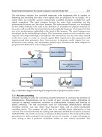
as shown in Figure 1. In vitro hydrophobins are able to self-assemble at
hydrophilic/hydrophobic interfaces into an amphipathic membrane, resulting in the change
of nature of surfaces.
Hydrophobins have been split in two groups, class I and class II, based on the differences in
their hydropathy patterns, spacing of aminoacids between the eight conserved cysteine
residues and properties of the aggregates they form (Linder et al. 2005). Class I
hydrophobins generate very insoluble assemblies, which can only be dissolved in strong
acids (i.e. 100% trifluoroacetic acid) and form rodlet structures outside the fungal cell wall.
Assemblies of Class II can be more easily dissolved in ethanol or sodium dodecyl sulphate
Organic-inorganic Interfaces for a New Generation of Hybrid Biosensors
313
and form assemblies that lack a distinct rodlet morphology. Despite these morphological
differences, no obvious distinction between the functions of class I and class II
hydrophobins within the fungal life cycle has yet emerged.
Fig. 1. Schematic of HFB role in fungal hyphae growth.
2.1 Hydrophobins structures
In order to provide a complete molecular description of hydrophobins, two complementary
points of view have to be considered: the structure of non-assembled hydrophobins and the
features of the assembled form. The structure of a protein is characterized in four ways: the
primary structure is the order of the different amino acids in a protein chain, whereas the
secondary structure consists of the geometry of chain segments; the main types of secondary
structure are two, called the α-helix and the β-sheets. The tertiary structure describes how
the full three dimensional arrangement of the chains and all its side groups, revealing how a
protein folds in on itself, and finally the quaternary structure of a protein describes how
different protein chains hook up with each other.
HFBs of both types I and II, although share quite a low sequence similarity, feature a clear
signature, namely eight Cys residues in a characteristic pattern. In this pattern, the third and
fourth as well as the sixth and seventh Cys residues are always adjacent in the sequence. In
the protein folded state, this special pattern gives rise to four disulfide bonds spanning over
the entire structure of the protein.
Class I HFBs consist of a four-stranded β-barrel core, an additional two-stranded β-sheet
and two sizeable disordered regions, as it can be seen in Figure 2. Notably, the charged
residues are localized at one side of the surface of the protein. This strongly suggests that the
water-soluble form is amphipathic (Zampieri et al., 2010). This structure is consistent with its
ability to form an amphipathic polymer.
Class II HFBs consist of a core with a β-barrel structure (Fig. 2), nevertheless do not contain
the two disordered loops. Furthermore, the additional two-stranded β-sheet in class I
hydrophobins is replaced with an α-helix, in the same region (Kwan, A.H.Y et al., 2006;
Hakanpää, J, et al., 2006). One side of the monomer surface contains only aliphatic side
chains. This creates a hydrophobic patch which constitutes 12% of the total surface area
(situated on the top of the structure showed in figure 2). The protein surface is otherwise
mainly hydrophilic, and thus the surface is segregated into a hydrophobic and a hydrophilic
part. This amphiphilic structure governs the properties of class II hydrophobins, such as
surface activity and surface adsorption.
2.2 The assembly process
The characteristic property of HFBs is adsorption at hydrophobic-hydrophilic interfaces, at
which they form amphiphilic films (Wessels J.G.H, et al., 2007; Wösten H.A.B, et al., 2007).
The interface can occur between solid and liquid, liquid and liquid or liquid and vapour. In
early studies, hydrophobins were found to self-assemble into aggregates and form various
Biosensors – Emerging Materials and Applications
314
Class I Class II
Fig. 2. HFBs structures.
types of self-assembled structures. Rodlets were first observed on the outer surface of spores
from Penicillium (Sassen et al., 1967; Hess et al.,1968) and Aspergillus (Hess et al., 1969;
Ghiorse and Edwards, 1973).
Class I HFBs at low concentration are in monomeric form, while at higher concentrations they
are mainly in a dimeric form (Wang, X et al., 2002; Wang, X et al., 2004). Self-assembly proceeds
through the formation of an intermediate form, the α- helical state (De Vocht, M.L et al., 2005,
Wang, X et al., 2005). Upon transfer to the β-sheet state, the content of β-sheet structures
increases. This is accompanied by the formation of a mechanically stable protein film.
However, during this transition the proteins forms nanometric wide fibrils, which are known
as rodlets. SE measurements have shown that the film is about 3 nm thick (Wang, X et al.,
2005). This and the fact that the diameter of the β-barrel of the protein is approximately 2.5 nm
suggest that the rodlets could be formed by a molecular monolayer (Kwan, A.H.Y et al., 2006).
The charged patch on the protein surface would face the hydrophilic side of the interface,
while the hydrophobic diametrically opposite site would face the hydrophobic side of the
interface. This arrangement is consistent with the way other surface active molecules orient
themselves at hydrophilic-hydrophobic interfaces (Kwan, A.H.Y et al., 2006).
Like class I, class II HFBs exist as monomers at low concentration (Szilvay, G.R et al., 2006).
When the concentration is increased, they form dimers and, at higher concentrations,
tetramers (Torkkeli, M et al., 2002). The monomers have a higher affinity for surfaces than
for formation of oligomers (Linder et al. 2005; Szilvay, G.R et al., 2006). These oligomers
would dissociate at a hydrophilic-hydrophobic interface, which would result in the
formation of a film which consists of a monolayer of the class II HFB: a scheme of these
differences is reported in Figure 3. In contrast to class I, self-assembly of class II HFB at the
water-air interface is not accompanied by a change in secondary structure (Askolin, S. et al.,
2006), furthermore this layer is not rodlet-like as in the case of class I HFBs.
Moreover, as described above, the end state of class I HFBs is very stable and cannot be
dissociated by pressure, detergent or 60% ethanol. In contrast, the end form of class II HFBs
readily dissolves under these conditions.
Organic-inorganic Interfaces for a New Generation of Hybrid Biosensors
315
Fig. 3. Main differences between class I and class II HFBs.
3. Hydrophobins self-assembling on solid surface: methods and
characterizations
A simple technique used to induce the self-assembling of the HFB biofilm on a solid substrate
is the drop-deposition method, where the drop is a micro-litre volume of a liquid solution
containing the proteins in their monomeric state. Even if this kind of film casting is not a
perfectly controlled process, i.e. the protein concentration increases in an uncontrollable way
during solvent evaporation, it is possible, by using proper starting conditions of some
parameters, such as temperature, surface cleaning, and so on, to obtain reproducible results in
term of film thickness and surface wettability. By using this technique, different kind of
surfaces have been conditioned: in the next paragraph we report the main experiences in
worldwide laboratories on this subject. Here, we present the standard processes to obtain self-
assembled HFB biofilms and the main characterization methods we use. Biofilm drop casting
is normally used in our laboratory to give new functionalities to silicon surface: silicon, and
silicon related materials, is the most used solid support in the microelectronic industry. For
this reason, silicon is widely used in all the application of electro-optic and photonic devices.
At this aim, highly doped p
+
silicon wafer, <100> oriented, 0.003 Ω cm resistivity, 400 µm tick,
was cut into 2 x 2 cm
2
pieces. The silicon substrates were cleaned using the standard RCA
process and dried in a stream of nitrogen gas. RCA is based on a combination of two cleaning
steps, one using solutions of ammonium hydroxide/hydrogen peroxide/deionized water, and
the other using hydrochloric acid/hydrogen peroxide/deionized water, both at the
temperature of 80 °C. The samples were prepared by coating the silicon chips with 200 µl of
HFB solution (0.2 mg/ml of protein dissolved in an ethanol-deionized water (60/40 v/v)
mixture) for 1 h, drying for 10 min on a hot plate at 80°C, and then washing with the ethanol-
water mixture. The incubation process was repeated two times. Then, the samples were
treated for 10 min at 100°C in 2% Sodium Dodecyl Sulfate (SDS), so as to remove the protein
not assembled into the biofilm, and again washed in deionized water.
The Langmuir technique is the most accurate way to get mono-molecular films of amphiphilic
molecules. A known quantity of the material is spread on the free surface of a suitable liquid
(the subphase) contained in a trough. The quantity of the material has to be predetermined on
Biosensors – Emerging Materials and Applications
316
the base of the molecular and trough areas, in order to know the surface molecular
concentration. In doing this, one should take care to use a sufficiently low surface
concentration in order to have an interfacial film of non-interacting molecules (the so-called
gas phase). Moreover, as one desires to have all molecules at the interface, it is very important
to adjust the subphase pH in order to match the isoelectric point of the used protein.
Once the interfacial film is formed, the presence of movable barriers on the Langmuir trough
allows to compress the film in a controlled manner, varying in this way the surface molecular
density and consequently the surface tension. The latter can be measured with several
techniques, but the most used is the Wilhelmy plate tensiometer, consisting of a thin plate
made from glass, platinum or even paper, attached to a scale or balance via a thin metal wire.
Fig. 4. Scheme and reference angle for Wilhelmy equation.
The force on the plate due to wetting is measured via a microbalance and used to calculate
the surface tension (σ) using the Wilhelmy equation (see Figure 4):
ϑ
σ
cos
F
=
where
λ is the wetted perimeter of the Wilhelmy plate and θ is the contact angle between the
liquid phase and the plate. In practice the contact angle is rarely measured, instead either
literature values are used, or complete wetting (θ = 0) is assumed.
The most important feature of the interfacial film is the pressure Vs. area isotherm, obtained
by recording the surface pressure as a function of the trough area. If the number of
molecules present at the interface is known, as in the case of water insoluble amphiphiles,
the surface pressure can be plotted as a function of water surface available to each molecule.
This curve can reveal several details about the interfacial film properties, in particular phase
transitions and collapses of the molecular film. In such cases the steric factor plays a relevant
role in the film stability and the entire sequence of phases transitions can be sperimentally
observed (see Fig. 5).
a) b) c)
Fig. 5. Cartoon sequence of the possible molecular arrangement for an amphiphile
monolayer as function of the molecular density: a) gas phase; b) liquid-expanded phase; c)
condensed phase.
Organic-inorganic Interfaces for a New Generation of Hybrid Biosensors
317
When proteins are used for making a Langmuir film things can be quite different, as a
considerable number of other factors has to be took into account: the state of the protein
(folded or unfolded) and her shape (in general almost globular), the presence of multiple
hydrophobic patches. In figure 6 a typical isotherm for Vmh-2 from Pleorotus Ostreatus is
shown. It is evident that many features are missing with respect to the case of a fatty acid. In
particular only one critical point is present. If one keeps in mind the quasi-globular shape of
this protein can easily realize that the gas - expanded liquid – solid transitions are in some
way continuous, without any abrupt molecular rearrangement as in the case of an elongated
molecule.
0.02 0.04 0.06 0.08 0.10 0.12
0.00
0.01
0.02
0.03
0.04
0.05
Surface pressure (N/m)
Trough area (m
2
)
Fig. 6. Typical isotherm measured for the hydrophobin Vmh-2 during film formation.
If the used amphiphilic protein is even only partially soluble in water, although the isotherm
features still hold, one has to face with the problem to determine the number of molecules
present at the interface and hence the surface molecular density. This occurrence can be
made evident by isobaric measurements, in which the ratio between the trough surface S
and the initial trough surface S
0
is plotted as a function of time at a given constant surface
pressure value. The plot shown in Fig. 7 refers to Vmh2 HFB from the fungus Pleurotus
Ostreatus. The decreasing of the trough area in time is due to a surface molecular depletion
that could be ascribed both to a bare solubilisation of the film or to some more complex
process involving the creation of soluble assemblies. From the same plot one can also argue
that an increasing in the surface pressure has the effect of stabilizing the film, reducing the
molecular depletion ratio, as the decreasing in the curve slope with the increasing of the
surface pressure demonstrate.
One possible method for at least estimate the surface molecular concentration is the fitting of
the experimental isotherm with a suitable 2-D equation of state, leaving the surface density
as fitting parameter. In the simplest case holds a Vollmer-like equation of the kind.
coh
A
mkT
Π−
−
=Π
ω
(1)
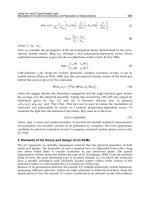
where Π is the surface pressure, k is the Boltzmann constant, T is the temperature, ω is the
limiting area of a molecule in the gaseous state, A is the area per molecule, Π
coh
is the
Biosensors – Emerging Materials and Applications
318
0 300 600 900 1200 1500
0,2
0,4
0,6
0,8
1,0
S/S
0
time (s)
0.003
0.007
0.014
Surface pressure (N/m)
Fig. 7. Isobaric measurement at different surface pressures for the Vmh-2 hydrophobin.
cohesion pressure accounting for the intermolecular interactions, and m is a parameter
accounting for the number of kinetically independent units (fragments or ions). The
parameter A is actually the inverse of the surface molecular density and can hence be
expressed as
n
S
A =
,
(2)
where S is the trough area and n the number of molecules present at the interface.
Once the Langmuir film has been characterized, it can be transferred onto a solid substrate
for applications or subsequent analyses. The most used methods are the vertical lift/dipping
of the substrate through the interfacial film (Langmuir-Blodgett technique) and the
horizontal plate lift from the interface (Langmuir-Shaeffer technique). When hydrophilic
subphases are used, the lift method allows to transfer the monolayer with his hydrophilic
side facing the substrate (also hydrophilic), leaving the hydrophobic side exposed to the air.
a) b) c)
Fig. 8. Schematic of Langmuir-Blodgett technique: with the barriers opened, in the gas
phase, the substrate is dipped in the subphase (a); then the film is compressed at the desired
surface pressure (b); finally the substrate is lifted through the interfacial film, dragging a
portion of it. In the meanwhile the closed-loop control closes the barriers in order to keep
constant the surface pressure (c).
Organic-inorganic Interfaces for a New Generation of Hybrid Biosensors
319
Alternatively, the film can be transferred with his hydrophobic side facing an hydrophobic
substrate by dipping the latter through the interfacial film. The monolayer side exposed to the
air in this case will be hydrophilic. In the Langmuir-Blodgett deposition technique, the trough
control system allows to perform the film transfer at constant surface pressure, closing the
barriers in order to compensate the surface molecular depletion due to the film transfer itself.
Fig. 9. Cartoon of ordered protein monolayer for Langmuir- Schaefer
The Langmuir-Shaeffer technique allows to remove a whole patch of the interfacial film at
once. Under the same initial conditions as above, this method allows the film sticking from
the hydrophobic side onto an hydrophobic substrate, leaving thus the hydrophilic side of
the monolayer exposed to the air. In this case, the closed-loop active surface pressure control
isn’t strictly required, providing that the monolayer is stable at the interface.
Fig. 10. Schematic of Langmuir-Shaeffer technique.
Sometimes it can be useful to lift the molecular film as self-standing, in order to eliminate
the interactions between molecules and an underlying substrate. This task is often
accomplished using metallic grids, of the kind used in TEM specimen preparation, featuring
a suitably fine mesh.
The HFBs film self-assembled on the silicon surface or by LB methods can be characterized
by means of several methods; the most common characterization techniques are the
spectroscopic ellipsometry, the atomic force microscopy, and the water contact angle.
3.1 Spectroscopic ellipsometry
Spectroscopic ellipsometry (SE) allows to determine the optical properties (i. e., the
refractive index n and extinction coefficient k) and the thickness of the HFB biofilm
assembled on a solid surface. The method is based on the measurement of the change in the
polarization state of the light over the spectral range after the reflection from the sample
surface. Ellipsometry measures the complex reflectance ratio (ρ) defined by:
tan
p
i
s
R
e
R
ρψ
Δ
== (3)
Biosensors – Emerging Materials and Applications
320
where R
p
and R
s
are the complex reflection coefficients of the light polarized parallel and
perpendicular to the plane of incidence. Thus, ψ and Δ are, respectively, the amplitude ratio
and the phase shift between s and p components of polarized light.
We have used a Jobin Yvon UVISEL-NIR phase modulated spectroscopic ellipsometer, at an
angle of incidence of 65° over the range 320-1600 nm with a resolution of 5 nm. The
properties of the biofilm have been extracted from the SE measurements using the analysis
software Delta Psi (Horiba Jobin Yvon).
The optical properties, n and k, as functions of the wavelength have been determined by
fitting the experimental results using the Tauc-Lorentz model, firstly proposed in 1996 by
Jellison and Modine as a new parameterization of the optical functions of amorphous
materials. The imaginary part of the dielectric function is based on the Lorentz oscillator
model and the Tauc joint density of states:
2
0
22222
2
0
()
1
()
0
g
AE C E E
E
EE CE
ε
−
=
−+
g
g
EE
EE
>
≤
(4)
The real part of the dielectric function is given by Kramers-Kronig integration:
2
1
22
2()
g
E
Pd
E
ξε ξ
εε
ξ
π
ξ
∞
∞
=+
−
(5)
These equations include five fitting parameters: the peak transition energy E
0
, the
broadening term C, the optical energy gap E
g
, the transition matrix element related A, and
the integration constant ε
∞
.
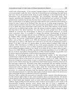
In Figure 11, n and k, as functions of the wavelength, are reported for the Vmh2 biofilm self-
assembled on silicon together with the values of the fitting parameters and the χ
2
.
400 600 800 1000 1200 1400 1600
1.385
1.390
1.395
1.400
0.00
0.01
0.02
0.03
0.04
0.05
n, Refractive Index
Wavelength (nm)
k, Extinction coefficient
E
g
=1.20 ± 0.07 eV
A=1.785
± 0.09 eV
E
0
=5.5 ± 0.2 eV
C=5.0
± 0.2 eV
ε∞=1.766 ± 0.02
χ
2
=0.05
Fig. 11. Optical properties, n and k, of the Vmh2 biofilm self-assembled on silicon surface as
functions of the wavelength.
Starting from these results, we can use the ellipsometric technique to estimate the thickness
of the biofilm depending on the concentration or on the post deposition washing procedure,
for example. We notice that before SDS washing the thickness of Vmh2 biofilm could be of
Organic-inorganic Interfaces for a New Generation of Hybrid Biosensors
321
tenths of microns, also depending on protein concentration in the starting solution. We have
verified that a step-by-step deposition allows the assembling of biofilms of increasing
thicknesses: after three consecutive depositions, for a total time of three hours, we have
obtained biofilms assembled on crystalline silicon up to 40 nm thick, that is, thicker than
those reported in literature. After hot SDS washing the biofilm is very much thinner: a value
of 3.91 ± 0.06 nm has been calculated modelling the HFB sample by a simple homogeneous
layer. We believe that this is the thickness of a monolayer of HFBs when self-assembled on
hydrophobic silicon: this value is consistent with a typical molecular size and comparable to
atomic force microscopy measurements. According to the above described model, the
washing step of the chip is strong enough to remove the proteins aggregates deposited on
the HFB monolayer that directly interacts with the hydrophobic silicon surface. This
behaviour points out the stronger interactions between the silicon surface and the HFB
monolayer with respect to those between the HFB aggregates and the HFB monolayer. The
experimental spectra Ψ and Δ, together with the calculated ones, are shown in Figure 12.
The persistence of Vmh2 biofilm on the silicon surface depends strongly on its chemical
nature: we have thus verified that the same deposition procedure on silicon dioxide, which
is a hydrophilic surface, does not give the same results in terms of biofilm chemical stability.
After washing the biofilm in hot SDS solution only sparse islands of protein biofilm can be
found on the silicon dioxide chip. This different behaviour can be ascribed to the greater
number of hydrophobic residues constituting the protein with the respect to those
hydrophilic.
400 600 800 1000 1200 1400 1600
15
20
25
30
150
160
170
180
Ψ
Wavelength (nm)
Ψ
fit Ψ
Δ
Silicon
HFB
Δ
fit Δ
χ
2
=0.07
Fig. 12. Measured and calculated spectra (Ψ, Δ) of the HFB biofilm self-assembled on a
silicon surface.
3.2 Atomic force microscopy
In the early 1980s the first Scanning Probe Microscope (SPM) has been introduced. It was
actually a Scanning Tunneling Microscope, exploiting the tunnelling current between the
surface sample and a very sharp conductive tip sweeping back and forth a few tens
nanometers over the sample surface. The electron tunnelling is a quantistic effect connected to
the probability for an electron to cross a gap between two conductive solids. Its development
was driven by the need of the semiconductors industry to characterize the semiconductor
inhomogeneities at the nanoscale, this issue becoming of increasing importance with the
Biosensors – Emerging Materials and Applications
322
progresses in the miniaturization of the electronic devices. A few years later, a similar
instrument was released, the Atomic Force Microscope (AFM) exploiting the van der Waals-
like interaction forces between the sample surface and the tip, instead of the tunnelling current
and allowing the imaging of non-conductive samples as well, down to the atomic scale.
In their essential parts, an AFM is made of a closed loop controlled scanning head and an
acquisition/digital signal processing board slotted in a PC running a devoted software.
Samples are under the form of small chips or thin films with x-y size ranging from the sub-
millimiter to a centimeter. The z size is usually in the order of a nm. The sample is placed at
the free end of a piezoelectric scanner; depending on the piezo characteristics, the scanned
area can range from a few tens nm
2
to ~10
4
sq. microns with a resolution down to a few
tenth nm. The AFM probes can feature pyramidal or conical shape, with a height in the
range of a few microns and a tip radius down to a couple nanometers; this makes a really
sharp tip, carring only a few atoms at the end! The tip is placed at the end of a flexible
cantilever; both the tip and the cantilever are usually made of silicon, although other
materials are now available, as diamond or carbon nanotubes.
When the tip is brought close to the sample surface, the van der Waals-like forces between
the sample surface and the tip cause the cantilever bending; if one knows the cantilever
spring constant, the bending measurements allows to deduce the interaction force. The
detection technique exploits the reflection of a laser beam from the upper side of the
cantilever; the bending is measured by the laser spot displacement over a two or four
quadrants photodetector.
An AFM can operate essentially in two different modes according to the sample surface-tip
distance range. When this distance is small, in the order of few Angstrom, one operates in
the repulsive region of the interaction potential and the operating mode is named contact
mode. On the contrary, when this distance is “large”, in the order of 1 – 10 nm , the attractive
region of the interaction potential is involved and the corresponding mode is named no-
contact mode. The difference between the contact and the no-contact mode is remarkable,
because of the difference in the range of the forces involved in the interaction between the
AFM tip and the sample surface; in the contact mode case, the repulsive forces are in the
order of tens of nN, al least one order of magnitude stronger than the case of the attractive
forces involved in the no-contact mode. Because of the extreme force weakness, the
detection technique used in no-contact AFM is very sensitive: driven by a piezo element, the
cantilever oscillates with a typical amplitude of few nm at a frequency slightly above the
resonance, while “flies over” the surface of the sample. The presence of Van der Waals and
other long-range attractive forces shifts the cantilever resonant frequency which, in turn,
causes the oscillation amplitude to decrease.
The NC-mode is useful in imaging soft samples, as is the case of biologic molecules, given
the weakness of the involved forces that prevents or minimizes the sample damage.
An evolution of the AFM no-contact operating mode is the tapping or intermittent mode.
In tapping mode the cantilever oscillates at its resonant frequency or slightly below. The
amplitude of oscillation typically ranges from 20 nm to 100 nm. Differently from the case of
no-contact mode, in which the tip never comes in contact with the sample surface, in the
tapping mode the tip lightly “taps” on the sample surface during scanning, contacting the
surface at the bottom of its oscillation.
Beside the bare imaging of soft samples, the AFM dynamical modes (NC and tapping) can
supply information about other features of the sample at the nano scale, as its wetting and
visco-elastic properties. In order to achieve such information, a fundamental measurement is
Organic-inorganic Interfaces for a New Generation of Hybrid Biosensors
323
the phase lag between the signal driving the cantilever piezo element and the cantilever
response. Then the experimental curves are fitted with a model describing the cantilever
dynamics. The recording of the phase lag at constant amplitude for each point of the
acquisition x-y grid will produce a phase image that closely relates to a dissipation map.
In our experience, a NanoScope V Multimode AFM (Digital Instruments/Veeco) was used
for the imaging of the HFB biofilm. The sample was imaged in air in tapping mode. The
scan frequency was tipically 1 Hz per line and images were flattened to a second order
polynomial.
The AFM images of the HFB silicon coated sample are reported in Figure 13; the formation
of a homogeneous biofilm can be observed in the phase image (right picture in Figure 13).
The AFM characterization also reveals the presence of rodlets-like structures on top of the
biofilm (Rodlet average height 4.11 ± 0.08 nm; Rodlet average width 23.9 ± 0.6 nm; Rodlet
average length 64 ± 3 nm; Mean roughness 3.32 nm).
Fig. 13. Height and phase atomic force microscopy images of silicon surface coated with
HFB biofilm.
3.3 Water contact angle measurements
The most common method for the determination of the surface wettability is the water
contact angle (WCA) measurement. The technique simple, quick, and cheap is based on the
analysis of the contact angle formed between a surface and a water droplet placed on it.
At the aim to determine the wettability of the HFB biofilm, we have used the sessile drop
method for WCA measurements on a OCA 30 – DataPhysics coupled with a drop shape
analysis software. Five measurements were analyzed for each sample.
The silicon surface, after the removal of the native oxide layer in hydrofluoridric acid, is
characterized by a WCA of (90.0±0.3)° (Figure 14 (A)). The presence of the HFBP biofilm
lowers the WCA down to (44±1)° (Figure 14 (B)): this interface is more hydrophilic due to
the assembly of the protein into a film with apolar groups disposed towards the
hydrophobic silicon and the polar groups on the other side.
4. Functional surfaces based on hydrophobins biofilms
The capability of hydrophobins to adhere to various surfaces was one of the first
observations among hydrophobins functions. An early finding on the behaviour of the class
I hydrophobin SC3 from Schizophyllum commune was that when it binds to for example
Biosensors – Emerging Materials and Applications
324
ABAB
Fig. 14. Water contact angle measurements of bare silicon (A) and silicon coated with HFB
(B).
Teflon, it can form a very insoluble layer (de Vocht et al. 2002). All hydrophobins adhere to
surfaces, but there is a difference in the binding characteristics (Askolin et al. 2006). While
class I members adhere very strongly, this is not seen for class II members which dissociate
more easily.
Glass has been one of the widely used substrates for protein chips. Qin et al (2007a) have
compared two methods for protein immobilization on glass slides, one is the traditional
method of silanization (Mezzasoma et al. 2002); the other is the coating with the class II
hydrophobin HFBI from Trichoderma reesei. The modified glass surfaces were characterized
with X-ray photoelectron spectroscopy (XPS), water contact angle measurement (WCA) and
immunoassay. The results have shown that HFBI coating can achieve the same result of
protein immobilization on glass slides as silanization did, or even better. Moreover the HFBI
coating does not require the complex procedure and strict surface cleanness as the
silanization needs. HFBI-coated surfaces can be used immediately or preserved for some
days prior to use. Therefore hydrophobin self-assemblies seems to be a simple and generic
way for protein immobilization on glass slides.
Mica and polydimethylsiloxane (PDMS) are other substrates used for patterning of
biomolecules. Freshly cleaved mica is a promising substrate for patterning applications,
because it is the most readily available surface with atomic-scale flatness. Biomolecules, such
as proteins, could not be immobilized on mica surfaces effectively without surface
modification. PDMS is a kind of soft polymer with attractive physical and chemical
properties: elasticity, optical transparency, flexible surface chemistry, low permeability to
water, low toxicity, and low electrical conductivity. Thus, it has been widely used in
microfluidic devices and microcontact printing technology. A simple method to modify both
mica and PDMS surfaces by HFBI for protein immobilization has been recently developed
(Qin et al. 2007b). XPS and WCA measurements illustrated that the wettability surface can
be changed from superhydrophobic (PDMS) or superhydrophilic (mica) to moderately
hydrophilic. The same surfaces, mica, glass, and PDMS, have been modified using the class I
hydrophobin HGFI from Grifola frondosa (Hou et al. 2009). The surface wettability was
efficiently changed by HGFI modification, as XPS and WCA measurements indicated.
Furthermore data showed that self-assembled HGFI has better stability than type II
hydrophobin HFBI: HGFI self-assembly was stable against rinsing by several solutions, i.e.
2% hot SDS solution and 60 vol.% ethanol.
Polystyrene and its variations are extensively used as solid supports to produce, for
example, polystyrene-microtitre plates and tubes in immunoassays. Wang et al. (2010a) have
Organic-inorganic Interfaces for a New Generation of Hybrid Biosensors
325
used the Class I hydrophobin HGFI to increase the hydrophilicity of polystyrene for
facilitating immobilization of biomolecules. The adsorption process of HGFI on the
polystyrene surfaces was studied by quartz crystal microbalance at different pH values, by
XPS, WCA measurements and AFM analyses. By self-assembling, hydrophobin easily
formed an intact charged film on the hydrophobic polystyrene that enhanced the
hydrophilicity of the polystyrene for a long time (Wang et al. 2010a).
Silicon is the most used solid support in all micro- and nanotechnologies developed for the
integrated circuits industry. For this reason, silicon is also used in many commercial
technological platforms for biomedical and biosensing applications. The anisotropic wet
micromachining of silicon, based on a water solution of potassium hydroxide (KOH), is a
standard fabrication process that is extensively exploited in the realization of very complex
microsystems such as cantilevers or membranes. A nanostructured self-assembled biofilm of
Vmh2, was deposited on crystalline silicon and since this procedure formed chemically and
mechanically stable layers of self-assembled proteins, the biomolecular membrane has been
tested as masking material in the KOH wet etch of the crystalline silicon. The process has
been monitored by SE and atomic force microscopy measurements. Because of the high
persistence of the protein biofilm, the hydrophobin-coated silicon surface is perfectly
protected during the standard KOH micromachining process (De Stefano et al. 2007). In
Figure 15 the optical photos of the two silicon samples after this treatment, are shown: on
the A image is clearly visible the etched surface whereas in the B image the HFB covered
surface is perfectly homogeneous.
Fig. 15. Optical images of the silicon surface after the KOH etching: (A) bare silicon; (B)
silicon coated with HFB.
This qualitative result is quantitative confirmed by ellipsometric and profilometric
measurements: an 8.40 (0.05) nm biofilm of hydrophobins is still optically detected by the
ellipsometer and the profilometer cannot detect any dig into the hydrophobins shielded
sample, as it can be seen in Figure 16.
Porous silicon (PSi), a silicon related material obtained by silicon dissolution in an
electrochemical cell in hydrofluoridic water solution, is really a versatile material owing to
its peculiar morphological, physical, and chemical properties, allowing the easy fabrication
of sophisticated optical multilayers, such as one-dimensional photonic crystals, by a simple
electrochemical etching process. The reflectivity spectrum of the photonic crystals shows
characteristic shapes, which are very useful in many applications, from biochemical sensing
to medical imaging. The major drawback of the ‘‘as etched’’ PSi is its chemical instability: it
has been shown that a PSi wafer can be dissolved under the physiological conditions that
are very often used in biological experiments. De Stefano et al. (2008) have covered the Psi
Biosensors – Emerging Materials and Applications
326
Fig. 16. Profilometric measurements of bare silicon (solid line) and silicon covered with HFB
(dash line).
surface with the highly stable and resistant biofilm resulting from the self-assembly of
Vmh2. The hydrophobin penetrates the whole stack, accumulating at the bottom and
modifies strongly the wettability of the PSi surface. Moreover, the protein membrane not
only protects the nanocrystalline material from basic dissolution in NaOH, but also leaves
unaltered the sensing ability of such an optical transducer, adding chemical stability, which
can be key in biomolecular experiments.
Gold is an excellent electric conductor with fine ductility and chemical inertness, thus an ideal
choice for developing bioelectronic devices. HFBI modification on smooth gold surfaces has
been proven to effectively enhance the surface hydrophilicity. The unmodified bare gold
surface exhibited a weak hydrophilicity, while the surface hydrophilicity was remarkably
improved after HFBI processing, as demonstrated by WCA measurements. The increase of
surface wettability was not affected by the presence of washing procedures (Zhao et al. 2009).
Carbon nanotubes (CNTs, hollow cylinders made of sheets of carbon atoms) have recently
emerged as building blocks of novel nanoscale structures and devices. CNTs have a variety
of electronic properties that can be exploited for a variety of applications. Nanotubes have
been functionalized to be biocompatible and to be capable of recognizing proteins (Shim et
al. 2002). Often this functionalization has involved noncovalent binding between a
bifunctional molecule and a nanotube in order to anchor a bioreceptor molecule with a high
degree of control and specificity. Furthermore, CNTs are commonly insoluble in all solvents
and usually form tangled network structures containing various impurities (Wu et al., 2010).
To overcome these limits, CNT surfaces are often tailored using either covalent (Wu et al.,
2007) or noncovalent modification (Hecht et al., 2006) strategies. A novel noncovalent
approach has been developed for the functionalization of multi-wall carbon nanotubes
(MWNTs, many layers to form concentric cylinders) using the class II hydrophobin, HFBI.
The HFBI–MWNTs nanocomposite was characterized by scanning electron microscopy
(SEM), transmission electron microscopy (TEM) and WCA. The hydrophobin HFBI
demonstrates to be an efficient solubilizing agent for MWNTs. The resulting HFBI–MWNTs
nanocomposite film with both merits of HFBI and MWNTs exhibited high hydrophilicity,
fast electron-transfer kinetics and excellent electrocatalytic activity (Wang et al. 2010b).
Organic-inorganic Interfaces for a New Generation of Hybrid Biosensors
327
The deposition of ceramic thin films from aqueous solutions at low temperature using
biopolymers as templates has attracted much attention due to economic and environmental
benefits. Titanium dioxide is one of the most attractive functional materials and shows a
wide range of applications across vastly different areas because of its unique chemical,
optical, and electrical properties. Santhia et al. (2010) deposited smooth, nanocrystalline
titania thin films by an aqueous deposition method on a hydrophobin film. Firstly, the
hydrophobins were self-assembled on a silicon substrate and characterized by XPS, AFM
and surface potential measurements. Hydrophobin-modified silicon substrates were then
used to deposit from aqueous solution at near-ambient conditions highly uniform, crack-
free nanocrystalline TiO
2
thin films. Nanoindentation tests showed the high resistance
against mechanical stress of the deposited titania films. The determined averaged hardness
value and the Young’s modulus highlight the compatibility of these films for coating
implants and other biomedical applications.
5. Biosensors based on organic/inorganic interfaces
In recent times, there has been an increasing interest in interfacing biological molecules with
nanomaterials and in understanding, controlling, and applying biomolecule-nanomaterial
interactions for sensing. If the biomolecule of interest is a protein, a critical issue is the
retention of its structure and activity on the nanoscale supports. It is of fundamental interest
to understand how nanomaterial properties affect the structure, activity, and stability of
conjugated proteins and identify optimal conditions to preserve functionality following
protein immobilization. A similar argument is also important in the case of antibodies: all
immunoassay based on the interaction between an antigen and its specific antibody
critically depend on how the antibodies are immobilized on solid supports. In particular, it
should be possible to bond the antibodies by their constant region, since the variable portion
should be free in order to recognize and link the antigen. In general, the adsorption of any
biomolecule to solid surfaces often induces structural changes that may affect the native
functionality. This is a frequently observed phenomenon, and the resulting changes in
structure, and function, can have profound consequences. When biomolecules are
immobilized on hydrophobic surface, they can suffer considerable denaturation and lose
biological activity due to strong hydrophobic interactions (Butler, 2004). In contrast,
biomolecules have less conformational change and retain functional activity when are
immobilized on hydrophilic surfaces, as the major driving force between biomolecules and
the hydrophilic surface is the electrostatic force (Goddard and Hotchkiss, 2007; Kaur et al.,
2004; Lubarsky et al., 2005). Moreover the strength and selectivity of protein-protein
interactions make proteins excellent candidates to serve as linkers to form ordered
structures (Wang 2010).
Taking into consideration the above mentioned evidences, self assembling proteins like
hydrophobins that have the remarkable property of adhering to almost any surface forming
stable amphiphylic films are very good candidates to easily manufacture stable, enzyme-
based catalytic surfaces for applications in biosensing. This approach has a bearing for
preparing stable enzyme-based catalytic surfaces in an easy, rapid, and reliable way. Within
very short times, without resorting to covalent chemistry, enzymes can be stably
immobilized on a solid surface. As a matter of fact several papers have been published
reporting the use of hydrophobins to immobilize proteins.
Biosensors – Emerging Materials and Applications
328
Palomo et al. (2003) reported the binding of Pleurotus ostreatus hydrophobins to a
hydrophilic matrix (agarose) to construct a support for noncovalent immobilization and
activation of lipases. Lipase immobilization on agarose-bound hydrophobins resulted in
increased lipase activity and stability. Its enantioselectivity was similar to that of lipases
interfacially immobilized on conventional hydrophobic supports.
Two redox enzymes, glucose oxidase from Aspergillus niger (GOX) and horseradish
peroxidase (HRP), were immobilized on glassy carbon electrodes coated with SC3 (Corvis
2005). It was shown that the immobilized GOX kept its activity on the 99th day of repeated
use while HRP was active on the 36th day after immobilization. The affinity for the substrate
was comparable for the immobilized and dissolved GOX and HRP, while the kinetic
measurements indicate that the enzyme specific activity is lower for the immobilized,
compared to the dissolved enzymes. The contact angle measurements suggest that the major
contribution to the interactions involved in the immobilization of the enzymes on the SC3
layers comes from polar amino acids.
The Vmh2 modified silicon surface has been tested by BSA immobilization: solutions
containing a rhodamine labelled BSA at different concentration, between 3 and 12 μM, have
been spotted on the HFB film (De Stefano et al. 2009). The labelled bioprobes have been
spotted also on other bare silicon samples, as a negative control on the possible aspecific
binding between the silicon and the probe. All the samples have been washed in deionized
water to remove the excess of biological matter and observed by the fluorescence
microscopy system. Under lamp illumination, we found that the fluorescence of silicon-
HFB-BSA system is brighter than the negative control. The fluorescence signal is also quite
homogeneous on the whole surface also after a overnight washing in deionized water,
which means that the strength of the affinity bond between the HFB and the BSA is enough
to conclude that the HFB-BSA system is very stable.
Protein immobilization on the Vmh2 biofilm on silicon has been also verified and analyzed
using an enzyme, POXC laccase. The enzymatic assay on the immobilized enzyme has been
performed by dipping the chip into the buffer containing the substrate and by following the
absorbance change during several minutes (pH 5 buffer, DMP as a substrate: a good trade-
off between stability and activity of the enzyme). A 30 µl drop of the enzyme solution (about
700 U/ml) has been deposited on protein modified chips (1cmx1cm) and, after several
washing, an activity between 0.1 and 0.2 U has been determined on each chip, resulting in
an immobilization yield of 0.5÷1%. This value is comparable to that one (7%) obtained in the
optimized conditions for laccase immobilization on EUPERGIT C 250L
©
(Russo et al. 2008).
Taking into account the specific activity of the free enzyme (430 Umg
-1
) and its molecular
mass (59 kDa), 0.5µg of laccase corresponds to about 8 pmol (5 x 10
12
molecules)
immobilized on each chip. A reasonable evaluation of the surface occupied by a single
protein molecule can be based on crystal structures of laccases. This surface should be 28 x
10
-12
mm
2
, considering the protein as a sphere with radius of 3 x 10
-6
mm. On this basis, the
maximum number of laccase molecule on each chip should be 3 x 10
12
. These data indicate
that the number of active immobilized laccase molecules on each chip is of the same order of
magnitude than the maximum expected. Laccase assays have been repeated on the same
chip after 24 and 48 hours in the same conditions. About one half of the activity has been
lost after one day, but no variation of the residual activity has been observed after the
second day. Moreover, comparison of these data with those of the free enzyme, stored at the
same temperature, showed that the immobilized enzyme is significantly more stable than
the free form.
Organic-inorganic Interfaces for a New Generation of Hybrid Biosensors
329
A class I hydrophobin, HGFI purified by Grifola frondosa, has been used in modifying
polystyrene wettability and also as a functional interface for immunofluorimetric assay. In
particular, time resolved fluorescence assay has been used for the quantitative
determination of carcinoembryonic antigen. A detection limit of 0.24 ng/mL is claimed by
authors (Wang et al. 2010).
Even if class II is less chemically stable, HFBI has been used in glucose biosensing once
decorated by multi wall carbon nanotube and glucose oxidase (Wang et al. 2010). A
detection limit of 8.2 μM has been reached by amperometric measurements.
HFBI has also been used in coating gold surfaces for applications in electrochemical
biosensing (Zhao et al. 2009). Choline oxidase has been immobilized on the gold electrode
functionalized by HFBI and a very large current response as been registered on exposure to
a choline substrate.
More futuristic applications of organic/inorganic interfaces based on hydrophobins on solid
support can be forecasted by thinking at the new hydrophobins molecules that can be
obtained by fusion proteins. Recently, HFBII, another hydrophobin from Trichoderma reesei,
was employed as molecular carrier: it was tagged genetically onto a functional protein
molecule, a Maltose binding protein (MBP), in order to construct a molecular interface.
HFBII fusion proteins were intermingled with native HFBII molecules at the optimal ratio:
the superfluous native HFBII molecules acted as nanospacers, resulting in the formation of a
tight self-organized protein layer on both an air/water interface and a solid surface
(Asakawa et al. 2009).
6. Conclusion
Beside improvement in transduction systems, readout electronics, and integrated
microfluidics, the next generation of biosensors requires strong advances in fabrication and
control of the organic/inorganic interfaces which are the first key issue to be developed for
achieving reliable commercial devices. Understanding the fundamentals physico-chemical
properties of these interfaces is the first step towards more performing instrumentation. The
role of self-assembling, natural amphiphilic biomolecules, as hydrophobins are, could be of
great impact on this multidisciplinary field of science and technology. The results obtained
in research and industrial laboratories, reported in this chapter, are very promising,
especially in biosensing applications. Hydrophobins –inorganic interfaces could be a kind of
paradigm for the development of bio/non-bio based innovative devices.
7. References
Askawa, H., Tahara, S., Nakamichi, M., Takehara, K., Ikeno, S., Linder, M.B., Haruyama, T.
(2009) The amphiphilic protein HFBII as a genetically taggable molecular carrier for
the formation of a self-organised functional protein layer on a solid surface,
Langmuir 25 (16), 8841-8844.
De Vocht, M.L.; Reviakine, I.; Ulrich, W.P.; Bergsma-Schutter, W.; Wosten, H.A B.; Vogel,
H.; Brisson, A.; Wessels, J.G.H.; & Robillard, G.T. (2002) Self-assembly of the
hydrophobin SC3 proceeds via two structural intermediates. Protein Science 11:
1199-1205.
Askolin, S.; Linder, M.; Scholtmeijer, K.; Tenkanen, M.; Penttilä, M.; de Vocht, M.L.; &
Wösten, H. A. B. (2006) Interaction and comparison of a class I hydrophobin from
Biosensors – Emerging Materials and Applications
330
Schizophyllum commune and class II hydrophobins from Trichoderma reesei.
Biomacromolecules 7: 1295-301.
Qin, M.; Hou, S.; Wang, L.K.; Feng, X.Z.; Wang, R.;. Yang, Y.L.; Wang, C.; Yu, L.; Shao, B.; &
Qiao M.Q. (2007a) Two methods for glass surface modification and their
application in protein immobilization. Colloids and surfaces. B, Biointerfaces 60: 243-9.
Jellison, G.E., Jr; Modine, F.A. (1996) Parameterization of the optical functions of amorphous
materials in the interband region. Applied Physics Letters, 69, 371.
Jellison, G.E., Jr; Modine, F.A. (1996) Erratum: ‘‘Parameterization of the optical functions of
amorphous materials in the interband region’’ [Appl. Phys. Lett. 69, 371 (1996)]
Applied Physics Letters, 69, 2137.
Mezzasoma, L.; Bacarese-Hamilton, T.; Di Cristina, M.; Rossi, R.; Bistoni, F.; & Crisanti, A.
(2002) Antigen Microarrays for Serodiagnosis of Infectious Diseases. Clinical
Chemistry 48: 121-130
Qin, M.; Wang, L. K.; Feng, X. Z.; Yang, Y. L.; Wang, R.; Wang, C.; Yu, L.; Shao, B.; & Qiao,
M. Q. (2007b) Bioactive surface modification of mica and poly(dimethylsiloxane)
with hydrophobins for protein immobilization. Langmuir 23: 4465-71.
Hou, S.; Li, X.; Li, X.; Feng, X. Z.; Wang, R.; Wang, C.; Yu, L.; & Qiao, M. Q. (2009) Surface
modification using a novel type I hydrophobin HGFI. Analytical and Bioanalytical
Chemistry 394: 783-9.
Wang, Z.; Huang, Y.; Li, S.; Xu, H.; Linder, M.B.; & Qiao, M. (2010) Hydrophilic modification
of polystyrene with hydrophobin for time-resolved immunofluorometric assay.
Biosensors and Bioelectronics 26: 1074-9.
De Stefano, L.; Rea, I.; Armenante A.; Giardina, P.; Giocondo, M.; & Rendina I. (2007) Self-
assembled Biofilm of Hydrophobins Protect the Silicon Surface in the KOH Wet
Etch Process. Langmuir 23: 7920-2
De Stefano; L.; Rea, I.; Giardina, P.; Armenante, A.; & Rendina I. (2008) Protein modified
porous silicon nanostructures Advanced Materials 20: 1529–1533
Russo M.E., Giardina P., Marzocchella A., Salatino P., Sannia G. (2008) Enzimology and
Microbiology Biotechnology 42, 521–530
Zhao, Z.X.; Wang, H.C.; Qin, X.; Wang, X.S.; Qiao, M.Q.; Anzai, J.I.; & Chen, Q. (2009) Self-
assembled film of hydrophobins on gold surfaces and its application to
electrochemical biosensing. Colloids and surfaces. B, Biointerfaces 71: 102-6.
Shim, M.;, Shi Kam, N. W.; Chen, R.J.; Li, Y.; & Dai H (2002) Functionalization of carbon
nanotubes for biocompatibility and biomolecular recognition. Nano Letters 2:285-
288
Wu, H.C.; Chang, X.L., Liu, L., Zhao, F., Zhao, Y.L., (2010) Chemistry of carbon nanotubes in
biomedical applications. Journal of Materials Chemistry 20:1036–1052.
Wu, B.Y.; Hou, S.H.; Yin, F.; Zhao, Z.X.; Wang, Y.Y.; Wang, X.S.; & Chen, Q. (2007)
Amperometric glucose biosensor based on multilayer films via layer-by-layer self-
assembly of multi-wall carbon nanotubes, gold nanoparticles and glucose oxidase
on the Pt electrode. Biosensors and Bioelectronics 22: 2854–2860.
Kern, W., Ed., (1993) Handbook of Semiconductor Cleaning Technology; Noyes Publishing Park
Ridge, NJ, Chapter 1.
W. van der Vegt, H.C. van der Mei, H.A.B. Wösten, J.G.H. Wessels , H.J. Busscher (1996) A
comparison of the surface activity of the fungal hydrophobin SC3p with those of
other proteins. Biophysical Che
mistry 57 253-260
Organic-inorganic Interfaces for a New Generation of Hybrid Biosensors
331
S. O. Lumsdon, J. Green, B. Stieglitz (2005) Adsorption of hydrophobin proteins at
hydrophobic and hydrophilic interfaces. Colloids and Surfaces B: Biointerfaces 44 172–
178
H.A.B. Wösten, T.G. Ruardy, H.C. van der Mei, H.J. Busscher, J. G.H. Wessels (1995)
Interfacial self-assembly of a Schizophyllum commune hydrophobin into an
insoluble amphipathic protein membrane depends on surface hydrophobicity.
Colloids and Surfaces B: Biointerfaces 5 189 195
K. Kisko, G.R. Szilvay, E. Vuorimaa, H. Lemmetyinen, M.B. Linder, M. Torkkelia and R.
Serimaaa (2007) Self-assembled films of hydrophobin protein HFBIII from
Trichoderma reesei. Journal of Applied Crystallography 40, s355–s360
K. Kisko, G.R. Szilvay, E. Vuorimaa, H. Lemmetyinen, M.B. Linder, M. Torkkelia and R.
Serimaaa (2009) Self-Assembled Films of Hydrophobin Proteins HFBI and HFBII
Studied in Situ at the Air/Water Interface. Langmuir, 25, 1612-1619
Szilvay GR, Nakari-Setala T, and. Linder MB (2006) Behavior of Trichoderma reesei
hydrophobins in solution: Interactions, dynamics, and multimer formation.
Biochemistry, 45, 8590-8598
V. B. Fainerman and D. Vollhardt (1999) Equations of state for Langmuir monolayers with
two-dimensional phase transitions. Journal of Physical Chemistry B, 103, 145-150.
D. Vollhardt, V. B. Fainerman, and S. Siegel (2000) Thermodynamic and textural
characterization of DPPG phospholipid monolayers. Journal of Physical Chemistry B,
104, 4115-4121.
D. Vollhardt*, and V. B. Fainerman (2002) Temperature dependence of the phase transition
in branched chain phospholipid monolayers at the air/water interface. Journal of
Physical Chemistry B, 106, 12000-12005.
Houmadi S, Ciuchi F, De Santo MP, De Stefano L, Rea I, Giardina P, Armenante A, Lacaze E,
and Giocondo M. (2008) Langmuir-Blodgett Film of Hydrophobin Protein from
Pleurotus ostreatus at the Air-Water Interface Langmuir, 24 (22), 12953–12957.
Binnig, G., Rohrer, H., Gerber, C., and Weibel, E. (1982) Tunneling through a controllable
vacuum gap. Applied Physics Letters 40, 178.
Binnig, G., Quate, C. F., and Gerber, Ch. (1986) Atomic Force Microscope. Physical Review
Letters 56, 9, 930.
J. P. Cleveland, B. Anczykowski, A. E. Schmid, V. B. Elings (1998) Energy dissipation in
tapping-mode atomic force microscopy. Applied Physics Letters, 72, 2613–5
B. Anczykowski, D. Krüger, and H. Fuchs (1996) Cantilever dynamics in quasinoncontact
force microscopy: Spectroscopic aspects. Physical Review B 53, 23
J. Tamayo and R. García (1996) Deformation, Contact Time, and Phase Contrast in Tapping
Mode Scanning Force Microscopy. Langmuir, 12, 4430-4435
Burnham, N.A., Behrend, O.P., Oulevey, F., Gremaud, G., Gallo, P-J., Gourdony, D., Dupas,
E., Kuliky, A.J., Pollockz, H.M., and Briggsx, G.A.D. (1997) How does a tip tap?
Nanotechnology 8 67–75.
García, R., and San Paulo, A., (1999) Attractive and repulsive tip-sample interaction regimes
in tapping-mode atomic force microscopy. Physical Review B 60, 7, 4961.
Anczykowski, B., Gotsmann, B., Fuchs, H., Cleveland, J.P.,
Elings, V.B., (1999) How to
measure energy dissipation in dynamic mode atomic force microscopy - Applied
Surface Science 140 376–382.
Biosensors – Emerging Materials and Applications
332
Wang, X.; de Vocht, M.L.; de Jonge, J.; Poolman, B.; Robillard, G.T. (2002) Structural changes
and molecular interactions of hydrophobin SC3 in solution and on a hydrophobic
surface. Protein Science 11, 1172-1181.
De Vocht, M.L.; Reviakine, I.; Ulrich, W.P.; Bergsma-Schutter, W.; Wösten, H.A.B.; Vogel, H.;
Brisson, A.; Wessels, J.G.H.; Robillard, G.T. (2002) Self-assembly of the
hydrophobin SC3 proceeds via two structural intermediates. Protein Science, 11,
1199-1205.
Wang, X.; Shi, F.; Wösten, H.A.B.; Hektor, H.J.; Poolman, B.; Robillard, G.T. The SC3
hydrophobin self-assembles into a membrane with distinct mass transfer properties
(2005) Biophysical Journal, 88, 3434-3443.
Szilvay, G.R.; Nakari-Setälä, T.; Linder, M.B. (2006) Behavior of Trichoderma reseei
hydrophobins in solution: Interactions, dynamics and multimer formation.
Biochemistry, 45, 8590-8598.
Torkkeli, M.; Serimaa, R.; Ikkala, O.; Linder, M.B. (2002) Aggregation and self-assembly of
hydrophobins from Trichoderma reesei: Low-resolution structural models. Biophysical
Journal, 83, 2240-2247.
17
Porous Silicon-based
Electrochemical Biosensors
Andrea Salis
1
, Susanna Setzu
2
, Maura Monduzzi
1
and Guido Mula
2
1
Dipartimento di Scienze Chimiche, Università di Cagliari–CSGI and CNBS
2
Dipartimento di Fisica, Università di Cagliari
Italy
1. Introduction
There is a growing need of highly efficient compact devices for a wide range of applications
in several fields. Among the candidate materials, porous silicon (PSi) has attracted an
increasing research interest, apart from its obvious potentially straightforward integration
with standard Si technologies, thanks to its unique properties, and its present applications
span from biomedicine (Anglin et al. 2008) to biosensing, from photonics (Huy et al. 2009) to
photovoltaic devices (Xiong et al. 2010).
After its discovery (Uhlir 1956), porous silicon hasn’t attracted much attention until the
discovery of its room temperature luminescence properties (Canham 1990). However, the
porous silicon-based photonics with all-silicon light-emitting devices never showed, to date,
high enough efficiency for real applications. Nevertheless, many other applications have
since been the object of much research, thanks to the many advantages of porous silicon: the
flexibility of its formation process (Föll et al. 2002), the extensive tailoring of its structural
properties (Lehmann et al. 2000), the very large specific surface (Halimaoui 1993) and also
its biocompatibility, mandatory for both drug delivery devices and several biosensing
applications (Low et al. 2009; Park et al. 2009). The deep knowledge of silicon chemistry is
easily applicable to porous silicon (Buriak 2002; Salonen and Lehto 2008) and allows
functionalization of the internal pore surface for the chemical bonding of the biological
molecules of interest or for a better stabilization of the structure. The relevance for the field
of PSi biosensors of a well controlled surface chemistry has been detailed by Lees and
coworkers and by Kilian and coworkers (Lees et al. 2003; Kilian et al. 2009) and will be
treated more deeply later in this article.
The very large internal surface of porous silicon proved to be a great advantage since, like
other porous materials, it allows the bonding of active molecules over a large surface in a
small volume (e.g. a 20 µm thick PSi sample with a specific surface area of 500 m
2
/cm
3
may
offer 1 m
2
of developed surface with only 1 cm
2
of external surface), with a sensible increase
of the efficiency of the devices. Moreover, the large internal surface is important when there
is the need of dispersing the active molecules, as happened in the case of laser dye dispersed
in a PSi matrix (Setzu et al. 1999). Differently from other materials, however, PSi may be
easily prepared either in powder or wafer, depending on the specific application. This
allows for the fabrication of devices that can be dispersed in a given medium or that can be
reusable. Devices integrating PSi layers with specific enzymes or with molecules with
Biosensors – Emerging Materials and Applications
334
specific target allow the realization of label free biosensors. Examples of this kind of devices
have been demonstrated, for instance, for DNA sensing (Rong et al. 2008) and for
triglycerides quantitative measurements (Setzu et al. 2007).
Porous silicon was successfully used in the development of a quite large variety of new
biosensors, mainly using optical detection (Chan et al. 2001; Jane et al. 2009). It is surprising
that porous silicon electrochemical sensors didn’t get as much attention as the optical
sensors. This is likely due to the fact that much research efforts have been devoted to the
development of optical PSi devices in the field of optoelectronics. Then, a natural transfer of
this knowledge to the field of biosensing has occurred when PSi-based biosensors become
an interesting research field. However, the general field of electrochemical sensors (Bakker
and Telting-Diaz 2002; Privett et al. 2008) is the most developed sensor branch and also PSi
electrochemical sensors have been developed showing interesting characteristics and
sensitivity properties.
Several reviews describe the state of the art of PSi biosensors (Anglin et al. 2008; Jane et al.
2009; Kilian et al. 2009) mainly based on optical signal transduction. The remarkable
development of optical PSi biosensor has been triggered by the ability to modulate the
porous silicon refractive index in the etch direction, and therefore to tailor the optical
properties of the devices to one’s needs, that has stimulated the research in the field of signal
transduction by optical means. Optical transduction through either Fabry-Perot fringes,
microcavity resonators or rugate filters has been widely investigated (Lin et al. 1997; Chan et
al. 2001; Sailor 2007). There are several examples of quantitative determination of a given
DNA strain by using optical detection on a single or multiple-layer PSi sample (Chan et al.
2000; De Stefano et al. 2007). Other examples show in-body detection of drug release (Anglin
et al. 2008). Optical detection may be extremely useful when there is the need of a very high
sensitivity for small amount of molecules to be detected, since the refractive index of the
porous layer is highly affected by a change in the refractive index of the liquid in the pores
(Anderson et al. 2003). The measured sensitivity of the optical biosensors strongly depends
on the chosen optical structure and on the analyte (Haes and Van Duyne 2002; DeLouise et
al. 2005).
It is, however, rather surprising that very little research has been made in the field of
electrochemical porous silicon-based sensors even if electrochemical sensors have several
important advantages: low cost and high sensitivity, together with a low power requirement
and relatively simple detection instruments. Moreover they can be miniaturised more easily
than optical biosensors. All these considerations make particularly worthwhile to review the
advantages of PSi electrochemical biosensors. This is the main aim of the present chapter.
2. Porous silicon formation, oxidation, functionalisation, and biomolecules
immobilisation
2.1 Formation process and main properties of porous silicon
Porous silicon samples are mainly produced by an electrochemical etch in the dark of a bulk
single crystalline silicon substrate (Lehmann 1996). Fig. 1 shows a typical electrochemical
cell used for PSi formation (Fig. 1a). A PSi sample is also shown (Fig. 1b). There are also
various alternative preparation methods (Kolasinski 2005), i.e. chemical vapour etching (Ben
Jaballah et al. 2005), metal-assisted etching (Harada et al. 2001; Chattopadhyay et al. 2002),
and stain etching (Steckl et al. 1993; Ünal et al. 2001).
Porous Silicon-Based Electrochemical Biosensors
335
Fig. 1. Electrochemical cell used of PSi formation (a) and a PSi sample (b).
These methods give, at present, less reproducible results with respect to electrochemical
etch, even though stain etch PSi is already commercially available. The etching solutions
are prepared using HF, ethanol and pure water in different concentrations. The
concentration of HF in the solution is one of the parameters controlling the structural
properties of the samples and gives different porosities and pores’ densities for a given
current density used in the fabrication process. The HF concentration is also a
fundamental parameter for the porosity range available. Halimaoui (Halimaoui 1993)
studied the PSi layer porosities as a function of the applied current density for different
HF concentrations, and observed that the porosity range between the lowest and highest
current densities available for the porous layer formation varied for different HF
concentrations. It has also been demonstrated that the PSi characteristics depend on the
HF concentration of the etching solution (Dian et al. 2004; Kumar et al. 2009) from pores
shape to density. In particular, Dian and coworkers showed that, for the same formation
current density, varying the HF concentration leads to layers with different characteristics
and porosities whose variations may reach about 30% in their experimental conditions.
Kumar and coworkers studied the variations of the physical and electronic properties of
PSi layers prepared using etching solutions with various HF contents by means of a
combination of volumetric sorption isotherms, visual colour observation,
photoluminescence, scanning electron microscopy, and Raman spectroscopy.
2.2 PSi layers morphology and design
Critical parameters for the defining of the PSi layers pores morphology are the doping type
and the doping level of the crystalline silicon substrates used for the preparation of the
samples (Föll et al. 2002). These parameters affect the kind of porosity, starting from the
pores’ diameter that can span from nanopores (a few nm) to mesopores (a few tens of nm to
a few hundreds of nm) up to macropores (a few µm).
Only the nanoporous p- and p
+
-type porous silicon show room temperature
photoluminescence (Cullis et al. 1997). p
+
and n
+
-type PSi are mesoporous and suitable for
immobilisation of bio-macromolecules with a few nm diameter, while p-type PSi, whose
pore diameter is of the order of a few nm, is suitable only for very small molecules.
Macroporous n-type PSi may accommodate larger molecules, whose size depends on the
pores’ diameter. It can be prepared with pores in the 100 nm – few µm range, depending on
