Biosensors for Health Environment and Biosecurity Part 4 potx
Bạn đang xem bản rút gọn của tài liệu. Xem và tải ngay bản đầy đủ của tài liệu tại đây (3.55 MB, 35 trang )
Nanobiosensor for Health Care
97
The oxidase-based amperometric biosensors previously relied on the immobilization of
oxidase enzymes on the surface of various electrodes. However, electron transfer efficiency
of redox enzymes is poor in the absence of mediator, because enzyme active sites are deeply
embedded inside the protein. The sensitivity of resulted biosensors can be significantly
improved by the immobilization of mediators in the matrices. Among the different
mediators described in the literature, ferrocene (Fc) and its derivatives, first reported by
Cass et al. (Cass et al., 1984), have proved to be the most efficient electron transfers for the
GOx enzymatic reaction. There are a lot of cases about ferrocene (Fc) and its derivatives
introduced to enzyme biosensor as the mediator. However, leakage has been a main
problem for the entrapment of mediators due to their low molecular weight in polymer
matrices. In order to prevent the leakage of mediator, mediator can be linked covalently
with polymer or with high molecular weight compounds before immobilization on the
surface of electrode. Gorton et al. (Gorton et al., 1990) studied ferrocene-containing siloxane
polymer modified electrode surface with a poly (ester-sulfuric acid) cation-exchanger to
improve the stability of the mediator. Another alternative method is to synthesize a few Fc
derivatives with specific functional groups (Jönsson et al., 1989, Foulds & Lowe, 1988), but
the preparation methods are complicated. For instance, Jönsson et al. (Jönsson et al., 1989)
used hydroxymethyl Fc and anthracene carboxylic acid to synthesize anthracene substituted
ferrocene. The other alternative method to increase the stability of Fc and its derivatives is
the formation of inclusion complex with cyclodextrin (CD), a class of torpidly shaped
cycloamyloses with a hydrophilic outer surface and a hydrophobic inner cavity, which
makes the dissolubility of Fc decrease. Several investigations have been made to study the
characterization of interacting Fc–CD system and their roles. Liu et al. (Liu et al., 1998)
developed the sensitive biosensor for glucose by immobilizing glucose oxidase in β-
cyclodextrin via cross-linking and by including ferrocene in the cavities of dextrin polymer
via host–guest reaction. Zhang et al. (Zhang et al., 2000) successfully used ferrocene with β-
cyclodextrin to prepare β-CD/Fc inclusion complex modified carbon paste electrode. The
water-soluble inclusion complex of 1,1-dimethylferrocene with (2- hydroxypropyl)-β-CD
has been used in bioelectrocatalysis (Bersier et al., 1991). Gold nanoparticles were capped by
inclusion complex between mono- 6-thio-β-cyclodextrin and ferrocene through –SH, which
resulted into stable fixation of ferrocene on the surface of gold nanoparticles (Chen & Diao,
2009). Then, the glucose biosensors were constructed by using GNPs/CD–Fc as the building
block. The composite nanoparticles showed excellent efficiency of electron transfer between
the GOx and the electrode for the electrocatalysis of glucose. The sensor (GNPs/CD–
Fc/GOD) showed a relatively fast response time (5 s), low detection limit (15 µM, S/N = 3),
and high sensitivity (ca. 18.2 mA.M
−1
.cm
−2
) with a linear range of 0.08–11.5 mM of glucose.
The excellent sensitivity was possibly attributed to the presence of the GNPs/CD–Fc film
that can provide a convenient electron tunneling between the protein and the electrode. In
addition, the biosensor demonstrated high anti-interference ability, stability and natural life.
The good stability and natural life can be attributed to the following two aspects: on the one
hand, the fabrication process was mild and no damage was made on the enzyme molecule,
on the other hand, the GNPs possessed good biocompatibility that could retain the
bioactivity of the enzyme molecules immobilized on the electrode.
In comparison with spherical nanoparticles, one-dimensional (1-D) nanomaterials,
especially nanowires, possess a number of unique physical and electronic properties that
endow them with new and important activities. The excellent properties of nanowires are
due to several beneficial features arising from their shape anisotropy on the electrochemical
Biosensors for Health, Environment and Biosecurity
98
reaction at electrodes: (i) facile pathways for the electron transfer by reducing the number of
interfaces between the nanoparticle catalysts and (ii) effective surface exposure to work as
active catalytic sites in the electrode–electrolyte interface. It has been reported that enzymes
can be adsorbed onto these nanostructures, because these materials provide large surface
area for enzyme loading and friendly microenvironment to stabilize the immobilized
enzymes. Recent results suggest the possibility of incorporating large numbers of nanowires
into large-scale arrays and complex hierarchical structures for high-density biosensors,
electronics, and optoelectronics. Biosensors based on nanowires showed improved signal-to-
noise ratios, high faradaic current density, fast electron-transfer rate, enhanced sensitivities,
better detection limit. Recently, increasing research interest in biosensor filed has been
focused on composite materials based on 1-D materials and noble metal nanoparticles with
a synergistic effect. Materials for such purposes include carbon nanotubes, carbon
nanofibers, redox mediators and metal nanoparticles.
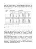
Fig. 3. Schematic illustration of sensing mechanism for electrocatalytic glucose on the
GNPs/CD–Fc/GOD modified platinum electrode surface (Chen & Diao, 2009).
For example, coupling carbon nanofibers with palladium nanoparticles resulted in a
remarkable improvement of the electroactivity of the composite materials towards reduction
of H
2
O
2
and oxidation of β-nicotinamide adenine dinucleotide in reduced form (NADH)
(Huang et al., 2008). Zou et al. reported a glucose biosensor based on electrodeposition of
platinum nanoparticles onto multiwalled carbon nanotubes (Zou et al., 2008). Wu et al.
constructed a glucose biosensor based on multi-walled carbon nanotubes and GNPs by
layer-by-layer self-assembly technique (Wu et al., 2007). Taking advantage of the nanowires
and GNPs, a novel glucose biosensor was developed, based on the immobilization of
glucose oxidase (GOx) with cross-linking in the matrix of bovine serum albumin (BSA) on a
Pt electrode, which was modified with gold nanoparticles decorated Pb nanowires (GNPs-
Nanobiosensor for Health Care
99
PbNWs) (Wanga et al., 2009). Pb nanowires (PbNWs) were synthesized by an l-cysteine-
assisted self-assembly route, and then gold nanoparticles (GNPs) were attached onto the
nanowire surface through –SH–Au specific interaction. The synergistic effect of PbNWs and
GNPs made the biosensor exhibit excellent electrocatalytic activity and good response
performance to glucose. In pH 7.0, the biosensor showed the sensitivity of
135.5µA.mM
−1
.cm
−2
, the detection limit of 2 µM (S/N = 3), and the response time <5 s with a
linear range of 5–2200 µM. Furthermore, the biosensor exhibits good reproducibility, long-
term stability and relative good anti-interference.
Fig. 4. TEM images of (a) GNPs, (b) GNPs-PbNWs (Wanga et al., 2009).
6.2 Cholesterol biosensors
Cholesterol is a fundamental parameter in the diagnosis of coronary heart disease,
arteriosclerosis, and other clinical (lipid) disorders and in the assessment of the risks of
thrombosis and myocardial infarction. The clinical analysis of cholesterol in serum samples
is important in the diagnosis and prevention of a large number of clinical disorders such as
hypertension, cerebral thrombosis and heart attack. Hence, it is important to develop a
reliable and sensitive biosensor which can permit a suitable and rapid determination of
cholesterol. Ideally, the total cholesterol concentration in a healthy person’s blood should be
less than 200 mg/dL (<5.17 mM). The borderline high is defined as 200–239 mg/dL (5.17–
6.18 mM), and the high value is defined as above 240 mg/dL (≥6.21 mM) (Shen & Liu, 2007).
Different analytical methods have been used for the determination of cholesterol for
instance colorimetric, spectrometric and electrochemical methods. Among these methods,
electrochemical detection of cholesterol has achieved significant attention due to the rapid
determination, simplicity, and low cost. Thus, amperometric biosensors are more attractive
due to their low detection limit and enzyme stabilization can be easily achieved. Especially,
the enzyme based cholesterol sensors have gained special focus taking the advantages of
good stability, high sensitivity and wide linear range they hold a leading position among the
presently available biosensor systems. Recently, many scientists and biologists focused on
the preparation of newer nanocomposite with good biocompatibility that could be the
Biosensors for Health, Environment and Biosecurity
100
promising matrices for enzyme immobilization which can enhance the selectivity and
sensitivity of the biosensors. Among the natural biocompatible macromolecules, chitosan
(CS) is the biodegradable polymer obtained from marine versatile biopolymer-chitin. CS
fibers situate apart from all other biodegradable natural fibers in several inherent properties
such as outstanding biocompatibility, non-toxicity, biodegradability, high mechanical
strength, fast metal complexation and hydrophilicity for enzyme immobilization. CS
nanofibers (NFs) have remarkable characteristic such as exceptionally minute pore size with
very outsized surface area-to-volume proportion, high porosity and diameters of the fiber
was in nanometer scale. These properties of CSNFs hold fine enzyme immobilization
scaffold and it was exploited for biosensor applications. These interesting matrices provide
high surface area for high enzyme loading and compatible micro-environment helping
enzyme stability. Besides, CS provides direct contact between enzyme active site and
electrode. Enzyme immobilization is currently the gigantic increasing subject of
considerable interest because the use of enzyme is frequently inadequate due to their
availability in tiny quantity, instability, high cost and the limited possibility of economic
recoveries of these bio-catalysts from an effective response unify. For a good enzyme
immobilization, biocompatibility is the one of the most important key requisite that benefits
the enzymatic bio-transformations to construct the biosensors. So, increase the
biocompatibility of the support, various surface modification protocol have often been used
such as adsorption, coating, self-assembly and graft polymerization. Among these
techniques, it is relatively graceful and efficient to directly bind natural bio-macromolecules
on the support surface to form a bio-mimetic compatible layer for enzyme immobilization.
In the recent years, there is a trend to use nanostructured materials as supports for enzyme
immobilization, since the large surface area to volume ratio of nanosize materials can
effectively improve to the loading enzyme per unit to volume ratio of support and the
excellent catalytic efficiency of the immobilized enzyme. Both nanofibers and nanoparticles
were explored for this purpose. Recent developments in the field of nanobiotechnology,
metal nanoparticles (MNPs) find numerous applications. Among the MNPs, GNPs be
widely used for the catalytic and biological application. GNPS provides adequate micro-
environment to enhance DET between biomolecule and electrode. In the fabrication of a
cholesterol biosensor, cholesterol oxidase (ChOx) is most commonly used as the biosensing
element. Cholesterol oxidase catalyzes the oxidation of cholesterol to H
2
O
2
and cholest-4-en-
3-one in the presence of oxygen. The enzymatic reaction in the use of cholesterol oxidase
(ChOx) as a receptor can be described as follows:
ChOx
Cholesterol + O
2
→ Cholest-4 −en−3−one + H
2
O
2
The electro-oxidation current of hydrogen peroxide is detected after application of a suitable
potential to the system. The major problem for amperometric detection is the overestimation
of the response current due to interferences such as ascorbic acid. This problem can be
overcome by using a combination of two or three enzymes, which are more selective for the
analyte of interest (Bongiovanni et al., 2001) or by devising techniques to eliminate or reduce
the interference. A novel amperometric cholesterol biosensor was fabricated by the
immobilization of ChOx (cholesterol oxidase) onto the chitosan nanofibers/gold
nanoparticles (designated as CSNFs/AuNPs) composite network (NW) (Gomathia et al.,
2010). The fabrication involves preparation of chitosan nanofibers (CSNFs) and subsequent
electrochemical loading of gold nanoparticles. Field emission scanning electron microscopy
Nanobiosensor for Health Care
101
(FE-SEM) was used to investigate the morphology of CSNFs (sizes in the range of 50–100
nm) and spherical GNPs. The CSNF–GNPs/ChOx biosensor exhibited a wide linear
response tocholesterol (concentration range of 1–45 µM), good sensitivity (1.02 µA/µM), low
response time (5 s) and excellent long term stability. The combined existence of GNPs within
CSNFs NW provides the excellent performance of the biosensor towards the electrochemical
detection of cholesterol.
Fig. 5. Fabrication of CSNF–GNPs/ChOx biosensor electrode (Gomathia et al., 2010).
Many researchers have reported the inclusion of metal nanoparticles with a catalytic effect
in polymer modified electrodes to decrease the overpotential applied to the amperometric
biosensors (Safavi et al., 2009, Hrapovic et al., 2004, Ren et al., 2005, Huang et al., 2004).
Amperometric cholesterol biosensors based on carbon nanotube–chitosan–platinum–
cholesterol oxidase nanobiocomposite was fabricated for cholesterol determination at an
applied potential of 0.4 V (Tsai et al., 2008). To improvethe selectivity of the biosensor,
Gopalana et al. reported the construction of a cholesterol biosensor by monitoring the
reduction current of H
2
O
2
at −0.05 V (Gopalana et al., 2009). Bimetallic alloys are widely
used in catalysis and sensing fields. Owing to the interaction between two components in
bimetallic alloys, they generally show many favorable properties in comparison with the
corresponding monometallic counterparts, which include high catalytic activity, catalytic
selectivity, and better resistance to deactivation. Among various bimetallic alloys, gold–
platinum (AuPt) alloy is very attractive. It has excellent catalysis and resistance to
deactivation due to the high synergistic action between gold and platinum (Xiao et al., 2009).
Owing to these advantages of bimetallic nanoparticles, it becomes significant to develop
AuPt nanoparticles for application in electrochemical sensors with appropriate
characteristics such as high sensitivity, fast response time, wide linear range, better
Biosensors for Health, Environment and Biosecurity
102
selectivity, and reproducibility. An electrodeposition method was applied to form gold–
platinum (AuPt) alloy nanoparticles on the glassy carbon electrode (GCE) modified with a
mixture of an ionic liquid (IL) and chitosan (Ch) (AuPt–Ch–IL/GCE). AuPt–Ch–IL/GCE
electrocatalyzed the reduction of H
2
O
2
and thus was suitable for the preparation of
biosensors. Cholesterol oxidase (ChOx) was then, immobilized on the surface of the
electrode by cross-linking ChOx and chitosan through addition of glutaraldehyde
(ChOx/AuPt–Ch–IL/GCE) (Safavia & Farjamia, 2011). The fabricated biosensor exhibited
two wide linear ranges of responses to cholesterol in the concentration ranges of 0.05–6.2
mM and 6.2–11.2 mM. The sensitivity of the biosensor was 90.7 µA.mM
−1
.cm
−2
and the limit
of detection was 10 µM of cholesterol. The response time was less than 7 s. The Michaelis–
Menten constant (Km) was found as 0.24 mM. The effect of the addition of 1 mM ascorbic
acid and glucose was tested on the amperometric response of 0.5 mM cholesterol and no
change in response current of cholesterol was observed.
Fig. 6. Schematic illustration of preparation procedures of ChOx/AuPt–Ch–IL/GCE (Safavia
& Farjamia, 2011).
6.3 Tyrosinase biosensors
Phenolic compounds often exist in the wastewaters of many industries, causing problems
for our living environment. Many of them are very toxic, showing adverse effects on
animal and plants. Therefore, the identification and quantification of such compounds are
very important for environment monitoring. Some methods are available for the phenolic
compound assay, including gas or liquid chromatography and spectrophotometry
(Chriswell et al. 1975, Poerschmann et al., 1997). However, demanding sample
pretreatments, low sensitivities, and time-consuming manipulations limit their practical
applications. A great amount of effort has been devoted to the development of simple and
effective analytical methods for the determination of phenolic compounds. Among them,
amperometric biosensor based on tyrosinase has been shown to be a very simple and
convenient tool for phenol assay due to its high sensitivity, effectiveness, and simplicity
(Wang et al., 2002, Dempsey et al., 2004, Rajesh et al., 2004, Xue & Shen, 2002, Zhang et al.,
2003, Wang et al., 2000a, Yu et al. 2003, Campuzano et al., 2003, Tatsuma & Sato, 2004). The
immobilization of tyrosinase is a crucial step in the fabrication of phenol biosensor. The
earlier reports on the immobilization methods included polymer entrapment (Wang et al.,
2002, Dempsey et al., 2004), electropolymerization (Dempsey et al., 2004, Rajesh et al., 2004),
sol–gels (Rajesh et al., 2004, Yu et al. 2003), self-assembled monolayers (SAMs)1
(Campuzano et al., 2003, Tatsuma et al., 2004), and covalent linking (Anh et al., 2002, Rajesh
et al., 2004a). However, some of these immobilizations are relatively complex, requiring the
use of solvents that are unattractive to the environment and result in relatively poor stability
Nanobiosensor for Health Care
103
and bioactivity of tyrosinase. Recent years have seen increased interest in searching for
simple and reliable schemes to immobilize enzymes. The biocompatible nanomaterials have
their unique advantages in enzyme immobilization. They could retain the activity of
enzyme well due to the desirable microenvironment, and they could enhance the direct
electron transfer between the enzyme’s active sites and the electrode (Gorton et al., 1999, Jia
et al., 2002). In spite of the big amount of literature on tyrosinase electrochemical biosensors,
two general limitations need to be solved yet in order to improve their practical usefulness.
One of them concerns the stability of the biosensors. Although many efforts have been made
to improve the useful lifetime and reusability of tyrosinase electrodes, searching for
appropriate microenvironments for retaining the biological activity of the enzyme, its
inherent instability provokes that this useful lifetime is too short for practical applications in
many cases. On the other hand, the low concentration levels of phenolic compounds that
should be detected due to their classification as priority pollutants, requires that the
tyrosinase biosensors are capable to achieve a high sensitivity. The aim of this work is the
design of a new tyrosinase bioelectrode able to improve significantly these important
analytical characteristics with respect to previous designs. The new bioelectrode design is
based on the combination of the advantageous properties of a graphite–Teflon composite
electrode matrix for the immobilization of enzymes, and the use of colloidal gold
nanoparticles. In this new design, both the enzyme tyrosinase and gold nanoparticles are
incorporated into the composite electrode matrix by simple physical inclusion. The use of
graphite–Teflon composite pellets for the construction of enzyme electrodes has been
extensively reported (Serra et al., 2002, GuzmanVazquez de Pradaet al., 2003, Pena et al.,
2001). The resulting bioelectrodes are easily renewable by polishing and allow incorporation
of biomolecules and other modifiers with no covalent attachments, thus making the
electrode fabrication procedure easy, fast and cheap. On the other hand, electrochemical
biosensors created by coupling biological recognition elements with electrochemical
transducers based on or modified with gold nanoparticles are playing an increasingly
important role in biosensor research over the last few years (Yanez-Sedeno & Pingarron,
2005). So, colloidal gold allows proteins to retain their biological activity upon adsorption
(Doron et al., 1995, Brown et al., 1996, Mena et al., 2005) and modification of electrodes with
this type of nanoparticles provides a microenvironment similar to that of the redox proteins
in native systems, reducing the insulating effect of the protein shell for the direct electron
transfer through the conducting tunnels of gold nanocrystals (Liu et al., 2003a). Surface
morphology of gold nanoparticles, and the interaction between the nanoparticles and the
electrode surface, are significant factors which contribute to improve the electrical contact
between the redox protein and the electrode material (Shipway et al., 2000). In this context,
biosensors based on the immobilization of enzymes on gold nanoparticles for the
determination of hydrogen peroxide, nitrite, glucose and phenols (Tang & Jiang, 1998, Xiao
et al., 2000, Gu et al., 2001, Liu & Ju, 2002, Jia et al., 2002, Liu & Ju, 2003, Liu et al., 2003b,
Xiao et al., 2003, Carralero-Sanz et al., 2005) have been recently reported.
The preparation of a tyrosinase biosensor based on the immobilization of the enzyme onto a
glassy carbon electrode modified with electrodeposited gold nanoparticles (Tyr-nAu-GCE)
was reported (Carralero-Sanz et al., 2005). The enzyme immobilized by cross-linking with
glutaraldehyde retains a high bioactivity on this electrode material. Under the optimized
working variables (a Au electrodeposition potential of −200mV for 60 s, an enzyme loading
of 457 U, a detection potential of −0.10V and a 0.1 mol. L
−1
phosphate buffer solution of pH
7.4 as working medium) the biosensor exhibited a rapid response to the changes in the
Biosensors for Health, Environment and Biosecurity
104
substrate concentration for all the phenolic compounds tested: phenol, catechol, caffeic acid,
chlorogenic acid, gallic acid and protocatechualdehyde. A R.S.D. of 3.6% (n = 6) was
obtained from the slope values of successive calibration plots for catechol with the same
Tyr-nAu-GCE with no need to apply a cleaning procedure to the biosensor. The useful
lifetime of one single biosensor was of at least 18 days, and a R.S.D. of 4.8% was obtained for
the slope values of catechol calibration plots obtained with five different biosensors. The
Tyr-nAu-GCE was applied for the estimation of the phenolic compounds content in red and
white wines. A good correlation of the results (r = 0.990) was found when they were plotted
versus those obtained by using the spectrophotometric method involving the Folin–
Ciocalteau reagent.
Fig. 7. Cyclic voltammograms for 2.0×10
−4
mol.L
−1
solutions of catechol (a) and caffeic acid
(b), at: (1) Tyr-nAu-GCE; (2) Tyr-GCE; (3) Au-GCE; (4) GCE; v = 25mVs−1. Supporting
electrolyte: 0.05 mol.L
−1
phosphate buffer (pH 7.4) (Carralero-Sanz et al., 2005).
The design of a new tyrosinase biosensor with improved stability and sensitivity was
reported (Carralero-Sanz et al., 2006). The biosensor design is based on the construction of a
graphite–Teflon composite electrode matrix in which the enzyme and colloidal gold
nanoparticles are incorporated by simple physical inclusion. The Tyr–Au
coll
–graphite–Teflon
biosensor exhibited suitable amperometric responses at −0.10 V for the different phenolic
compounds tested (catechol; phenol; 3,4-dimethylphenol; 4-chloro-3-methylphenol; 4-
chlorophenol; 4- chloro-2-methylphenol; 3-methylphenol and 4-methylphenol). The limits of
detection obtained were 3 nM for catechol, 3.3 µM for 4- chloro- 2-methylphenol, and
approximately 20 nM for the rest of phenolic compounds. The presence of colloidal gold
into the composite matrix gives rise to enhanced kinetics of both the enzyme reaction and
the electrochemical reduction of the corresponding o-quinones at the electrode surface, thus
allowing the achievement of a high sensitivity. The biosensor exhibited an excellent
renewability by simple polishing, with a lifetime of at least 39 days without apparent loss of
the immobilized enzyme activity. The usefulness of the biosensor for the analysis of real
Nanobiosensor for Health Care
105
samples was evaluated by performing the estimation of the content of phenolic compounds
in water samples of different characteristics.
A highly efficient enzyme-based screen printed electrode (SPE) was obtained by using
covalent attachment between 1-pyrenebutanoic acid, succinimidyl ester (PASE) adsorbing
on the graphene oxide (GO) sheets and amines of tyrosinase-protected gold nanoparticles
(Tyr-Au) (Song et al., 2010). Herein, the bi-functional molecule PASE was assembled onto
GO sheets. Subsequently, the Tyr-Au was immobilized on the PASE-GO sheets forming a
biocompatible nanocomposite, which was further coated onto the working electrode surface
of the SPE. Attributing to the synergistic effect of GO-Au integration and the good
biocompatibility of the hybrid-material, the fabricated disposable biosensor (Tyr-Au/PASE-
GO/SPE) exhibited a rapid amperometric response (less than 6 s) with a high sensitivity and
good storage stability for monitoring catechol. This method shows a good linearity in the
range from 8.3×10
-8
to 2.3×10
-5
M for catechol with a squared correlation coefficient of
0.9980, a quantitation limit of 8.2×10
-8
M (S/N = 10) and a detection limit of 2.4×10
-8
M (S/N
= 3). The Michaelis-Menten constant was measured to be 0.027 mM. This disposable
tyrosinase biosensor could offer a great potential for rapid, cost-effective and on-field
analysis of phenolic compounds.
Fig. 8. Assembling process of Tyr-Au/PASE-GO on SPE (Song et al., 2010).
6.4 Urease biosensors
Kidneys perform key roles in various body functions, including excreting metabolic waste
products such as urea from the bloodstream, regulating the hydrolytic balance of the body,
and maintaining the pH of body fluids. The level of urea in blood serum is the best
measurement of kidney function and staging of kidney diseases. The normal urea level in
serum ranges from 15 to 40 mg/dL (i.e., 2.5–7.5 mM). An increase in urea concentration
causes renal failure such as acute or chronic urinary tract obstruction with shock, burns,
dehydration, and gastrointestinal bleeding, whereas a decrease in urea concentration causes
hepatic failure, nephritic syndrome, and cachexia. Therefore, there is an urgent need to
develop a device that rapidly monitors urea concentration in the body. Most existing urea
Biosensors for Health, Environment and Biosecurity
106
biosensors utilize urease (Urs) as the sensing element. The available Urs on the electrode
surface hydrolyzes urea into NH
4
+
and HCO
3
−
ions. The concentration of urea is measured
by monitoring the librated ions using a transducer such as amperometric, potentiometric,
optical, thermal, or piezoelectric. Although various urea biosensors that use a range of
transducers have been studied extensively, the Urs-based amperometric urea biosensor is
considered one of the most promising approaches because it offers fast, simple, and low-cost
detection. The response time of such a biosensor is directly associated with the hydrolysis
rate of urea on the electrode surface; therefore, rapid production of NH
4
+
ions on the
electrode will lead to a highly sensitive biosensor. It is well established that the performance
of biosensors greatly depends on the physicochemical properties of the electrode materials,
enzyme immobilization procedure, and enzyme concentration on the electrode surface.
Many electrode materials have been used to fabricate urea biosensors. However, there is an
ongoing demand for new types of electrode materials that can provide the Urs enzyme with
better stability and performance for in vitro urea measurement. In this context, the use of
nanomaterials to fabricate biosensors is one of the most exciting approaches because
nanomaterials have a unique structure and high surface-to-volume ratio. The surfaces of
nanomaterials can also be tailored in the molecular scale in order to achieve various
desirable properties. Many attempts have been made to fabricate a third-generation
biosensor with self-assembly technology; however, these approaches were based on planar
self-assembly that may only offer limited available surface area on the electrode, which can
compromise the performance of the biosensor. Meanwhile, gold nanoparticles have played
an increasingly important role for biosensor applications over the last decade.
Gold nanoparticles can (1) provide a stable surface for the immobilization of biomolecules
without compromising their biological activities and (2) permit direct electron transfer from
the redox biomolecules to the bulk electrode materials, thereby enhancing the
electrochemical sensing ability. For example, Shipway et al. systematically studied the new
electronic, photoelectronic, and sensoring systems that used gold nanoparticle
superstructures on the electrode surface (Shipway et al., 2000). In addition, previous studies
indicated that biological macromolecules such as enzymes can generally retain their
enzymatic and electrochemical activity after being immobilized onto the gold nanoparticles
(Brown et al., 1996, Xiao et al., 1999). Anamperometric biosensor was fabricated for the
quantitative determination of urea in aqueous medium using hematein, a pH-sensitive
natural dye (Tiwari et al., 2009). The urease (Urs) covalently immobilized onto an electrode
made of gold nanoparticles functionalized with hyperbranched polyester-BoltronR H40
(H40–Au) coated onto an indium–tin oxide (ITO) covered glass substrate. The covalent
linkage between the Urs enzyme and H40–Au nanoparticles provided the resulting enzyme
electrode (Urs/H40–Au/ITO) with a high level of enzyme immobilization and excellent
lifetime stability. The biosensor based on Urs/H40–Au/ITO as the working electrode
showed a linear current response to the urea concentration ranging from 0.01 to 35 mM. The
urea biosensor exhibited a sensitivity of 7.48nA/mMwith a response time of 3 s. The
Michaelis–Menten constant for the Urs/H40–Au/ITO biosensor was calculated to be
0.96mM, indicating the Urs enzyme immobilized on the electrode surface had a high affinity
to urea.
A renewable potentiometric urease inhibition biosensor based on self-assembled gold
nanoparticles has been developed for the determination of mercury ions (Yang et al., 2006a).
Nanobiosensor for Health Care
107
Fig. 9. Schematic presentation of the [A] preparation of hyperbranched gold (H40–Au)
nanoparticles and [B] fabrication of H40–Au/ITO and Urs/H40–Au/ITO electrodes(Tiwari
et al., 2009).
Gold nanoparticles were chemically adsorbed on the PVC-NH2 matrix membrane pH
electrode surface containing N,Ndidecylaminomethylbenzene (DAMAB) as a neutral carrier
and urease was then immobilized on the gold nanoparticles. The linear range of
determination of Hg
2+
was 0.09–1.99 µmol.L
−1
with a detection limit of 0.05 µmol.L
−1
. The
advantages of self-assembled immobilization are low detection limit, fast response and ease
regeneration. The assembled gold nanoparticles and inactive enzyme layers denatured by
Hg
2+
can be rinsed out via a saline solution with acid and alkali successively. This sensor is
generally of great significance for inhibitor determination, especially in comparison with
expensive base transducers.
Biosensors for Health, Environment and Biosecurity
108
Fig. 10. TEM of gold nanoparticles with different size: 12 nm (a), 20 nm (b) and 35 nm (c)
(Yang et al., 2006a).
6.5 Acetylcholinesterase biosensors
Carbamate and organophosphate pesticides have come into widespread use in agriculture
because of their high insecticidal activity and relatively low environmental persistence.
However, overuse of these pesticides results in pesticide residues in food, water and
environment, and leads to a severe threat to human health due to their high toxicity to
acetylcholinesterase (AChE), which is essential for the functioning of the central nervous
system in humans. For these reasons, it has great significance to develop a fast, reliable and
inexpensive analytical method for determination of trace amounts of these pesticides.
Common analytical techniques for determination of these compounds, such as gas and
liquid chromatography are sensitive, reliable and precise. However, these methods require
expensive instrumentation, complicated pretreatment procedure and professional operators,
which limit their application for real-time detection of these compounds. In order to
simplify procedure and decrease cost, enzyme based biosensors could be a reliable and
promising alternative to classical methods because of their simple fabrication, easy
operation, high sensitivity and selectivity. It is well known that acetylthiocholine chloride
(ATCl) can be catalytically hydrolyzed by AChE to thiocholine (TCh), which could be
electrochemically oxidized at special potential. The hydrolysis reaction of ATCl would be
inhibited by carbamate and organophosphorous pesticides, because AChE could
irreversibly combine with these pesticides, which results in AChE inactivation to give low
TCh concentration and low oxidation current. Therefore, based on the inhibition of
carbamate and organophosphate pesticides on the AChE activity, the concentrations of
Nanobiosensor for Health Care
109
pesticides would be monitored by measuring the electrochemical oxidation peak current of
TCh. The key aspect in construction of this kind of biosensor is the immobilization of AChE
on the solid electrode surface with high electron transfer rate and bioactivity. In order to
settle it, a variety of matrix materials have been employed, among them, GNPs have
attracted enormous interest in the fabrication of electrochemical biosensors for possessing
conductive sensing interface, catalytic properties and conductivity properties. Moreover,
GNPs can provide an environment similar to that of proteins in a native system and allow
protein molecules more freedom in orientation, which will reduce the insulating property of
protein shell and facilitate the electron transfer through the conducting tunnel of GNPs.
Gold nanoparticles were synthesized in situ and electrodeposited onto Au substrate (Dua et
al., 2008). The GNPs modified interface facilitates electron transfer across self-assembled
monolayers of 11-mercaptoundecanoic acid (MUA). After activation of surface carboxyl
groups with 1-ethyl-3-(3-dimethylaminopropyl) carbodiimide and N-hydroxysuccinimide,
the interface displayed good stability for immobilization of biomolecules. The immobilized
acetylcholinesterase (AChE) showed excellent activity to its substrate, leading to a stable
AChE biosensor. Under the optimal experimental conditions, the inhibition of malathion on
AChE biosensor was proportional to its concentration in two ranges, from 0.001 to 0.1
µg.mL
−1
and from 0.1 to 25 µg.mL
−1
, with detection limit of 0.001 µg.mL
−1
. The simple
method showed good reproducibility and acceptable stability, which had potential
application in biosensor design.
Fig. 11. Principle of GNPs served as mediator for electron transfer across SAMs for AChE
biosensor design (Dua et al., 2008).
GNPs are particularly attractive for fabricating electrochemical sensors and biosensor.
However, GNPs are inherently instable and apt to agglomerate. In order to settle this
problem, it is necessary to use protective agents. SF is a natural protein, which can be
Biosensors for Health, Environment and Biosecurity
110
extracted from silkworm cocoon. Due to the unique properties of SF with thermal stability,
nontoxicity, low cost and biocompatibility, it is widely used as a substrate for enzyme
immobilization. Furthermore, GNPs could be in situ produced by the reduction of SF at
room temperature, in which SF acts as both reducing agent and protector. It has been
demonstrated that GNPs and SF could interact to form a bioconjugate, and this kind of
GNPs–SF colloid possessed a stable and highly dispersed nature. A sensitive and stable
amperometric biosensor for the detection of methyl paraoxon, carbofuran and phoxim had
been developed based on immobilization of acetylcholinasterase (AChE) on gold
nanoparticles and silk fibroin (SF) modified platinum electrode (Yin et al., 2009). The SF
provided a biocompatible microenvironment around the enzyme molecule to stabilize its
biological activity and effectively prevented it from leaking out of platinum electrode
surface. In the presence of acetylthiocholine chloride (ATCl) as a substrate, GNPs promoted
electron transfer reaction at a lower potential and catalyzed the electrochemical oxidation of
thiocholine (TCh), thus increasing detection sensitivity. Under optimum conditions, the
inhibition percentages of methyl paraoxon, carbofuran and phoxim were proportional to
their concentrations in the range of 6x10
-11
–5x10
-8
M, 2x10
-10
–1x10
-7
M and 5x10
-9
–2x10
-7
M,
respectively. The detection limits were found to be 2x10
-11
M for methyl paraoxon, 1x10
-10
M
for carbofuran and 2x10
-9
M for phoxim. Moreover, the fabricated biosensor had good
reproducibility and acceptable stability. The biosensor is a promising new tool for pesticide
analysis.
A novel interface embedded in situ gold nanoparticles (GNPs) in chitosan hydrogel was
constructed by one-step electrochemical deposition in solution containing tetrachloroauric
(III) acid and chitosan (Du et al., 2007a). This deposited interface possessed excellent
biocompatibility and good stability. The immobilized AChE, as a model, showed excellent
activity to its substrate and provided a quantitative measurement of organophosphate
pesticides involved in the inhibition action. Operational parameters, including the
deposition time, tetrachloroauric (III) acid concentration have been optimized. Under the
optimal electrodeposition, an amperometric sensor for the fast determination of malathion
and monocrotophos, respectively was developed with detection limit of 0.001 µg.mL
-1
. The
simple method showed good fabrication reproducibility and acceptable stability, which
provided a new avenue for electrochemical biosensor design.
6.6 Horseradish peroxidase
Over the last years, considerable efforts have been devoted to the development of
horseradish peroxidase (HRP, EC 1.11.1.7, H
2
O
2
oxidoreductase)-based mediatorless
electrochemical biosensors for the fast, simple, selective and accurate quantification of H
2
O
2
.
This interest is justified by the industrial, chemical and biomedical applications of this
oxidant compound. In addition, H
2
O
2
constitutes a relevant biochemical mediator in many
cellular processes, as well as a by-product of several oxidases with analytical applications.
Different strategies has been described for connecting the catalytic active site of HRP with
electrode surfaces, in order to construct such kind of third generation H
2
O
2
biosensors in
which the direct electron transfer between the enzyme and the electrode is allowed without
the use of any natural or artificial redox mediator. Among these methods, it should be
highlighted the use of electroconductive polymers (Zhaoyang et al., 2006, Luo et al., 2006,
Mala Ekanayake et al., 2009), metal nanoparticles (Zhaoyang et al., 2006, Luo et al., 2006,
Nanobiosensor for Health Care
111
Mala Ekanayake et al., 2009, Jeykumari et al., 2008, Schumb et al., 1995, Ferreira et al., 2004,
Alonso Lomillo et al., 2005, Pingarron et al., 2008), redox polymers and sol–gel materials
(Wang et al., 2000, Jia et al., 2005, Garca et al., 2007), DNA (Song et al., 2006) and carbon
nanotubes (Jeykumari et al., 2008) as wiring materials for HRP. On the other hand, the
immobilization strategy to be employed is another key factor to consider in the design of an
enzyme biosensor. This approach should favor the maintenance of the active enzyme
conformation as well as provide a favorable hydrophilic microenvironment around the
biocatalyst in order to contribute to the best catalytic performance of the enzyme (Song et
al., 2006, Villalonga et al., 2007). In this regard, it has been previously reported the
preparation of highly active and stable biocatalysts by the polyelectrostatic immobilization
of enzymes in polysaccharide-coated supports (Gomez et al., 2006). In addition, several ionic
polysaccharides such as sodium alginate (Camacho et al., 2007, Ionescu et al., 2006, Cosnier
et al., 2004) and chitosan and its derivatives (Qin et al., 2006, Li et al., 2008), have been
successfully used as coating materials for preparing robust enzyme biosensors. Horseradish
peroxidase was cross-linked with cysteamine-capped Au nanoparticles and further
immobilized on sodium alginate-coated Au electrode through polyelectrostatic interactions
(Chico et al., 2009). The electrode was employed for constructing a reagentless
amperometric biosensor for H
2
O
2
. The electrode showed linear response (poised at -400 mV
vs. Ag/AgCl) toward H
2
O
2
concentration between 20 µM and 13.7 mM at pH 7.0. The
biosensor reached 95% of steady-state current in about 15 s, and its sensitivity was 40.1
mA/M.cm
2
. The detection limit of the enzyme-based electrode was determined as 3 µM, at a
signal-to-noise ratio of three. The electrode retained 97% of its initial analytical response
after 1 month of storage at 4 ºC in 50 mM sodium phosphate buffer, pH 7.0. The stability of
the biosensor was significantly reduced when it was incubated in high ionic strength
solutions, retaining only 44% of its initial response after 1 month of storage at 4 ºC in 1 M
NaCl ionic strength in 50 mM sodium phosphate buffer, pH 7.0.
The preparation of horseradish peroxidase (HRP)-GNPs-silk fibroin (SF) modified glassy
carbon electrode (GCE) by one step procedure was reported (Yina et al., 2009). The enzyme
electrode showed a quasi-reversible electrochemical redox behavior with a formal potential
of −210mV (vs. SCE) in 0.1M phosphate buffer solution at pH 7.1. The response of the
biosensor showed a surface-controlled electrochemical process with one electron transfer
accompanying with one proton. The cathodic transfer coefficient was 0.42, the electron
transfer rate constant was 1.84 s
−1
and the surface coverage of HRP was 1.8×10
−9
mol.cm
−2
.
The experimental results indicated that GNPs–SF composite matrix could not only steadily
immobilize HRP, but also efficiently retain its bioactivity. The biosensor displayed an
excellent and quick electrocatalytic response to the reduction of H
2
O
2
.
A novel method for fabrication of horseradish peroxidase (HRP) biosensor has been
developed by self-assembling gold nanoparticles on thiol-functionalized poly(styrene-co-
acrylic acid) (St-co-AA) nanospheres (Xu et al., 2004). At first, a cleaned gold electrode was
immersed in thiol-functionalized poly(St-co-AA) nanosphere latex prepared by emulsifier-
free emulsion polymerization of St with AA and function with dithioglycol to assemble the
nanospheres, then gold nanoparticles were chemisorbed onto the thiol groups. Finally,
horseradish peroxidase was immobilized on the surface of the gold nanoparticles. The
sensor displayed an excellent electrocatalytical response to reduction of H
2
O
2
without the
aid of an electron mediator. The sensor was highly sensitive to hydrogen peroxide with a
detection limit of 4.0 µmol.L
−1
, and the linear range was from 10.0 µmol.L
−1
to 7.0 mmol.L
−1
.
Biosensors for Health, Environment and Biosecurity
112
The biosensor retained more than 97.8% of its original activity after 60 days of use.
Moreover, the studied biosensor exhibited good current repeatability and good fabrication
reproducibility.
Fig. 12. Steady-state amperometric responses of electrodes to the reduction of H
2
O
2
in the
stirring PB under elimination of oxygen: (a) the non-modified gold electrode; (b) the latex
modified electrode; (c) the gold nanoparticle modified electrode before HRP addition; (d)
the gold nanoparticle modified electrode after HRP addition; (e) the latex modified electrode
after HRP addition; Applied potential, −200mV; supporting electrolyte, 100 mmol.L
−1
pH 7
(Xu et al., 2004).
A one-step method for fabrication of horseradish peroxidase (HRP) biosensor has been
developed (Di et al., 2005). The gold nanoparticles and HRP were simultaneously embedded
in silica sol–gel network on gold electrode surface in the presence of cysteine. The
immobilized HRP exhibited direct electrochemical behavior toward the reduction of
hydrogen peroxide. The heterogeneous electron transfer rate constant was evaluated to be
7.8 s
−1
. The biosensor displayed an excellent elctrocatalytic response to the reduction of H
2
O
2
without any mediator. The calibration range of H
2
O
2
was from 1.6 µmol.L
−1
to 3.2 mmol.L
−1
and a detection limit of 0.5 µmol.L
−1
at a signal-to- noise ratio of 3. The biosensor exhibited
high sensitivity, rapid response and long-term stability.
The design and development of a screen printed carbon electrode (SPCE) on a polyvinyl
chloride substrate as a disposable sensor is described (Tangkuaram et al., 2007). Six
configurations were designed on silk screen frames. The SPCEs were printed with four inks:
silver ink as the conducting track, carbon ink as the working and counter electrodes,
silver/silver chloride ink as the reference electrode and insulating ink as the insulator layer.
Selection of the best configuration was done by comparing slopes from the calibration plots
generated by the cyclic voltammograms at 10, 20 and 30mM K
3
Fe(CN)
6
for each
configuration. The electrodes with similar configurations gave similar slopes. The 5
th
configuration was the best electrode that gave the highest slope. Modifying the best SPCE
configuration for use as a biosensor, horseradish peroxidase (HRP) was selected as a
biomaterial bound with gold nanoparticles in the matrix of chitosan (HRP/GNP/CHIT).
Biosensors of HRP/SPCE, HRP/CHIT/SPCE and HRP/GNP/CHIT/SPCE were used in the
amperometric detection of H
2
O
2
in a solution of 0.1M citrate buffer, pH 6.5, by applying a
potential of −0.4 V at the working electrode. All the biosensors showed an immediate
Nanobiosensor for Health Care
113
response to H
2
O
2
. The effect of HRP/GNP incorporated with CHIT
(HRP/GNP/CHIT/SPCE) yielded the highest performance. The amperometric response of
HRP/GNP/CHIT/SPCE retained over 95% of the initial current of the 1
st
day up to 30 days
of storage at 4 ºC. The biosensor showed a linear range of 0.01–11.3 mM H
2
O
2
, with a
detection limit of 0.65 µM H
2
O
2
(S/N = 3). The low detection limit, long storage life and
wide linear range of this biosensor make it advantageous in many applications, including
bioreactors and biosensors.
6.7 DNA biosensors
DNA biosensors for the detection of nucleic acid sequences have attracted ever increasing
interests in connection with highly demanding research efforts directed to gene analysis,
clinical disease diagnosis, or even forensic applications (Service, 1998, Butler, 2006, Staudt,
2001, Farace et al., 2002, Reisberga et al., 2006). Various techniques including optical,
electrochemistry, surface plasmon resonance spectroscopy, and quartz crystal microbalance,
etc have been well developed for DNA detection (Rosi & Mirkin, 2005, Gerion et al., 2003,
Drummond et al., 2003, He et al., 2000). Among them, electrochemistry offer great
advantages such as simple, rapid, low-cost and high sensitivity (Lao et al., 2005). A key issue
faced with any DNA hybridization biosensor is the immobilization amount and accessibility
of probe DNA for hybridization recognition (Moses et al., 2004, Lowe et al., 2003, Ding et al.,
2008, Ostatná et al., 2005). Increasing the immobilization amount and controlling over the
molecular orientation of probe DNA would markedly improve the performance properties
of DNA biosensor. It has been well elaborated that the immobilization amount and the
molecular orientation of probe single-stranded DNA could remarkably influence the
operational performance of DNA electrochemical biosensor (Liu et al., 2008, Basuray et al.,
2009). Therefore, numerous different immobilization strategies have been proposed and
employed aimed at improving the link stability between DNA and transducer surface
(Cederquist et al., 2008), or increasing the amountof immobilized DNA (Liu et al., 2005), and
sometimes simplifying the immobilization procedure (Kjllman et al., 2008). In order to
achieve this goal, nanomaterials could be used as an elegant solution for the control of DNA
immobilization and hybridization. For a decade, metal nanoparticles have shown huge
potential in the fields of biosensing, diagnostics and molecular therapeutics because of its
excellent optical and electrical properties (Brown et al., 1996, Bao et al., 2003, Ma et al., 2004,
Kidambi et al., 2004). Owing to the large surface area and biocompatibility with biosystem,
gold nanoparticles have been shown as a good candidate for enhancement of DNA
immobilization and hybridization and they have been directly linked onto the biosensor
surface via various strategies such as covalent linking, electrodeposition, electroless
deposition, sol–gel, etc (Li et al., 2007, Yamada et al., 2003, Zhao et al., 2007, Jena& Raj, 2007).
The self-assembly of GNPs on the electrode surface could be easily achieved via the use of a
bi-functional chemical linking agent such as 1,6-hexanedithiol, cysteamine. Although these
self-assembly methods are very simple and rapid, the formed monolayer on the electrode
surface are usually insulated or could not offer a good electrical conductivity between GNPs
and electrode surface, which is not especially favorable for the fabrication of electrochemical
sensor or biosensor.
A novel protocol for development of DNA electrochemical biosensor based on GNPs
modified glassy carbon electrode (GCE) was proposed (Li et al., 2011), which was carried
out by the self-assembly of GNPs on the mercaptophenyl film (MPF) via simple
Biosensors for Health, Environment and Biosecurity
114
electrografting of in situ generated mercaptophenyl diazonium cations. The resulting MPF
was covalently immobilized on GCE surface via C–C bond with high stability, which was
desirable in fabrication of excellent performance biosensors. Probe DNA was self-assembled
on GNPs through the well-known Au–thiol binding. The recognition of fabricated DNA
electrochemical biosensor toward complementary single-stranded DNA was determined by
differential pulse voltammetry with the use of Co(phen)
3
3+
as the electrochemical indicator.
Taking advantage of amplification effects of GNPs and stability of MPF, the developed
biosensor could detect target DNA with the detection limit of 7.2×10
−11
M, which also
exhibits good selectivity, stability and regeneration ability for DNA detection. DNA
biosensor which was based on the self-assembly of GNPs on the mercapto-
diazoaminobenzene monolayer modified electrode was also reported (Liet al., 2010a). The
mercapto-diazoaminobenzene monolayer was obtained by covalent immobilization of 4-
aminothiophenol (4-ATP) molecules onto another 4-ATP monolayer functionalized gold
electrode bydiazotization-coupling reaction. The DNA immobilization and hybridization on
the GNPs modified electrode was further investigated. The prepared GNPs–ATP–diazo-
ATP film demonstrated efficient electron transfer ability for the electroactive species toward
the electrode surface due to a large conjugated structure of the mercapto-
diazoaminobenzene monolayer. The recognition of fabricated electrochemical DNA
biosensor toward complementary single-stranded DNA was determined by differential
pulse voltammetry with the use of Co(phen)
3
3+
as an electrochemical indicator. A linear
detection range for the complementary target DNA was obtained from 3.01×10
−10
to
1.32×10
−8
M with a detection limit of 9.10×10
−11
M. The fabricated biosensor also possessed
good selectivity and could be regenerated easily.
Fig. 13. Schematic representation of the fabrication of DNA biosensor (Li et al., 2011).
Colloidal gold nanoparticles and carboxyl group-functionalized CdS Nanoparticles (CdS
NPs) were immobilized on the Au electrode surface to fabricate a novel electrochemical
Nanobiosensor for Health Care
115
DNA biosensor (Du et al., 2009). Both GNPs and CdS NPs, well known to be good
biocompatible and conductive materials, could provide larger surface area and sufficient
amount of binding points for DNA immobilization. DNA immobilization and hybridization
were characterized with differential pulse voltammetry (DPV) by using
[Co(phen)
2
(Cl)(H
2
O)]Cl·2H
2
O as an electrochemical hybridization indicator. With this
approach, the target DNA could be quantified at a linear range from 2.0×10
−10
to 1.0 ×10
−8
M, with a detection limit of 2.0×10
−11
M by 3σ. In addition, the biosensor exhibited a good
repeatability and stability for the determination of DNA sequences.
Layers for DNA immobilization Detection limit (mol.L
−1
)
Au and CdS NPs (Du et al., 2009) 2.0×10
−11
LBL Au NPs and MWCNTs (Ma et al., 2008) 7.5×10
−12
CNTs (Niu et al., 2011) 1.4×10
−10
Pt NPs and CNTs (Zhu et al., 2005) 1.0×10
−11
ZrO
2
/SWNTs/PDC/GCE (Yang et al., 2007) 1.38 × 10
−12
Multilayer gold nanoparticles (Tsai et al., 2005) 1×10
−11
Conducting polyaniline nanotube (Chang et al., 2007) 3.759×10
−14
CdS nanoparticles and polypyrrole (Peng et al., 2006) 1×10
−9
Table 4. The performance comparison of various fabricated DNA biosensors (Du et al.,
2009).
7. Conclusion
Nanotechnology has been widely and successively applied in the field of sensing of drugs
and biological molecules. The most important example of nanosensors are gold
nanoparticles (GNPs) which offer many advantages, such as large surface-to-volume ratio,
high surface reaction activity and strong adsorption ability to immobilize the desired
biomolecules, good microenvironment for retaining the activity of enzyme, excellent
catalytic effects on many important chemical reactions, their catalytic effect is highly size-
dependent thus, the unique active sites and electronic states of GNPs can lead to their
anomalous catalytic activity.
The biomaterials to be sensed include a large variety of materials such as:
1. Glucose and cholesterol which are largely attributed to the human health and the food
industry.
2. Phenolic compounds whose identification and quantification are very important for
environment monitoring.
3. Some carbamate and organophosphate pesticides which affect food, water and
environment, and leads to a severe threat to human health.
4. H
2
O
2
whose quantification is justified by the industrial, chemical and biomedical
applications of this oxidant compound. In addition, H
2
O
2
constitutes a relevant
biochemical mediator in many cellular processes, as well as a by-product of several
oxidases with analytical applications.
5. DNA and nucleic acids sequences detection which are directed to gene analysis, clinical
disease diagnosis, or even forensic applications.
Biosensors for Health, Environment and Biosecurity
116
8. Copyright permissions
Final version after cut& edit Licence no. according to “Copyright Clearance Center”
Fig1 (
Table1 Not published
Fig2 Not published
Table2 2634100245226
Table3 2634101412860
Fig3 2633701365374
Fig4 2633701475869
Fig5 2634161086239
Fig6 2633710689524
Fig7 2633710837251
Fig8 2634150357126
Fig9 2633710969058
Fig10 2633711095490
Fig11 2633711408445
Fig12 2633720091311
Fig13 2633720380564
Table4 2634110691438
9. References
Alonso Lomillo, M.A.A.; Ruiz, J.G. & Pascual, F.J.M. (2005). Biosensor based on platinum
chips for glucose determination. Analytical Chimica Acta, Vol.547, No.2, pp.209–
214.
Anh, T.M.; Dzyadevych, S.V.; Soldatkin, A.P.; Chien, N.D.; Renault, N.J. & Chovelon, J.M.
(2002). Development of tyrosinase biosensor based on pH-sensitive field-effect
transistors for phenols determination in water solutions. Talanta, Vol.56, No.4,
pp.627–634.
Atta, N.F. & Abdel-Mageed, A.M. (2009). Smart electrochemical sensor for some
neurotransmitters using imprinted sol-gel films. Talanta, Vol.80, pp.511-518.
Atta, N.F. & El-Kady, M.F. (2009a). Poly(3-methylthiophene)/palladium sub-micro-
modified sensor electrode. Part II: Voltammetric and EIS studies, and analysis of
catecholamine neurotransmitters, ascorbic acid and acetaminophen. Talanta,
Vol.79, pp.639-647.
Atta, N.F. & El-Kady, M.F. (2010a). Novel poly(3-methylthiophene)/Pd nanoparticles
sensor: Synthesis, characterization and its application to the simultaneous analysis
Nanobiosensor for Health Care
117
of dopamine and ascorbic acid in biological fluid. Sensors & Actuators B: Chemical,
Vol.145, pp.299-310.
Atta, N.F.; Galal, A. & Ahmed, R.A. (2011a). Direct and simple electrochemical
determination of morphine at PEDOT modified Pt electrode. Electroanalysis,
Vol.23, pp,1-10.
Atta, N.F.; Galal, A. & Ahmed, R.A. (2011b). Poly(3,4-ethylene-dioxythiophene) electrode for
the selective determination of dopamine in presence of sodium dodecyl sulfate.
Bioelectrochemistry, Vol.80, pp.132-141.
Atta, N.F.; Galal, A. & El-Kady, M.F. (2009b). Palladium nanoclusters-coated poly(furan) as
a novel sensor for catecholamine neurotransmitters and paracetamol. Sensors &
Actuators B: Chemical, Vol.141, pp.566-574.
Atta, N.F.; Galal, A. & El-Kady, M.F. (2010b). Simultaneous determination of
catecholamines, uric acid and ascorbic acid at physiological levels using poly(N-
methylpyrrole)/Pd-nanoclusters sensor. Analytical Biochemistry, Vol.400, pp.78-88.
Atta, N.F.; Galal, A.; Abu-Attia, F.M. & Azab, S.M. (2010c). Carbon paste gold nanopatricles
sensor for the selective determination of dopamine in buffered solutions. Journal of
Electrochemical Society, Vol.157, pp.F116-F123.
Atta, N.F.; Galal, A.; Abu-Attia, F.M. & Azab, S.M. (2011c). Characterization and
electrochemical investigations of micellar/drug interactions. Electrochimica Acta,
Vol.56, pp.2510-2517.
Atta, N.F.; Hamed, M.M. & Abdel-Mageed, A.M. (2010d). Computational investigation and
synthesis of a sol-gel imprinted materials for sensing application of some
biologically active molecules. Analytical Chimica Acta, Vol.667, pp.63-70.
Bao, L.L.; Mahurin, S.M.; Haire, R.G. & Dai, S. (2003). Silver-Doped Sol−Gel Film as a
Surface-Enhanced Raman Scattering Substrate for Detection of Uranyl and
Neptunyl Ions. Analytical Chemistry, Vol.75, No.23, pp.6614–6620.
Barbadilloa, M.; Caseroa, E.; Petit-Domíngueza, M.D.; Vázquezb, L.; Parientea, F. &
Lorenzoa, E. (2009). Gold nanoparticles-induced enhancement of the analytical
response of an electrochemical biosensor based on an organic–inorganic hybrid
composite material. Talanta, Vol.80, No.2, pp.797–802.
Basuray, S.; Senapati, S.; Aijian, A.; Mahon, A.R. & Chang, H. (2009). Shear and AC Field
Enhanced Carbon Nanotube Impedance Assay for Rapid, Sensitive, and Mismatch-
Discriminating DNA Hybridization. ACS Nano, Vol.3, pp.1823–1830.
Bersier, P.M.; Bersier, J. & Klingert, B. (1991). Electrochemistry of cyclodextrins and
cyclodextrin inclusion complexes. Electroanalysis, Vol.3, No.6, pp.443-455.
Bongiovanni, C.; Ferri, T.; Poscia, A.; Varalli, M.; Santucci, R. & Desideri, A. (2001). An
electrochemical multienzymatic biosensor for determination of cholesterol.
Bioelectrochemistry, Vol.54, No.1, pp.17–22.
Brown, K.R.; Fox, A.P. & Natan, M.J. (1996). Stereoselective Total Synthesis of Natural (+)-
Streptazolin via a Palladium-Catalyzed Enyne Bicyclization Approach. Journal of
the American Chemical Society, Vol.118, No.5, pp.1154–1157.
Butler, J.M. (2006). Genetics and genomics of core short tandem repeat loci used in human
identity testing. Journal of Forensic Science, Vol.51, pp.253–265.
Camacho, C.; Matas, J.C.; Garca, D.; Simpson, B.K. & Villalonga R. (2007). Amperometric
enzyme biosensor for hydrogen peroxide via Ugi multicomponent
reaction. Electrochemistry Communications, Vol.9, No.7, pp.1655–1660.
Campuzano, S.; Serra, B.; Pedrero, M.; Javier, Manuel de Villena, F. & Pingarron, J.M. (2003).
Amperometric flow-injection determination of phenolic compounds at self-
Biosensors for Health, Environment and Biosecurity
118
assembled monolayer-based tyrosinase biosensors. Analytical Chimica Acta ,
Vol.494, pp.187–197.
Carralero-Sanz, V.; Mena, M.L.; Gonzalez-Cortes, A.; Yanez-Sedeno, P. & Pingarron, J.M.
(2006). Development of a high analytical performance-tyrosinase biosensor based
on a composite graphite–Teflon electrode modified with gold nanoparticles.
Biosensors and Bioelectronics, Vol.22, No.5, pp.730–736.
Carralero-Sanz, V.; Mena, M.L.; Gonzalez-Cortes, A.; Yanez-Sedeno, P. & Pingarron, J.M.
(2005). Development of a tyrosinase biosensor based on gold nanoparticles-
modified glassy carbon electrodes: Application to the measurement of a
bioelectrochemical polyphenols index in wines. Analytical Chimica Acta, Vol.528,
No.1, pp.1–8.
Cass A.E.G.; Davis G.; Francis G.D.; Hill H.A.O.; Aston W.J.; Higgins I.J.; Plotkin E.V.; Scott,
L.D.L. & Turner, A.P.F. (1984). Ferrocene-mediated enzyme electrode for
amperometric determination of glucose. Analytical Chemistry, Vol.56, No.4,
pp.667-671.
Cederquist, K.B.; Golightly, R.S. & Keating, C.D. (2008). Molecular Beacon−Metal Nanowire
Interface: Effect of Probe Sequence and Surface Coverage on Sensor Performance.
Langmuir, Vol.24, No.16, pp.9162–9171.
Chang, H.X.; Yuan, Y.; Shi, N.L. & Guan, Y.F. (2007). Electrochemical DNA Biosensor Based
on Conducting Polyaniline Nanotube Array. Analytical Chemistry, Vol.79, No.13,
pp.5111–5115.
Chen, M. & Diao, G. (2009). Electrochemical study of mono-6-thio-β-cyclodextrin/ferrocene
capped on gold nanoparticles: Characterization and application to the design of
glucose amperometric biosensor. Talanta, Vol.80, No.2, pp.815–820.
Chen, X.; Jia, J. & Dong, S. (2003). Organically Modified Sol-Gel/Chitosan Composite Based
Glucose Biosensor. Electroanalysis, Vol.15, No.7, pp.S608–612.
Chico, B.; Camacho, C.; Pérez, M.; Longo, M.A.; Sanromán, M.A.; Pingarrón, J.M. &
Villalonga, R. (2009). Polyelectrostatic immobilization of gold nanoparticles-
modified peroxidase on alginate-coated gold electrode for mediatorless biosensor
construction. Journal of Electroanalytical Chemistry, Vol.629, No.1-2, pp.126–132.
Chriswell, C.D.; Chang, R.C. & Fritz, J.S. (1975). Chromatographic determination of phenols
in water. Analytical Chemistry, Vol.47, No.8, pp.1325–1329.
Clark, L.C. & Lyons, C. (1962). Electrode systems for continuous monitoring in cardiovas-
cular surgery. Annals of the New York Academy of Sciences, Vol.102, No. 1, pp.
29–45.
Cosnier, S.; Mousty, C.; de Melo, J.; Lepellec, A.; Novoa, A.; Polyak, B. & Marks, R.S. (2004).
Organic Phase PPO Biosensors Prepared by Multilayer Deposition of Enzyme and
Alginate Through Avidin-Biotin Interactions. Electroanalysis, Vol.16, No.24,
pp.2022–2029.
Dai, X.; Nekrassova, O.; Hyde, M.E. & Compton, R.G. (2004). Anodic Stripping Voltammetry
of Arsenic(III) Using Gold Nanoparticle-Modified Electrodes. Analytical
Chemistry, Vol.76, No.19, pp.5924-5929.
Dempsey, E.; Diamond, D. & Collier, A. (2004). Development of a biosensor for endocrine
disrupting compounds based on tyrosinase entrapped within a poly(thionine) film.
Biosensors and Bioelectronics, Vol.20, No.2, pp.367– 377.
Di, J.; Shen, C.; Peng, S.; Tu, Y. & Li, S. (2005). A one-step method to construct a third-
generation biosensor based on horseradish peroxidase and gold nanoparticles
embedded in silica sol–gel network on gold modified electrode. Analytical Chimica
Acta, Vol.553, No.1-2, pp.196–200.
Nanobiosensor for Health Care
119
Ding, C.F.; Zhao, F.; Zhang, M.L. & Zhang, S.S. (2008). Hybridization biosensor using 2,9-
dimethyl-1,10-phenantroline cobalt as electrochemical indicator for detection of
hepatitis B virus DNA. Bioelectrochemistry, Vol.72, No.1, pp.28–33.
Doron, A.; Katz, E. & Willner, I. (1995). Organization of Au-colloids as monolayer films onto
ITO-glass surfaces: Application of the metal-colloid films as base interfaces to
construct redox-active and photoactive self-assembled monolayers. Langmuir,
Vol.11, pp.1313–1317.
Drummond, T.G.; Hill, M.G. & Barton, J.K. (2003). Electrochemical DNA sensors. Nature
Biotechnology, Vol.21, pp.1192–1199.
Du, D.; Ding, J.; Cai, J. & Zhang, A. (2007a). One-step electrochemically deposited interface
of chitosan–gold nanoparticles for acetylcholinesterase biosensor design. Journal of
Electroanalytical Chemistry, Vol.605, No.1, pp.53–60.
Du, P.; Li, H.; Mei, Z. & Liu, S. (2009). Electrochemical DNA biosensor for the detection of
DNA hybridization with the amplification of Au nanoparticles and CdS
nanoparticles. Bioelectrochemistry, Vol.75, No.1, pp.37–43.
Du, Y.; Luo, X.; Xu, J. & Chen, H. (2007b). A simple method to fabricate a chitosan-gold
nanoparticles film and its application in glucose biosensor. Bioelectrochemistry,
Vol.70, No.2, pp.342–347.
Dua, D.; Ding, J.; Cai, J.; Zhang, J. & Liua, L. (2008). In situ electrodeposited nanoparticles for
facilitating electron transfer across self-assembled monolayers in biosensor design.
Talanta, Vol.74, No.5, pp.1337–1343.
Esumi, K.; Takei, N. & Yoshimura, T. (2003). Antioxidant-potentiality of gold–chitosan
nanocomposites. Colloids and Surfaces B: Biointerfaces, Vol.32, No.2, pp.117–125.
Farace, G.; Lillie, G.; Hianik, T.; Payne, P. & Vadgama, P. (2002). Reagentless biosensing
using electrochemical impedance spectroscopy. Bioelectrochemistry, Vol.55, No.1-2,
pp.1–3.
Ferreira, S.M.; Lerner, S.F.; Brunzini, R.; Evelson, P.A. & Llesuy, S.F. (2004). Oxidative stress
markers in aqueous humor of glaucoma patients. American Journal of
Ophthalmology, Vol.137, No.1, pp.62–69.
Foulds, N.C. & Lowe, C.R. (1988). Immobilization of glucose oxidase in ferrocene-modified
pyrrole polymers. Analytical Chemistry, Vol.60, No.22, pp.2473-2478.
Garca, A.; Peniche-Covas, C.; Chico, B.; Simpson, B.K. & Villalonga, R. (2007). Ferrocene
Branched Chitosan for the Construction of a Reagentless Amperometric Hydrogen
Peroxide Biosensor. Macromolecular Bioscience, Vol.7, No.4, pp.435–439.
Gerion, D.; Chen, F.; Kannan, B.; Fu, A.; Parak, W.J.; Chen, D.J.; Majumdar, A. & Alivisatos,
A.P. (2003). Room-Temperature Single-Nucleotide Polymorphism and Multiallele
DNA Detection Using Fluorescent Nanocrystals and Microarrays. Analytical
Chemistry, Vol.75, No.18, pp.4766–4772.
Gomathia, P.; Ragupathy, D.; Choic, J.H.; Yeumc, J.H.; Leeb, S.C.; Kimb, J.C.; Leed, S.H. &
Ghima, H.D. (2010). Fabrication of novel chitosan nanofiber/gold nanoparticles
composite towards improved performance for a cholesterol sensor. Sensors and
Actuators B: Chemical, In press.
Gomez, L.; Ramrez, H.L.; Villalonga, M.L.; Hernndez, J. & Villalonga, R. (2006).
Immobilization of chitosan-modified invertase on alginate-coated chitin support
via polyelectrolyte complex formation. Enzyme and Microbial Technology, Vol.38,
No.1-2, pp.22–27.
Gopalana, A.I.; Leea, K.P. & Ragupathya, D. (2009). Development of a stable cholesterol
biosensor based on multi-walled carbon nanotubes–gold nanoparticles composite
Biosensors for Health, Environment and Biosecurity
120
covered with a layer of chitosan–room-temperature ionic liquid network.
Biosensors and Bioelectronics, Vol.24, No.7, pp.2211–2217.
Gorton, L.; Karan, H.I.; Hale, P.D.; Inagaki, T.; Okamoto, Y. & Skotheim, T.A. (1990). A
glucose electrode based on carbon paste chemically modified with a ferrocene-
containing siloxane polymer and glucose oxidase, coated with a poly(ester-sulfonic
acid) cation-exchanger. Analytical Chimica Acta, Vol.228, pp.23-30.
Gorton, L.; Lindgren, A.; Larsson, T.; Munteanu, F.D.; Ruzgas, T. & Gazaryan, I. (1999).
Direct electron transfer between heme-containing enzymes and electrodes as basis
for third generation biosensors. Analytical Chimica Acta , Vol.400, pp.91–108.
Gu, H.; Yu, A. & Chen, H. (2001). Direct electron transfer and characterization of
hemoglobin immobilized on a Au colloid–cysteamine-modified gold electrode.
Journal of Electroanalytical Chemistry, Vol.516, No.1-2, pp.119–126.
GuzmanVazquez de Prada, A.; Pena, N.; Mena, M.L.; Reviejo, A.J. & Pingarron, J.M. (2003).
Graphite–Teflon composite bienzyme amperometric biosensors for monitoring of
alcohols. Biosensors and Bioelectronics, Vol.18, No.10, pp.1279–1288.
He, L.; Musick, M.D.; Nicewarner, S.R.; Salinas, F.G.; Benkovic, S.J.; Natan, M.J. & Keating,
C.D. (2000). Colloidal Au-Enhanced Surface Plasmon Resonance for Ultrasensitive
Detection of DNA Hybridization. Journal of the American Chemical Society,
Vol.122, No. 32, pp.9071–9077.
Hrapovic, S.; Liu, Y.; Male, K.B. & Luong J.H.T. (2004). Electrochemical Biosensing Platforms
Using Platinum Nanoparticles and Carbon Nanotubes. Analytical Chemistry,
Vol.76, No.4, pp.1083–1088.
Huang, H.; Yuan, Q. & Yang, X. (2004). Preparation and characterization of metal–chitosan
nanocomposites. Colloids & Surfaces B: Biointerfaces, Vol.39, No.1-2, pp.31–37.
Huang, J.S.;Wang, D.W.; Hou, H.Q. & You, T.Y. (2008). Electrospun Palladium
Nanoparticle-Loaded Carbon Nanofibers and Their Electrocatalytic Activities
towards Hydrogen Peroxide and NADH. Advanced Functional Materials, Vol.18,
No.3, pp.441–448.
Ionescu, R.E.; Abu-Rabeah, K.; Cosnier, S.; Durrieu, C.; Chovelon, J.M. & Marks, R.S. (2006).
Amperometric Algal Chlorella vulgaris Cell Biosensors Based on Alginate and
Polypyrrole-Alginate Gels. Electroanalysis, Vol.18, No.11, pp.1041–1046.
Jena, B.K. & Raj, C.R. (2007). Ultrasensitive nanostructured platform for the
electrochemicalsensing of hydrazine. Journal of Physical Chemistry: C, Vol.111,
pp.6228–6232.
Jeykumari, D.R.S. & Narayanan, S.S. (2008). Fabrication of bienzyme nanobiocomposite
electrode using functionalized carbon nanotubes for biosensing applications.
Biosensors and Bioelectronics, Vol.23, No.11, pp.1686–1693.
Jia, J.; Wang, B.; Wu, A.; Cheng, G.; Li, Z. & Dong, S. (2002). A Method to Construct a Third-
Generation Horseradish Peroxidase Biosensor: Self-Assembling Gold
Nanoparticles to Three-Dimensional Sol−Gel Network. Analytical Chemistry,
Vol.74, No.9, pp. 2217–2223.
Jia, N.; Zhou, Q.; Liu, L.; Yan, M. & Jiang, Z. (2005). Direct electrochemistry and
electrocatalysis of horseradish peroxidase immobilized in sol–gel-derived tin
oxide/gelatin composite films. Journal of Electroanalytical Chemistry, Vol.580,
No.2, pp.213–221.
Jönsson, G.; Gorton, L. & Pettersson, L. (1989). Mediated electron transfer from glucose
oxidase at a ferrocene-modified graphite electrode. Electroanalysis, Vol.1, No.1,
pp.49-54.