New Perspectives in Biosensors Technology and Applications Part 5 ppt
Bạn đang xem bản rút gọn của tài liệu. Xem và tải ngay bản đầy đủ của tài liệu tại đây (2.28 MB, 30 trang )
2.2 The sensitivity of the SPR biosensor
Δ
Δ
Δ
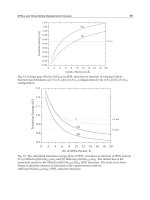
2.3 The optimization of the gold layer thickness
2.4 Sensitivity of the model
p
p
p
s
p
Require:
p
y p
y <
p p y <
while p do
p p
p
y p
<
p p y <
p p
p
end while
p p
<
3. Some metaheuristic optimization methods for plasmonics
p
3.1 Evolutionary method: a selective method
p
p
Require:
Require:
Require:
while > do
∑
∑
if then
else if then
end if
end while
Require:
Require:
Require:
while > do
∑ ∑
if then
else if then
end if
end while
p
p
p
p
p
p
<
3.2 Particle Swarm method: a collaborative method
p
p v
v
v p p p p
p
p v
p p
v
p
p
<
p
v
p
<
p
p
3.3 Numerical study of the selective and collaborative methods
>>
>
www.nanonatenna.eu
4. Conclusion
5. References
New Perspectives in Biosensors Technology and Applications
128
assay formats (Yen, 2004; Blasse, 1994). This review is composed of four sections, and is
intended to summarize the recent advances in luminescent semiconductor QDs and rare
earth UCNs for biosensing application. In the first section, the production mechanism, size-
dependent luminescence, spectral characteristics, bioconjugation technology and potential
biosensing application of semiconductor QDs are comprehensively reviewed; In the second
section, the controlled synthesis, characterization, luminescence mechanism, and biosensing
application of rare-earth UCNs are systemically introduced; In the third section, the
comparative assessment of advantage and limitation of semiconductor QDs and rare earth
UCNs in biosensing application are discussed; In the fourth section, the concluding remarks
and perspective for semiconductor QDs and rare-earth UCNs in biosensing application are
presented.
2. Semiconductor QDs as fluorescent labels for biosensing application
2.1 Concept of semiconductor QDs
Semiconductors have a filled band called the “valence band” and an empty band known as
the “conduction band”. At nanoscale dimension, the normally collective electronic
properties of semiconductors become severely distorted and the electrons tend to follow the
“particle in-a-box” model accounting for approximated band structure (Murray, 1993). From
a quantum mechanical point of view, when a semiconductor is irradiated with light of
photon energy (h
ν
) higher than E
g
, an electron will be promoted from the valence to the
conduction band, leaving a “hole” or “absence of an electron” in the valence band. Thus,
this “hole” is assumed to be a “particle” with its particular effective mass and positive
charge. The bound state of the electron-hole pair is called an “exciton” (Brus, 1984). The
exciton can be considered a hydrogen-like system, and a Bohr approximation of the atom
can be used to calculate the spatial separation of the electron–hole pair of the exciton by Eq. (1):
2
2
r
h
r
me
ε
π
= (1)
where r is the radius of the sphere, defined by the 3-D separation of the electron-hole pair, ε
is the dielectric constant of the semiconductor, m
r
is the reduced mass of the electron-hole
pair, h is Planck’s constant, and e is the charge on the electron. For many semiconductors,
the masses of the electron and hole have been determined by ion cyclotron resonance and
are generally in the range 0.1-3 m
e
(m
e
is the mass of the electron). For typical semiconductor
dielectric constants, the calculation suggests that the electron-hole pair spatial separation is
1-10 nm for most semiconductors (Gaponenko, 1998).
Because the physical dimensions of a QD can be smaller than the exciton diameter, the QD is
a good example of the “particle-in-a-box” calculations of undergraduate physical chemistry.
In those calculations, the energies of the particle in the box depend on the size of the box. In
the QD, the bandgap energy becomes size-dependent (Alivisatos, 1996; Gaponenko, 1998;
Zhang, 1997; Weller, 1993; Murphy, 2002).
2.2 Optical properties of QDs
QDs are nearly spherical semiconductor particles with diameters on the order of 1-10 nm,
containing roughly 200-10,000 atoms. When semiconductor QDs are smaller than their
Biosensing Based on Luminescent Semiconductor
Quantum Dots and Rare Earth Up-conversion Nanoparticles
129
exciton Bohr radii, the quantum confinement and size-dependent effects make QDs have
unique optical properties (Fig. 1): (1) single excitation, multi-emission and size-dependent;
(2) large stokes shift, narrow and symmetrical fluorescence peak; (3) visible light range
fluorescence and resistance to photobleaching; (4) superior signal brightness. In addition,
changing QD surface functional groups, luminescent properties and stability are greatly
improved and more conducive to the coupling of biological molecules. For conventional dye
molecules, their narrow excitation spectrum makes the simultaneous excitation difficult in
most cases, and their broad emission spectrum may cause a long tail at red wavelengths;
while for semiconductor QDs, the absorbance onset and emission maximum shift to higher
energy with the decrease of particle sizes (Alivisatos, 1996). The excitation tracks the
absorbance, resulting in a tunable fluorescence that can be excited efficiently at any
wavelength shorter than the emission peak, and therefore the characteristic narrow and
symmetric spectrum can be realized regardless of the excitation wavelength (Bruchez, 1998).
Fig. 1. Size-dependent optical properties of QDs. (a) Surface color of suspensions in toluene
in visible light; (b) Schematic diagram of band gap and emission color as a function of
particle size; (c) Light emission of suspensions in toluene when excited with UV light;
(d) Fluorescence spectra of the QDs samples (from left to right are respectively representative
2.2, 2.9,4.1 and 7.3nm QDs). (Feldmann, 2010; Mansur, 2010; Smith, 2008).
Due to their unique optical properties, semiconductor QDs can be used as fluorescent labels
for biological detection. In order to establish the utility of QDs for biological sensing
application, mouse 3T3 fibroblast cells were labeled with green and red emitting CdSe/CdS
nanocrystals. The green and red labels were spectrally resolved to the eye clearly under the
excitation of a single light source by a laser scanning confocal microscope. Nonspecific
labeling of the nuclear membrane by both the red and green probes resulted in a yellow
color [Fig. 2(a)]. The intensity of the fluorescein drops quickly to autofluorescence levels,
whereas the intensity of the QDs drops only slightly. Comparatively, the red QD labels are
20 times as bright, 100 times as stable against photobleaching [Fig. 2(b)] (Bruchez, 1998).
In general, QDs synthesized in nonpolar solutions using aliphatic coordinating ligands are
only soluble in nonpolar organic solvents, which are not suit for biological application.
New Perspectives in Biosensors Technology and Applications
130
Fig. 2. Schematic diagrams of dual-color labeling and photostability. (a) The mouse 3T3
fibroblasts were labeled with dual color. (b) Sequential scan photostability comparison of
fluorescein-phalloidin-labeled actin fibers compared with nanocrystal-labeled actin fibers
(Bruchez, 1998).
Fig. 3. (a) Scheme of a CdSe/ZnS QD covalently coupled to a protein; (b) Luminescent
images obtained from the original QDs; (c) mercapto-solubilized QDs; (d) Time-resolved
photobleaching curves for the original QDs, solubilized QDs and dye R6G (Chan, 1998).
Moreover, QDs have a huge surface/volume ratio, which makes them extremely unstable in
solution because of the high surface energy. Hence, any route chosen to synthesize QDs
should consider the stabilization of the QDs by minimizing the surface energy via “capping”
and avoiding further structure growth (Weaver, 2009). Warren and coworkers presented a
valuable way to solve this problem by coating CdSe QDs with higher bandgap materials
such as ZnS shell in order to increase the photostability and luminescence properties of
CdSe QDs (Chan, 1998). When reacting with CdSe/ZnS QDs in chloroform, the mercapto
group binds to a Zn atom, and the free carboxyl group is available for covalent coupling to
various biomolecules such as proteins by cross-linking to reactive amine groups [Fig. 3(a)]
(Hermanson, 1996). A comparison of color luminescence images were obtained from the
original QDs, water soluble QDs and protein-conjugated QDs [Figs. 3(b) and (c)], which
(a)
Biosensing Based on Luminescent Semiconductor
Quantum Dots and Rare Earth Up-conversion Nanoparticles
131
indicated that the optical properties of QDs remain unchanged after solubilization and
conjugation. The photophysical properties of QD conjugates with rhodamine 6G (R6G) was
also studied. The emission of mercapto-CdSe is somehow weaker than that of single QDs,
which is nearly 100 times as stable as R6G against photobleaching [Fig. 3(d)].
2.3 Synthesis of biocompatible QDs
The most common method for synthesizing water-soluble QDs is coated with a monolayer
of hydrophilic thiols, typically mercaptoacetic acid (MAA), to replace the hydrophobic
trioctylphosphine oxide (TOPO) coating on QDs (Chan, 1998; Hood, 2002; Duncan, 2006).
But, when the MAA replace the TOPO coating on QDs, the QDs become instable
accompanied by significant decreases in the quantum yield to 7% compared with TOPO-
coated QDs (Kim, 2004). To overcome this problem, Alivisatos and coworkers developed an
effective route to coat QDs with a cross-linked silica shell, which can be readily modified
with a variety of organic functionalities such as primary amines, carboxylic acids or thiols
(Gerion, 2001). The coated QDs were very stable and retained 60-80% of the quantum yield
of the original QDs. Gao and coworkers developed another effective method to synthesize
CdSe/ZnS QDs stabilized by a coordinating ligand (TOPO) and an amphiphilic polymer
coating through hydrophobic attraction (Gao, 2004). Because of the strong hydrophobic
interactions between TOPO and polymer hydrocarbon, the two layers bonds to each other
and form a hydrophobic protection structure that resists hydrolysis and enzymatic
degradation even under complex in vivo conditions. In most designs of the amphiphilic
polymers, carboxylic acids provide solubility in water and can be utilized as
Fig. 4. Schematic diagrams of biocompatible QDs.
chemical handles for conjugation to primary amines in proteins through water soluble cross-
linking reagents such as 1-ethyl-3-(3-dimethylaminopropyl) carbodiimide (EDAC).
Similarly, many of these QDs also may be modified to contain polyethylene glycol (PEG) to
decrease surface charge and increase colloidal stability (Fig. 4) (Dubertret, 2002 ; Smith, 2006;
You, 2007; Bagwe, 2003).
2.4 Biosensing based on biocompatible QDs
On the base of the synthetic methods of biocompatible QDs, water-soluble QDs can be
covalently or electrostatically bound to a biological target, which have also acted as a new
class of sensor. If the QDs encapsulated in amphiphilic polymers and PEG conjugated to
antibodies, it would yield specificity for a variety of antigens. In addition, QDs cross-linked
to other small molecule ligands, inhibitors, peptides, or aptamers can bind with many
different cellular receptors and targets (Fig. 5) (Lidke, 2004; Xing, 2007). Gao and coworkers
developed multifunctional nanoparticle probes based on semiconductor QDs for prostate
New Perspectives in Biosensors Technology and Applications
132
cancer targeting and imaging (Gao, 2004). The probes with passive and active tumor
targeting behaviors were produced. This new class QDs probes contain an amphiphilic tri-
block copolymer for in vivo protection, targeting-ligands for tumor antigen recognition and
multiple PEG molecules for improved biocompatibility and circulation [Fig. 5(a)]. In the
passive mode, antigenic tumors produce vascular endothelial growth factors, which can
hyper-permeabilize the tumor-associated neovasculatures and cause the leakage of
circulating macromolecules and small particles, leading to macromolecule or nanoparticle
accumulation [Fig. 5(b)] (Gao, 2004; Duncan, 2003; Jain, 1999, 2001). While for active tumor
targeting, antibody-conjugated QDs can track a prostate-specific membrane antigen
(PSMA), which could be selected as an attractive target for imaging and therapeutic
intervention of prostate cancer [Fig. 5(b)] (Gao, 2004; Schulke, 2003). This study opens new
possibilities for ultrasensitive and simultaneous imaging of multiple biomarkers involved in
cancer metastasis and invasion.
Fig. 5. Schematic illustration of bioconjugated QDs for in vivo cancer targeting and imaging.
(a) Structure of a multifunctional QD probe; (b) Permeation and retention of QD probes via
leaky tumor vasculatures (passive targeting) and high affinity binding of QD-antibody
conjugates to tumor antigens (active targeting) (Gao, 2004).
Subsequently, Wu and coworkers demonstrated the use of 535QD-IgG and red 630QD-
streptavidin to detect Her2 on the cell surface and nuclear antigens in the nucleus of SK-BR-
3 cells. When the sample was observed under a fluorescence microscope, 630QD-labeled
(red) nuclear antigens and 535QD-labeled (green) membrane-associated Her2 were visible
simultaneously (Wu, 2003). This indicated that QDs conjugated to different secondary
detection reagents can effectively detectect two cellular targets in the same cell. These results
demonstrated the practicality of QDs in biological cellular real time and dynamic state
imaging fields.
Fluorescence resonance energy transfer (FRET) is most commonly utilized in biosensors for
detecting maltose (Medintz, 2003), aptamers (Hansen, 2006), 2,4,6-trinitrotoluene (Goldman,
2005), toxins (Goldman, 2004), and DNA (Zhang, 2005). Because of their high sensitivity,
good reproducibility, and real-time monitoring capabilities, QDs are usually acted as
fluorescence donors and make up of FRET with organic dyes. Medintz and coworkers
designed a maltose sensor [Fig. 6(a)], in which an organic dye QSY-9 (fluorescence acceptor)
was first conjugated to β-cyclodextrin (β-CD), then bonded to maltose binding protein
(MBP), and at last β-CD-QSY-9/MBP complex was attached to the 560 QDs (fluorescence
donors) surface through a peptide His-tag (Medintz, 2003). The optimized sensor contained
10 copies of β-CD-QSY9 bound to the QD complex, where 75% of the QD fluorescence was
quenched by QSY-9. When free maltose was added, it would displace the β-CD-QSY9.
Biosensing Based on Luminescent Semiconductor
Quantum Dots and Rare Earth Up-conversion Nanoparticles
133
Moreover, the displacement of β-CD-QSY-9 with maltose could result in QD fluorescence
increasing about 3-fold. This technique can be used to achieve the sensing of maltose.
However, due to the uncertainty in the distance between the QDs and acceptors, some
limitations in this sensor were arisen. In order to overcome the limitations, another maltose
sensor was architected [Fig. 6(b)], in which 10 copies of Cy3 labeled MBP were first
incorporated on the 530QDs surface, followed by binding of the Cy3.5 labeled β-CD, at last
β-CD-Cy3.5/MBP-Cy3 complex was bound to QD through a peptide His-tag and Cy3.5
fluorescence emitted through a two-step FRET process. Sufficient fluorescence energy was
initially transferred from the 530QD to MBP-Cy3, and the minimized emission energy of
Cy3 was then transferred to β-CD-Cy3.5. When free maltose was added, the displacement of
β-CD-Cy3.5 with maltose resulted in fluorescence increasing from Cy3 and concomitantly
fluorescence decreasing from Cy3.5. The results demonstrate that the appropriately
designed QD complexes with peptide immobilization tags can be used in determining small
molecule concentrations in the 100 nM-10 μM range (Medintz, 2003)
Another biosensor based on combination of QDs and multi-walled carbon nanotubes (CNT)
makes the detection of DNA and antigen more quickly and simply. Cui et al reported a
highly selective, ultrasensitive, fluorescent detection method for DNA and antigen based on
self-assembly of multi-walled carbon nanotubes (CNT) and CdSe QDs via oligonucleotide
hybridization (Cui, 2008). This method could achieve the detection limit of 0.2 pM DNA
molecules and 0.01 nM antigen molecules, and the novel detection system not only can be
used for multicomponent detection and antigen-antibody immunoreaction, but also has
great potential in photoelectrical biosensing application.
(a)
(b)
Emission
Maltose
Excitation
FRET quenching
β-cyclodextrin-
QSY-9
Emission Emission
FRET 1
FRET 1
FRET 2
Excitation
Maltose
His-tag
His-tag
Fig. 6. QD based maltose nanosensor. (a) β-CD-QSY-9/MBP complex bound to QD through
a peptide His-tag; (b) β-CD-Cy3.5/MBP-Cy3 complex bound to QD through a peptide His-
tag (Zhou, 2007).
Recent advances in single-molecule detection, aptameric sensors with the surface
functionalizing QDs hold exciting promise for many potential applications. Zhang et al
New Perspectives in Biosensors Technology and Applications
134
developed a single-QD-based aptameric sensor through (FRET) between 605QD and Cy5
and Iowa Black RQ (Zhang, 2009). This aptamertic sensor can recognize cocaine through
both signal-off and signal-on modes, indicating the higher sensitivity and more extremely
low sample consumption than that of the aptameric sensors established before. With the
development of aptamers for small molecules, nucleic acids, metal ions, and proteins, this
single-QD-based aptameric sensor might find wide application in forensic analysis,
environmental monitoring, and clinic diagnostics (Zhang, 2009).
Fig. 7. Schematic illustrations of optical coding and DNA hybridization assays using QD-
tagged beads (a) Optical coding based on wavelength and intensity multiplexing. Large
spheres represent polymer microbeads, in which small multicolor QDs are embedded
according to predetermined intensity ratios; (b) DNA hybridization assays using QD-tagged
beads. Probe oligos were conjugated to the beads by crosslinking, and target DNA molecules
were detected with a blue fluorescent dye such as Cascade Blue (Han, 2001).
Han and coworkers synthesized monodispersed CdSe/ZnS QDs with fluorescence emission
at three primary colors (red, green and blue) (Han, 2001). Through embedding different-
sized QDs into polymeric microbeads at precisely controlled intensity ratios [Fig. 7(a)],
multicolor-tagged beads were prepared. Code readout is accomplished by measuring the
fluorescence spectra of single beads. Their fluorescence emission wavelength can be
continuously tuned by changing the particle size, and a single wavelength can be used for
simultaneous excitation of different-sized QDs (Alivisatos, 1996; Han, 2001, Nirmal, 1999).
On the other hand, molecular probes (A-E) may be attached to the bead surface for
biological binding and recognition, such as DNA-DNA hybridization. In order to
demonstrate the use of QD-tagged beads for DNA hybridization, oligonucleotide probes
were conjugated to the beads by cross-linking. Target DNA molecules are directly labeled
with a blue fluorescent dye such as Cascade Blue [Fig. 7(b)] (Han, 2001). Optical
spectroscopy at the single-bead level yields both the coding and the target signals.
Moreover, each color code corresponds to a specific DNA sequence. The coding signals
can identify the DNA sequence, whereas the target signal can indicate the abundance of
that sequence. A surprising finding is that the number of codes increases exponentially
when multiple wavelengths and intensities are used simultaneously, for example, a
Biosensing Based on Luminescent Semiconductor
Quantum Dots and Rare Earth Up-conversion Nanoparticles
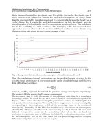
135
3-color/10-intensity scheme yields 999 codes, whereas a 6-color/10-intensity scheme has a
theoretical coding capacity of about one million. In general, n intensity levels with m
colors generate (n
m
-1) unique codes (Han, 2001). If every code corresponds one type
specific DNA sequence, it would be labeled every biomolecules with fluorescent code.
This system made by multicolor-tagged QDs will completely change the ability of human
identifying gene.
3. Rare-earth up-converting nanoparticles as fluorescent labels for
biosensing application
3.1 Up-converting luminescence mechanism
Up-conversion (UC) refers to nonlinear optical processes characterized by the successive
absorption of two or more pump photons via intermediate long-lived energy states followed
by the emission of the output radiation at a shorter wavelength than the pump wavelength.
UC processes are mainly divided into three broad classes: excited state absorption (ESA),
energy transfer up-conversion (ETU), and photon avalanche (PA). All these processes
involve the sequential absorption of two or more photons (Fig. 8).
In the case of ESA, the excitation takes the form of successive absorption of pump photons
by a single ion. This is the basic process of up-conversion [Fig. 8(a)]. If the excitation energy
is resonant with the transition from ground level G to excited metastable level E1, the
phonon absorption occurs and populates E1 from G in a process known as ground state
absorption (GSA). A second pump photon that promotes the ion from E1 to higher-lying
state E2 results in UC emission, corresponding to the E2-G optical transition (Wang, 2009).
ETU is similar to ESA in that both processes utilize sequential absorption of two photons to
populate the metastable level. The essential difference between ETU and ESA is that the
excitation in ETU is realized through energy transfer between two neighboring ions. In an
ETU process, each of two neighboring ions can absorb a pump phonon of the same energy,
thereby populating the metastable level E1 [Fig. 8(b)]. A non-radiative energy transfer
process promotes one of the ions to upper emitting state E2, while the other ion relaxes back
to ground state G. The dopant concentration that determines the average distance between
the neighboring dopant ions has a strong influence on the UC efficiency of an ETU process
(Wang, 2009).
The phenomenon of PA was first discovered by Chivian and co-workers in Pr
3+
-based
infrared quantum counters (Chivian, 1979). PA-induced UC features an unusual pump
mechanism that requires pump intensity above a certain threshold value. The PA process
starts with population of level E1 by non-resonant weak GSA, followed by resonant ESA to
populate upper visible-emitting level E2 [Fig. 8(c)]. After the metastable level population is
established, the cross-relaxation energy transfer or ion pair relaxation occurs between the
excited ion and a neighboring ground state ion, resulting in both ions occupying the
intermediate level E1. The two ions readily populate level E2 to further initiate cross-
relaxation and exponentially increase level E2 population by ESA, producing strong UC
emission as an avalanche process (Wang, 2009).
The UC luminescent efficiency in these three processes varies considerably. ESA is the least
efficient UC process. Efficient UC is possible in PA with metastable, intermediate levels that
can act as a storage reservoir for pump energy. However, the PA process suffers from a
number of drawbacks, including pump power dependence and slow response to excitation
(up to several seconds) due to numerous looping cycles of ESA and cross-relaxation