báo cáo hóa học: " Patterns of pulmonary dysfunction in asbestos workers: a cross-sectional study" docx
Bạn đang xem bản rút gọn của tài liệu. Xem và tải ngay bản đầy đủ của tài liệu tại đây (564.86 KB, 7 trang )
Abejie et al. Journal of Occupational Medicine and Toxicology 2010, 5:12
/>Open Access
RESEARCH
© 2010 Abejie et al; licensee BioMed Central Ltd. This is an Open Access article distributed under the terms of the Creative Commons
Attribution License ( which permits unrestricted use, distribution, and reproduction in
any medium, provided the original work is properly cited.
Research
Patterns of pulmonary dysfunction in asbestos
workers: a cross-sectional study
Belayneh A Abejie
1
, Xiaorong Wang
2
, Stefanos N Kales
3
and David C Christiani*
3
Abstract
Background: Restrictive patterns of pulmonary function abnormalities associated with asbestos exposure are well
described. Studies are less consistent, however, regarding the association of asbestos inhalation with airway
dysfunction and obstructive impairment.
Methods: We compared pulmonary function test results between 277 chrysotile exposed workers (22% non-smokers)
and 177 unexposed controls (50.3% non-smokers). Information on exposure and smoking were collected using a
standardized questionnaire. Standardized spirometric and DCLO Measurement methods were utilized. CXRs were read
based on ILO pneumoconiosis guidelines.
Results: Asbestos exposed subjects had significantly reduced FVC, FEV1, FEV1/FVC and DLCO. Restricting the analysis
to non-smokers, asbestos workers still had about 3% lower FEV1/FVC ratio than controls, but this difference did not
reach statistical significance. Among exposed workers, the presence of radiographic evidence of asbestosis further
lowered FVC and DLCO but not FEV1/FVC compared to asbestos exposure without radiographic asbestosis.
Additionally, smoking asbestos workers had significantly lower DLCO compared to non-smoking workers.
Conclusion: Asbestos exposure, especially when radiographic evidence of interstitial fibrosis from asbestosis is
present, leads to significant decreases in FVC, FEV1 and the DLCO. However, asbestos exposure alone is not significantly
associated with a reduction of the FEV1/FVC. Smoking-asbestos workers had significantly lower DLCO than their non-
smoking counterparts. Whether asbestos interacts with smoking additively or synergistically on DLCO needs further
investigation. Similarly, further studies are needed to assess the progression and clinical significance of asbestos
induced airway dysfunction.
Introduction
The association of a restrictive pulmonary function with
interstitial lung disease is well described [1-12]. However,
the results of studies examining obstructive airway
impairment in asbestos- exposure are not entirely consis-
tent. Such investigations of airway function have been
conducted in animal models, clinical series, and epidemi-
ological surveys.
In 1982 Begin observed small and large airway disease
in sheep with tracheal installation of high concentrations
of chrysotile asbestos [13]. He further demonstrated that
asbestos airway disease appears to be dose dependent
[14]. In 1985, Filipenko et al found thickened membra-
nous and respiratory bronchioles in Guinea Pigs [15].
Similarly, Bellis observed small air way lesions in lung
autopsies [16]. Dumortier reported small airway patho-
logic changes in Guinea pigs after amosite exposure in
1990 [17]. However, whether asbestos can induce clini-
cally significant obstruction in non-smoking human pop-
ulations remains somewhat controversial. Additionally,
because occupational exposure is often to mixed-mineral
dust, rather than only to asbestos, the ability to extrapo-
late from animal studies to humans is limited.
Harless observed that chrysotile exposed workers
developed abnormal FEF25-75 and nitrogen washout
curves [18]. Consequently, Rodriguez-Roisin and his col-
leagues found flow volume curve abnormalities sugges-
tive of small air way lesions [19]. Similarly, Begin found
evidence of diminished flows at low lung volumes in non-
smoking chrysotile workers [20], and Becklake observed
an obstructive pattern of reduction in spirometry in
groups with high dust exposure[21]. Later, Griffith et al
demonstrated airway disease in a non-smoking cohort of
* Correspondence:
3
Harvard School of Public Health, Boston, MA, USA
Full list of author information is available at the end of the article
Abejie et al. Journal of Occupational Medicine and Toxicology 2010, 5:12
/>Page 2 of 7
asbestos workers [22]. Kilburn and Warshaw observed a
reduction in FEV1, FEV1/FVC ratio, and an increase in
RV/TLC, an obstructive pattern [23]. Wang et al showed
significant decrease in FEF25-75% in older asbestos
workers [12].
However; earlier studies [12,24-31] did not support the
relationship of asbestos exposure to obstructive lung dys-
function. Similarly, in 1994, Miller et al did not observe
strong evidence for obstructive impairment on 2611 par-
ticipants (20% non-smokers) [32]. Still other studies have
shown mixed PFT abnormalities [33-35]. Furthermore, in
most of the studies, especially those conducted before the
mid-nineties, either small sample size and/or the effect of
smoking were limitations making interpretation difficult.
Other areas of concern in most of the studies included
the use of FEF, a less stable measure of obstruc-
tion[12,22,33,36], incomparable or absent control groups,
[18,23,32], lack of DLCO measurement [32,33], single
chest -x-ray reader [23], and use of unadjusted FEV1/
FVC and RV/TLC ratios[23]. To provide additional infor-
mation, we compared the pattern of pulmonary dysfunc-
tion in asbestos workers using spirometric and DLCO
measurements in a relatively large groups of chrysotile
exposed subjects and controls without asbestos exposure.
Methods
Study Population
As a part of a study on the respiratory health status of
dust exposed workers, chrysotile factory workers were
surveyed in 1989. The workers came from a factory in
which asbestos textile products were manufactured in
Southwest China. Their pulmonary examination
included clinical evaluation, chest radiography, and
spirometry and diffusion capacity (DLCO) measure-
ments. Subject selection was restricted to male workers
with direct asbestos exposure for at least 2 years, but no
overt neuromuscular and clinical cardiopulmonary disor-
ders other than pneumoconiosis at the time of survey.
Invitations for participation covered all current workers,
and retired workers who were living close to the asbestos
factory. Retired workers living far from the factories were
not included for logistic reasons. Women were not
included because they comprised a very small number.
Study subjects were not exposed to other fibers or dust
except asbestos.
Control groups were drawn from employees of the elec-
tronic industry located in the same geographic area as the
asbestos factory. Selection was restricted to male workers
with at least 2 years work history, no history of asbestos
or any other dust exposure, and no overt cardiopulmo-
nary and neuromuscular problems. The study was
approved by the Human Subjects Committee of the West
China University Medical School.
Exposure Assessment
The factory was established in 1950. Since the 1970s
some engineering control measures were in place, but in
most cases the area sample concentration range still
exceeded 2 mg/m
3
, the Chinese maximum allowable con-
centration at that time. Workers did not use personal pro-
tective equipment. During their stay in the plant,
employees changed job types frequently and did not hold
the same job title for long period of time. Therefore, the
individual cumulative duration of work in exposed areas
was used as surrogate measure of total asbestos exposure.
Clinical Evaluation
Using a Chinese standardized respiratory questionnaire,
which was based on Medical Research Council Question-
naire [37], face- to- face interviews of both exposed and
control groups was conducted by two physicians. Infor-
mation was gathered on demographic data, occupational
history, smoking habits and respiratory symptoms. Spe-
cial attention was given to job title and beginning and end
dates at each job in occupational history. Smoking was
quantified in pack years and also categorized in to 3 qual-
itative groups defined as follows. Current smokers were
those who were currently smoking or had quit smoking
less than 3 months before the time of interview; ex-smok-
ers as those who quit smoking at least 3 months prior to
the interview and non-smokers as those who had never
smoked more than 20 packs of cigarettes in their life time
or no more than 1 cigarette per day for one or more years.
Pack years were defined as the number of packs (one pack
= 20 cigarettes) multiplied by the number of years
smoked.
Radiographic Evaluation
Posterior-Anterior (PA) chest-x-rays (CXR) on full inspi-
ration and standing position were done at least once for
each asbestos worker and were read by panels for pneu-
moconiosis and emphysema. Panel members include pul-
monologists, radiologists and occupational health
experts. Readers were blinded to PFT values and the CXR
findings were based on the consensus of at least two
experts. The 1986 Chinese Roentgeno-Diagnositc criteria
of pneumoconiosis, established based on the 1980 inter-
national labor organization (ILO) classification of pneu-
moconiosis, were used to grade the severity of asbestosis.
Stage 0, I, and II correspond to ILO stages (0/-to 1/0), (1/
1 to 2/3) and (3/2 to 3/+) respectively. Stage III represents
ILO large opacities with categories A, B, and C. Radio-
graphic asbestosis was defined as perfusion densities
stage I (1/1) or greater in persons with a history of asbes-
tos exposure. There is a good agreement between the
Chinese Roentgeno-Diagnosis criteria of pneumoconio-
sis and ILO CXR system [38]. Emphysema was diagnosed
Abejie et al. Journal of Occupational Medicine and Toxicology 2010, 5:12
/>Page 3 of 7
and graded radiographically as none, mild, moderate or
severe by panel member consensus.
Spirometric and DCLO Measurements
A 9-L water- sealed spirometer (Godart Pulontest, NV,
The Netherlands) was used to measure FVC and FEV1
following ATS guidelines [39]. Participants did not smoke
for at least one hour before the test. At least three accept-
able efforts were obtained in each participant while wear-
ing nose clips in standing position. Care was taken to
maintain expiration for at least 6-seconds or until flow
plateau was observed. The largest values of FEV1 and
FVC were chosen for analysis. Single breath diffusion
capacity for carbon monoxide (DLCO) test was per-
formed based on Epidemiology Standardization Project
protocol [40]. Subjects were in sitting position during the
test and the breath hold time was 10-seconds. For sub-
jects with FVC of 2 or more liters (L), the washout vol-
ume was 1 L and for those with FVC of less than 2 L, the
washout volume was 0.5 L. A pulmonary gas analyzer
(GC-1, Shanghai, China) was used for gas analysis. DLCO
was calculated using inspired volume, breath hold time,
and CO and helium concentrations. Measured values
(except FEV1/FVC ratios) corrected for body tempera-
ture, ambient pressure and saturated water vapor were
expressed as the percentage of predicted values calcu-
lated with equations that considered age, height and gen-
der derived from the Chinese general population. The
same team of technicians conducted the tests in both the
exposed and control groups using same equipment and
procedures. Although PFT technicians were not blinded
to exposure status (because testing was conducted on
worksite), they were not aware of the clinical and radio-
graphic characteristics of each participant. Similar to
Ohar et al [41], mutually exclusive predictive value per-
centages were used to define PFT patterns as follows.
Normal: FVC ≥80%, and FEV1/FVC ≥70%; Restrictive:
FVC < 80% and FEV1/FVC ≥70%; Obstructive: FVC
≥80% and FEV1/FVC < 70%; and Mixed: FVC <80% and
FEV1/FVC <70%.
Statistical Methods
The mean values of baseline characteristics were
obtained from the SAS proc means procedure (SAS 9
version). Multiple regression techniques were utilized to
analyze the relationships of exposure and other indepen-
dent variables with pulmonary function test values. With
regard to smoking, pack-years rather than yes/no was
included in the regression models. In all analyses, a p
value less than 0.05 (two sided) was considered signifi-
cant. SAS software (Version 9.1, Cary, NC) was used for
all statistical data analyses.
Results
Two hundred seventy seven asbestos workers and 177
control subjects were included in the study (Table 1). The
participation rates for exposed and control subjects were
not different: 77% and 80% respectively. The asbestos
workers were significantly older than the controls. Smok-
ing was more frequent among asbestos workers, and they
also had smoked a greater number of pack years. Among
the asbestos workers, 36% had radiographic changes con-
sistent with emphysema, 31% with asbestosis, and 15%
had CXR findings consistent with both asbestosis and
emphysema.
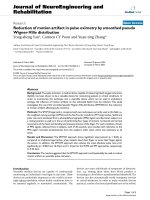
As shown in figure 1, more than 80% of the controls
had normal pulmonary function compared to only half of
the asbestos workers had normal pulmonary function.
Consequently, the proportions of subjects with obstruc-
tive, restrictive and mixed patterns of pulmonary dys-
Table 1: Basic Characteristics
Variable Exposed (N = 277) Controls (N = 177) P-value
Age, mean(SD) 55.1 (12.0) 37.2 (10.1) .0001
Height, mean(SD) 162.5 (5.9) 167.0 (5.8) .0001
Exposure year, mean(SD) 16.7 (9.3)
Pack year, mean(SD) 27.9 (18.3) 10.8 (8.3) .0001
FEV1, mean(SD) 96.6 (22.0) 103.9 (13.9) .0001
FVC, mean(SD) 96.3 (17.0) 102.6 (12.4) .0001
FEV1/FVC, mean(SD) 71.3 (12.1) 78.3 (8.0) .0001
DLCO, mean(SD) 97.1 (21.6) 124.6 (18.8) .0001
Ever smokers (%) 78.0 49.7
Asbestosis (%) 30.7
Emphysema (%) 35.7
Asbestosis emphysema (%) 15.2
Abejie et al. Journal of Occupational Medicine and Toxicology 2010, 5:12
/>Page 4 of 7
function in the exposed group were all higher than the
corresponding proportions in the control group.
Although the PFT values except the FEV1/FVC ratio
were adjusted for age and height, we also included age in
the regression analysis as there was significant age differ-
ence between the exposed and control groups (Table 2).
Because we were interested in examining the effect of
mere asbestos exposure (without CXR evidence of asbes-
tosis) on patterns of pulmonary, we excluded patients
with radiographic asbestosis from the model. After
accounting for age and smoking, asbestos exposure was
significantly associated with restrictive pattern of pulmo-
nary dysfunction and decreased DLCO. However, asbes-
tos exposure was not significantly associated with FEV1/
FVC ratio. Similar results were found when FEV1, FVC,
and DLCO were regressed on exposure status and pack
year without including age in the model (not shown).
As shown in Table 3, when PFT values were regressed
on exposure status in non-smokers only, asbestos expo-
sure was significantly associated with low FEV1, FVC and
DLCO percent predicted values after accounting for age.
In addition, the results indicate that non-smoking asbes-
tos workers had close to 3% less FEV1/FVC ratios com-
pared to non -smoker control workers of similar age and
height. This last relationship, however, was not statisti-
cally significant. For FEV1, FVC and DLCO, the results
were similar when age was excluded from the regression
model as the PFT values were adjusted for age (analysis
not shown).
To compare the effect of radiographic asbestosis on
PFT values as opposed to asbestos exposure (without
asbestosis), we performed regression analysis of PFT val-
ues on asbestos exposed subjects only (Table 4). We
removed patients with radiographic emphysema in this
analysis to avoid the possible confounding effect of
emphysema. As expected, individuals with radiographic
asbestosis had significantly lower FVC and DLCO values
than those asbestos exposed individuals without asbesto-
sis. However, there was no significant difference in the
FEV1/FVC ratio between these two groups.
Finally, to examine the effect of smoking per see on
DLCO and FVC values, we performed regression analysis
on exposed subjects who do not have radiographic asbes-
tosis or emphysema (Table 5). As expected, pack-years of
smoking was significantly associated with FEV1 and
FEV1/FVC ratio. Similarly, the pack-years variable was
significantly associated with DLCO. Furthermore, pack-
years was negatively related to FVC, although not statisti-
cally significant.
Discussion
Our study supports, that asbestos exposure, with or with-
out radiographic asbestosis, contributes to obstructive
airway impairment. The proportion of asbestos exposed
subjects with obstructive pulmonary impairment was
about 2.5 times higher than that of the controls. However,
caution should be exercised in the interpretation of our
results, because different smoking habits may explain
some of the difference as more than 80% of the partici-
pants in exposed group were smokers compared to 50%
in controls. In a separate regression analysis, we found no
significant difference in FEV1/FVC ratios between non-
smoking asbestos workers and non-smoking controls, but
asbestos workers still had almost 3% lower FEV1/FVC
ratios compared to their corresponding controls (Table
3). We believe this difference did not reach statistical sig-
nificance due to limits of our available sample size. More-
over, the FEV1/FVC ratio generally reflects large airways
function, and the earliest asbestos lung lesions are peri-
Figure 1 Patterns of Pulmonary Dysfunction in Exposed and Con-
trol Groups. Exposed (N = 277). ᮀ Controls (N = 177).
51.6
33.8
7.3 7.3
81.4
14.7
3.4
0.5
0
10
20
30
40
50
60
70
80
90
normal obstructive restrictive mixed
Patterns of Pulmonary Dysfunction
Percent
Table 2: Regression Analysis: Control and Exposed Groups without Asbestosis (N = 369)
FEV1 FVC FEV1/FVCa DLCO
Age 0.09 (0.08)† 0.11 (0.07) -0.40 (0.05)* -0.20 (0.10)*
Exposure -6.69 (2.62)* -5.72 (1.81)* 0.25 (1.26) -19.30(2.71)*
Pack Year -0.14 (0.07)* -0.06 (0.05) -0.07 (0.04)* -0.173(0.07)
† = coefficient (standard error) a = adjusted for height * = significant (p < .05)
Abejie et al. Journal of Occupational Medicine and Toxicology 2010, 5:12
/>Page 5 of 7
bronchiolar, and abnormalities in this anatomic region of
the lung are not well-captured on standard pulmonary
function testing [39,42]. In addition to smoking, another
potential confounder was age. The asbestos workers were
significantly older than the controls. However, we believe
that we minimized any confounding by age. Age was
adjusted for twice (first using PFT prediction equation,
and again in multivariate regression analysis).
Our findings are in general agreement with past stud-
ies, finding excess obstruction among asbestos exposed
workers, but remaining inconclusive as to how much of
the effect is independent from smoking. Kilburn reported
significant differences in FEV1/FVC and RV/TLC
between non-smoking asbestos exposed subjects and
controls in 1994[23]. However, his study was criticized
for using unadjusted FEV1/FVC and RV/TLC ratios [43].
Several other studies showed similar results. For instance,
Harless [18] demonstrated airflow obstruction in 23
heavily exposed male asbestos workers. Garcia-Closas, M
and Christiani, DC reported mixed (restrictive-obstruc-
tive) patterns in a study of carpenters with pleural
plaques [34]. Similarly, airway dysfunction has been
reported in several other studies [18-20,22].
However, Miller did not observe significant differences
in FEV1/FVC and FEF25-75% among non-smoking
asbestos exposed subjects compared for duration of
exposure in 1994[32]. Similarly, Sue et al reported that
cigarette smoking, not asbestos, was the major contribut-
ing factor for the decline in FEV1/FVC ratio in asbestos-
exposed workers in 1985[28]. Earlier studies in the 1970's
did not support the claim that asbestos exposure was
associated with airway dysfunction [24,26,27,29]. How-
ever, most of these studies had serious limitations such as
the lack of unexposed controls and failure to control the
effect of smoking. The strengths of our study included the
use of unexposed controls from the same area and socio-
economic stratum, a detailed smoking history, and the
analysis of airway dysfunction, and the use of standard-
ized (ATS) pulmonary function testing and interpretation
criteria.
The proportion of subjects with restrictive impairment
in the exposed group was 2.2 times more than the corre-
sponding proportion of subjects in the control group.
The historical area sample concentrations, lack of expo-
sure control measures in the company and the average
duration of exposure (16.7 years) supports that the mag-
nitude of asbestos exposure was high. Our study shows
that asbestos exposure (without radiographic asbestosis)
is significantly associated with decreased FVC, FEV1 and
DLCO, consistent with previous studies [1-12]. The
reduced FVC does not necessarily indicate volume loss as
it could result from air trapping. In addition, the marked
DLCO reduction in exposed subjects favors interstitial
lung disease with alveolar involvement, since asbestos
does not cause emphysema. Similarly, one may argue that
pleural diseases might have contributed to the reduced
FVC and FEV1. However, as Miller, and Garcia-Closas
and Christiani pointed out, the association between dis-
crete pleural diseases (plaques), and restrictive impair-
ment is weak [32,34,35]. Given that pleural plaques are
rare with in less than 20 year of exposure [ATS2004], and
the average exposure of our study group was less than 11
years, pleural thickening is unlikely to explain our find-
ings. Furthermore, the marked difference in DLCO again
supports substantial early interstitial abnormalities that
are not detected by plain radiographs. The proportion of
subjects with a mixed pattern of pulmonary impairment
in exposed subjects was more than 14 times greater than
among the controls, which is also consistent with other
previous findings[34].
As expected, workers with radiographic asbestosis had
significantly lower FVC and DLCO values compared to
other exposed workers. However, the two groups were
similar in terms of FEV1/FVC ratio. This finding is con-
Table 3: Regression Analysis: Non-Smokers (N = 130)
FEV1 FVC FEV1/FVC a DLCO
Age 0.22 (0.12)† 0.18 (0.10) -0.38 (0.07) * 0.12 (0.17)
Exposure -11.67(3.69)* -6.58 (3.14)* -2.79(2.07) -31.87(5.11)*
† = coefficient (standard error) a = adjusted for height * significant(p < .05)
Table 4: Regression Analysis: Exposed Subjects without Emphysema (N = 175)
FEV1 FVC FEV1/FVCb DLCO
Asbestosis -1.85 (3.46)† -5.66(2.91)* 0.75 (1.6) -9.76 (3.93)*
Pack Year -0.14 (0.08) -0.03 (0.06) -0.10 (0.04)* -0.21 (0.09)*
† = coefficient (standard error) b = adjusted for age & height * = significant(p < .05)
Abejie et al. Journal of Occupational Medicine and Toxicology 2010, 5:12
/>Page 6 of 7
sistent with those of Kilburn and Warshaw findings [23].
The reason for lack of significant difference in FEV1/FVC
ratio between those asbestos exposed groups with and
without radiographic asbestosis is not clearly understood.
Some say airway dysfunction is not related to asbestos
fiber burden[22]. However, others observed small airway
dysfunction only in long-term exposure, [2,11,19] and
still others claim that low cumulative exposures are less
likely to produce airway abnormalities [5,27,44]. Other
explanations include enhanced elastic recoil in asbestosis
[ATS 2004] and increase lung radial traction by fibro-
sis[36].
Asbestos workers (without radiographic asbestosis or
emphysema) who smoked had significantly lower DLCO,
and not surprisingly lower FEV1 and FEV1/FVC ratio
compared to asbestos workers who did not smoke.
Among 21 studies, reviewed by Weiss, 11 showed an
additive positive interaction between smoking and asbes-
tos[45]. Similarly, Kilburn and Wright reported a syner-
gistic effect of smoking with asbestos in insulators and
Guinea pigs respectively [46,47]. However, Alfonso et al
reported no significant interaction between asbestos and
smoking[1].
Although the mechanism for asbestos related intersti-
tial pulmonary diseases is well described, the pathogene-
sis of asbestos-related disease obstructive airway diseases
is still unsolved. Begin et al reported peribrochial alveoli-
tis, in high dose crysotile asbestos exposed sheep and
fibrosis with obliteration and narrowing of the small air-
ways in lung biopsy of three asbestos workers in 1982 and
1983 respectively [1,13,48]. Wright and Churg demon-
strated sever diffuse airway pathology after studying
necropsy of 36 asbestos miners and their matched con-
trols in 1985[49]. Similarly, Filipenko et al demonstrated
significantly thickened non-cartilaginous airways in
amosite exposed guinea pigs in 1985[15]. On the other
hand Griffin et al claimed that mineral-dust airway dis-
ease is irritant phenomenon based on individual suscepti-
bility irrespective of dust burden[22].
Our study had several limitations: First, the asbestos
workers were significantly older than the controls. How-
ever, age was adjusted for in both the predictive equations
and our regression model. Second, unlike the radio-
graphic panel experts, the PFT technicians were not
blinded to the status of asbestos exposure. Other weak-
nesses include the lack of chest films in controls, the
absence of pleural radiographic information in asbestos
workers, and the lack of area or personal asbestos mea-
surements for exposure assessment. Nonetheless, these
limitations do not negate our findings of the lower pul-
monary function among the asbestos exposed workers.
In conclusion our study showed that asbestos exposure
with or without radiographic asbestosis is significantly
associated with reduced DLCO and restrictive lung
impairment. However, asbestos exposure was not signifi-
cantly associated with reducedFEV1/FVC. Among the
exposed workers, radiographic asbestosis was associated
with lower FEV1, FVC and DLCO values, but was not
associated with any further reduction in the FEV1/FVC
ratio. Finally smoking-asbestos exposed subjects had sig-
nificantly reduced DLCO compared to their non-smok-
ing counterparts. Further investigation is needed to
determine whether combined exposure to asbestos and
smoking act in an additive or synergistic fashion in reduc-
ing lung function, and to assess the progression and clini-
cal significance of asbestos-induced airway impairment.
Competing interests
The authors declare that they have no competing interests.
Authors' contributions
BA conceived the study hypothesis; conducted the data analysis; participated
in the interpretation of results and drafted the paper.
XW developed the study design; managed the data collection; and partici-
pated in the analysis and interpretation of the results.
SK participated in the interpretation of results, paper writing and editing.
DC supervised the analysis, interpretation of results and paper editing; raised
funding.
All the authors read and approved the final manuscript.
Acknowledgements
This study was supported by The National Institute for Occupational Safety and
Health T42 OH008416.
Author Details
1
University of California San Francisco School of Medicine, Fresno Medical
Education Program, Fresno, CA, USA,
2
School of Public Health and Primary
Care, The Chinese University of Hong Kong, SAR, China and
3
Harvard School of
Public Health, Boston, MA, USA
References
1. Alfonso HS, Fritschi L, de Klerk NH, Olsen N, Sleith J, Musk AW: Effects of
asbestos and smoking on the levels and rates of change of lung
function in a crocidolite exposed cohort in Western Australia. Thorax
2004, 59:1052-1056.
2. Becklake MR, Fournier-Massey G, Rossiter CE, McDonald JC: Lung function
in chrysotile asbestos mine and mill workers of Quebec. Arch Environ
Health 1972, 24:401-409.
Received: 17 March 2010 Accepted: 3 June 2010
Published: 3 June 2010
This article is available from: 2010 Abejie et al; licensee BioMed Central Ltd. This is an Open Access article distributed under the terms of the Creative Commons Attribution License ( which permits unrestricted use, distribution, and reproduction in any medium, provided the original work is properly cited.Journal of Occupational Medicine an d Toxicology 2010, 5:12
Table 5: Regression Analysis: Exposed groups without asbestosis and emphysema (N = 132)
FEV1 FVC FEV1/FVCa DLCO
Pack Year -0.21 (0.08)†* -0.12(0.08) -0.09 (0.05)* -0.25(0.07)*
† = coefficient (standard error) a = adjusted for age & height * = significant(p < .05)
Abejie et al. Journal of Occupational Medicine and Toxicology 2010, 5:12
/>Page 7 of 7
3. Delpierre S, Delvolgo-Gori MJ, Faucher M, Jammes Y: High prevalence of
reversible airway obstruction in asbestos-exposed workers. Arch
Environ Health 2002, 57:441-445.
4. Murphy RL Jr, Ferris BG Jr, Burgess WA, Worcester J, Gaensler EA: Effects of
low concentrations of asbestos. Clinical, environmental, radiologic and
epidemiologic observations in shipyard pipe coverers and controls. N
Engl J Med 1971, 285:1271-1278.
5. Nakadate T: Decline in annual lung function in workers exposed to
asbestos with and without pre-existing fibrotic changes on chest
radiography. Occup Environ Med 1995, 52:368-373.
6. Rom WN: Accelerated loss of lung function and alveolitis in a
longitudinal study of non-smoking individuals with occupational
exposure to asbestos. Am J Ind Med 1992, 21:835-844.
7. Rom WN, Casey KR, Parry WT, Mjaatvedt CH, Moatamed F: Health
implications of natural fibrous zeolites for the Intermountain West.
Environ Res 1983, 30:1-8.
8. Rom WN, Travis WD: Lymphocyte-macrophage alveolitis in
nonsmoking individuals occupationally exposed to asbestos. Chest
1992, 101:779-786.
9. Rom WN, Travis WD, Brody AR: Cellular and molecular basis of the
asbestos-related diseases. Am Rev Respir Dis 1991, 143:408-422.
10. Selikoff IJ, Hammond EC: Asbestos-associated disease in United States
shipyards. CA Cancer J Clin 1978, 28:87-99.
11. Wang XR, Yano E, Nonaka K, Wang M, Wang Z: Pulmonary function of
nonsmoking female asbestos workers without radiographic signs of
asbestosis. Arch Environ Health 1998, 53:292-298.
12. Wang XR, Yano E, Wang M, Wang Z, Christiani DC: Pulmonary function in
long-term asbestos workers in China. J Occup Environ Med 2001,
43:623-629.
13. Begin R, Masse S, Bureau MA: Morphologic features and function of the
airways in early asbestosis in the sheep model. Am Rev Respir Dis 1982,
126:870-876.
14. Begin R, Masse S, Sebastien P, Bosse J, Rola-Pleszczynski M, Boctor M, Cote
Y, Fabi D, Dalle D: Asbestos exposure and retention as determinants of
airway disease and asbestos alveolitis. Am Rev Respir Dis 1986,
134:1176-1181.
15. Filipenko D, Wright JL, Churg A: Pathologic changes in the small airways
of the guinea pig after amosite asbestos exposure. Am J Pathol 1985,
119:273-278.
16. Bellis D, Andrion A, Delsedime L, Mollo F: Minimal pathologic changes of
the lung and asbestos exposure. Hum Pathol 1989, 20:102-106.
17. Dumortier P, De Vuyst P, Strauss P, Yernault JC: Asbestos bodies in
bronchoalveolar lavage fluids of brake lining and asbestos cement
workers. Br J Ind Med 1990, 47:91-98.
18. Harless KW, Watanabe S, Renzetti AD Jr: The acute effects of chrysotile
asbestos exposure on lung function. Environ Res 1978, 16:360-372.
19. Rodriguez-Roisin R, Merchant JE, Cochrane GM, Hickey BP, Turner-
Warwick M, Clark TJ: Maximal expiratory flow volume curves in workers
exposed to asbestos. Respiration 1980, 39:158-165.
20. Begin R, Masse S, Cantin A, Menard HA, Bureau MA: Airway disease in a
subset of nonsmoking rheumatoid patients. Characterization of the
disease and evidence for an autoimmune pathogenesis. Am J Med
1982, 72:743-750.
21. Becklake MR: The mineral dust diseases. Tuber Lung Dis 1992, 73:13-20.
22. Griffith DE, Garcia JG, Dodson RF, Levin JL, Kronenberg RS: Airflow
obstruction in nonsmoking, asbestos- and mixed dust-exposed
workers. Lung 1993, 171:213-224.
23. Kilburn KH, Warshaw RH: Airways obstruction from asbestos exposure.
Effects of asbestosis and smoking. Chest 1994, 106:1061-1070.
24. Bader ME, Bader RA, Teirstein AS, Miller A, Selikoff IJ: Pulmonary function
and radiographic changes in 598 workers with varying duration of
exposure to asbestos. Mt Sinai J Med 1970, 37:492-500.
25. Bader ME, Bader RA, Tierstein AS, Selikoff IJ: Pulmonary function in
asbestosis: serial tests in a long-term prospective study. Ann N Y Acad
Sci 1965, 132:391-405.
26. Konietzko N, Muller M, Adam WE, Matthys H: [Research on mucociliary
clearence following administration of atrovent in healthy subjects and
patients with chronic bronchitis]. Wien Med Wochenschr Suppl 1974,
21:15-19.
27. Murphy RL Jr, Gaensler EA, Redding RA, Belleau R, Keelan PJ, Smith AA,
Goff AM, Ferris BG Jr: Low exposure to asbestos. Gas exchange in ship
pipe coverers and controls. Arch Environ Health 1972, 25:253-264.
28. Sue DY, Oren A, Hansen JE, Wasserman K: Lung function and exercise
performance in smoking and nonsmoking asbestos-exposed workers.
Am Rev Respir Dis 1985, 132:612-618.
29. Zitting A, Huuskonen MS, Alanko K, Mattsson T: Radiographic and
physiological findings in patients with asbestosis. Scand J Work Environ
Health 1978, 4:275-283.
30. Pearle JL: Smoking and duration of asbestos exposure in the
production of functional and roentgenographic abnormalities in
shipyard workers. J Occup Med 1982, 24:37-40.
31. Wang ML, Huang JQ: [Regression equations for estimating the
predicted value of lung function tests for physical labor of moderate
strength]. Zhonghua Jie He He Hu Xi Xi Ji Bing Za Zhi 1985, 8:284-286.
32. Miller A, Lilis R, Godbold J, Chan E, Wu X, Selikoff IJ: Spirometric
impairments in long-term insulators. Relationships to duration of
exposure, smoking, and radiographic abnormalities. Chest 1994,
105:175-182.
33. Dave SK, Ghodasara NB, Mohanrao N, Patel GC, Patel BD: The relation of
exposure to asbestos and smoking habit with pulmonary function
tests and chest radiograph. Indian J Public Health 1997, 41:16-24.
34. Garcia-Closas M, Christiani DC: Asbestos-related diseases in
construction carpenters. Am J Ind Med 1995, 27:115-125.
35. Glencross PM, Weinberg JM, Ibrahim JG, Christiani DC: Loss of lung
function among sheet metal workers: ten-year study. Am J Ind Med
1997, 32:460-466.
36. Kilburn KH, Warshaw R, Thornton JC: Asbestosis, pulmonary symptoms
and functional impairment in shipyard workers. Chest 1985,
88:254-259.
37. MRC: Questionnaire on respiratory symptoms. London, Medical
Research Council; 1986.
38. Hodous TK, Chen RA, Kinsley KB, Liu XT, McLaughlin JK, Chen JQ, Wu ZE,
Blot WJ: A comparison of pneumoconiosis interpretation between
Chinese and American readers and classifications. J Tongji Med Univ
1991, 11:225-229.
39. ATS: Standardization of spirometry 1987 update. Statement of the
American Thoracic Society. Am Rev Respir Dis 1987, 136:1285-1298.
40. Ferris BG: Epidemiology Standardization Project (American Thoracic
Society). Am Rev Respir Dis 1978, 118:1-120.
41. Ohar J, Sterling DA, Bleecker E, Donohue J: Changing patterns in
asbestos-induced lung disease. Chest 2004, 125:744-753.
42. ATS: Diagnosis and initial management of nonmalignant diseases
related to asbestos. Am J Respir Crit Care Med 2004, 170:691-715.
43. Jones RN, Glindmeyer HW, Weill H: Review of the Kilburn and Warshaw
Chest article airways obstruction from asbestos exposure. Chest 1995,
107:1727-1729.
44. Lilis R, Miller A, Godbold J, Chan E, Benkert S, Selikoff IJ: The effect of
asbestos-induced pleural fibrosis on pulmonary function: quantitative
evaluation. Ann N Y Acad Sci 1991, 643:162-168.
45. Weiss W: Smoking and pulmonary fibrosis.
J Occup Med 1988, 30:33-39.
46. Kilburn KH, Lilis R, Anderson HA, Miller A, Warshaw RH: Interaction of
asbestos, age, and cigarette smoking in producing radiographic
evidence of diffuse pulmonary fibrosis. Am J Med 1986, 80:377-381.
47. Wright JL, Tron V, Wiggs B, Churg A: Cigarette smoke potentiates
asbestos-induced airflow abnormalities. Exp Lung Res 1988, 14:537-548.
48. Begin R, Cantin A, Berthiaume Y, Boileau R, Peloquin S, Masse S: Airway
function in lifetime-nonsmoking older asbestos workers. Am J Med
1983, 75:631-638.
49. Wright JL, Churg A: Severe diffuse small airways abnormalities in long
term chrysotile asbestos miners. Br J Ind Med 1985, 42:556-559.
doi: 10.1186/1745-6673-5-12
Cite this article as: Abejie et al., Patterns of pulmonary dysfunction in asbes-
tos workers: a cross-sectional study Journal of Occupational Medicine and Tox-
icology 2010, 5:12