báo cáo hóa học:" Alendronate increases BMD at appendicular and axial skeletons in patients with established osteoporosis" docx
Bạn đang xem bản rút gọn của tài liệu. Xem và tải ngay bản đầy đủ của tài liệu tại đây (296.51 KB, 6 trang )
BioMed Central
Page 1 of 6
(page number not for citation purposes)
Journal of Orthopaedic Surgery and
Research
Open Access
Research article
Alendronate increases BMD at appendicular and axial skeletons in
patients with established osteoporosis
Ling Qin*
1,2
, Wingyee Choy
1
, Szeki Au
2
, Musei Fan
2
and Pingchung Leung
1,2
Address:
1
Department of Orthopaedics and Traumatology, The Chinese University of Hong Kong, Hong Kong SAR, China and
2
Hong Kong Jockey
Club Center for Osteoporosis Care and Control, The Chinese University of Hong Kong, Hong Kong SAR, China
Email: Ling Qin* - ; Wingyee Choy - ; Szeki Au - ;
Musei Fan - ; Pingchung Leung -
* Corresponding author
Abstract
Background: To identify high-risk patients and provide pharmacological treatment is one of the
effective approaches in prevention of osteoporotic fractures. This study investigated the effect of
12-month Alendronate treatment on bone mineral density (BMD) and bone turnover biochemical
markers in postmenopausal women with one or more non-traumatic fractures, i.e. patients with
established osteoporosis.
Methods: A total of 118 Hong Kong postmenopausal Chinese women aged 50 to 75 with low-
energy fracture at distal radius (Colles' fracture) were recruited for BMD measurement at lumbar
spine and non-dominant hip using Dual-Energy X-ray Absorptiometry (DXA). 47 women with
BMD T-score below -2 SD at either side were identified as patients with established osteoporosis
and then randomized into Alendronate group (n = 22) and placebo control group (n = 25) for BMD
measurement at spine and hip using DXA and distal radius of the non-fracture side by peripheral
quantitative computed tomography (pQCT), and bone turnover markers, including bone forming
alkaline phosphatase (BALP) and bone resorbing urinary Deoxypyridinoline (DPD). All
measurements were repeated at 6 and 12 months.
Results: Alendronate treatment significantly increased BMD, more in weight-bearing skeletons
(5.1% at spine and 2.5% at hip) than in non-weight bearing skeleton (0.9% at distal radius) after 12
months treatment. Spine T-score was significant improved in Alendronate group (p < 0.01) (from
-2.2 to -1.9) but not in control placebo group. The Alendronate treatment effect was explained by
significant suppression of bone turnover.
Conclusion: 12 months Alendronate treatment was effective to increase BMD at both axial and
appendicular skeletons in postmenopausal women with established osteoporosis.
Background
Our recent retrospective study shows that postmenopau-
sal women with low-energy Colles' fractures are associ-
ated with osteoporosis [1]. Similar studies are also
reported before that osteoporotic fracture is often seen in
high risk patients such as those with established oste-
oporosis, i.e. osteoporotic patients with one or more low-
energy fractures [2-4]. Osteoporotic fracture incurs high
morbidity, mortality and healthcare expenditure [3,5-7].
The current consensus for effective prevention of oste-
Published: 21 May 2007
Journal of Orthopaedic Surgery and Research 2007, 2:9 doi:10.1186/1749-799X-2-9
Received: 17 October 2006
Accepted: 21 May 2007
This article is available from: />© 2007 Qin et al; licensee BioMed Central Ltd.
This is an Open Access article distributed under the terms of the Creative Commons Attribution License ( />),
which permits unrestricted use, distribution, and reproduction in any medium, provided the original work is properly cited.
Journal of Orthopaedic Surgery and Research 2007, 2:9 />Page 2 of 6
(page number not for citation purposes)
oporotic fracture is to identify the high-risk patients and
put them on effective pharmacological intervention pro-
grams.
Anti-resorptive drugs such as Bisphosphonates have been
proven to be effective for treatment of osteoporosis and
fracture prevention in patients with osteoporosis [7-10].
The aim of this study was to evaluate effects of 12-month
Alendronate treatment in postmenopausal women with
established osteoporosis. Bone mineral density (BMD)
was used as the end-point and bone turnover biochemical
markers were evaluated for monitoring bone metabolism
in response to drug treatment effects.
Methods
In order to confirm our treatment effects, we identified
patients with established osteoporosis for treatment and
used bone mineral density (BMD) at both axial and
appendicular skeletons as the end-point for evaluations.
Subject recruitment and identification of patients with
established osteoporosis
One hundred eighteen postmenopausal women, aged
50–75 with one low-energy fracture at distal radius
(Colles' fracture) in the past 5 years, were concurrently
recruited form the hospital of the investigators as
described in our recent study [1]. Each author certifies that
his or her institution has approved the human protocol
for this investigation and that all investigations were con-
ducted in conformity with ethical principles of research,
and that informed consent was obtained. Exclusion crite-
ria were women under hormonal replacement therapy or
drug treatment known to affect bone metabolism, with
condition such as hypo- or hyperparathyroidism and
hypo- or hyperthyroidism, renal or liver disease. In order
to avoid the possible adverse effect of Alendronate on gas-
trointestinal tract, women with history of gastrointestinal
tract disease or chronic stomach disease were also
excluded [8,9]. All subjects had BMD measurement at
lumbar spine (L2-L4) and non-dominant hip (femoral
neck) by Dual-Energy X-ray Absorptiometry (DXA) (Nor-
land XR36, Norland Corporation, Fort Atkinson, WI,
USA). Patients with established osteoporosis were those
found to have T-score below -2 SD at spine or hip [1,6,11].
Finally, a total of around 40%, i.e. 47 subjects were iden-
tified and recruited as patients with established oste-
oporosis and randomized into Alendronate treatment
group (n = 22) and placebo control group (n = 25). Body
height and body weight were measured and body mass
index (BMI, kg/m
2
) was calculated.
Treatments
10 mg alendronate was used for subjects in Alendronate
group, together with 1200 mg calcium supplement per
day, as it dose was reported as an effective dose for the
same study population for Hong Kong Chinese [8,9].
Control group was given 'placebo tablets', i.e.1200 mg cal-
cium supplement per day. The intervention lasted 12
months.
Monitoring treatment effects
In order to investigate systemic treatment effects, clinical
important axial and appendicular skeletal sides prone to
osteoporotic fractures were selected for BMD measure-
ment, including areal BMD (g/cm
2
) at spine and hip
measured by DXA and volumetric BMD (g/cm
3
) of the
non-fracture distal radius using a multilayer peripheral
quantitative computed tomography (pQCT) (Densiscan
2000, Scanco Medical, Bassersdorf, Switzerland). For
pQCT measurement, a standard program with 16 tomo-
graphs was used. Thickness of each layer is 1 mm with 1.5
mm interval between each other. Trabecular BMD (tBMD)
in a core volume (central 50% area of total bone area) and
integral BMD (iBMD) within the total volume of the
ultradistal radius were obtained from the first ten distal
tomographs. Cortical BMD (cBMD) was obtained from
the pure cortical compartment of distal disphysis from the
six proximal tomographs. Technical details are described
previously [1,12]. Quality control scans for both DXA and
pQCT were performed daily with a manufacture-supplied
anthropometric phantom, which showed a precision
error of 1.2% for DXA and 0.3% for pQCT reported for the
same reference population [1,12].
Bone turnover biochemical markers
Biochemical markers were used to monitor the changes of
bone turnover after Alendronate treatment and compared
with placebo control group. These included serum bone
specific alkaline phosphatase (BALP) as a bone formation
marker by collecting the blood sample at the same day
time and urinary Deoxypyridinoline (DPD) as a bone
resorption marker by collecting urine as the first morning
void sample on the same day. Both blood and urine sam-
ples were then stored in -80 C freezer before biochemical
assay. BALP was measured with a specific lectin precipita-
tion method using autoanalyser (Abbott VP system) and
DPD was measured by commercial available ELISA kit
PYRILINKS-D (Metra Biosystem, USA) [8,13]. Serum
BALP and urinary DPD were measured at both baseline
and follow-up at 6 and 12 months.
Dropout and facture case
Both number and reason of dropout was recorded. Frac-
ture cases during 12-month treatment period was also
recorded and confirmed radiographically.
Statistics
Un-paired T-test was used to study the homogeneity on
the randomization of two groups at baseline. ANOVA was
used to detect the difference in BMD at both axial and
Journal of Orthopaedic Surgery and Research 2007, 2:9 />Page 3 of 6
(page number not for citation purposes)
appendicular skeletons at baseline, 6 and 12 months for
each group and between two groups. The statistical signif-
icance was set at p < 0.05. SPSS 11.0 statistical program
(444 North Michigan Avenue, Chicago, IL 60611, USA)
was used for data analysis.
Results
Randomization of subjects for two study groups
Table 1 shows the homogeneity in anthropometric varia-
bles (age, YSM, body height and weight, and BMI) and
DXA T-score compared between Alendronate group and
placebo control group before starting intervention. 39.8%
(47 out of 118) postmenopausal women with Colles' frac-
ture are identified as patients with established osteoporo-
sis using DXA T-score -2 SD for BMD measured at either
spine or hip.
BMD at baseline and its changes compared between two
study groups
Table 2 summarizes the baseline BMD and its changes at
6 and 12 months measured at spine and femoral neck by
DXA and at non-fracture distal radius by pQCT. The per-
centage difference is also shown in Figure 1. No difference
is shown for the baseline BMD between Alendronate
group and control group. There is a significant increase in
BMD in Alendronate group, with on average 5.1% at spine
and 2.5% at femoral neck after 12 months treatment.
Slight reduction of BMD is found at both spine (0.7%)
and femoral neck (0.1%) in placebo control group after
12 months intervention, however without statistical sig-
nificance (p > 0.05, for both). The percentage increase of
BMD at distal radius of the Alendronate group is milder as
compared with that found at spine and femoral neck after
12 months treatment, with 0.9%, 0.2% and 0.1% in
tBMD, iBMD and cBMD, respectively. Only the increase in
tBMD is found statistically significant as compared with
its baseline (p < 0.05). Control group shows no change or
slightly decreased BMD at distal radius (p > 0.05). DXA T-
score is improved in Alendronate group, on average from
-2.2 at baseline to -1.9 after 12-month intervention (p <
0.01).
Bone turnover biochemical markers
Table 3 summarizes bone forming serum alkaline phos-
phatase (BALP) and urinary Deoxypyridoline (DPD) in
both Alendronate group and control group at baseline
and their changes at 6-months and 12-months. The per-
centage difference is also shown in Figure 2. BALP and
DPD level decreases 39.9% and 42.6% respectively (p <
0.01 for both) in Alendronate group after 12 months
treatment while the control group shows 16.7% decrease
in BALP level (p < 0.01) and no change in DPD level. The
changes in both BALP and DPD are found significantly
different between Alendronate treatment and placebo
control group (p < 0.05 and p < 0.01, respectively).
Dropout and fracture cases
There are 13.6% (3 out of 22 subjects) and 4% (1 out of
25 subjects) patients dropped out in Alendronate and
control group during 12 months intervention, respec-
tively. The main reasons of dropout are due to loss contact
in follow up measurement. None of them dropped out
due to uncomfortable feeling of stomach development
after Alendronate treatment. Only one ankle fracture is
recorded in control group during 12-month intervention
as a result of fall.
Discussion
This study was designed to evaluate 12-month Alendro-
nate treatment effects on BMD in postmenopausal
women with established osteoporosis, i.e. patients with
both low-energy Colles' fracture and BMD values below -
2SD of DXA T-score measured either at spine or at hip and
with. The use of -2SD of DXA T-score as intervention
thresholds for drug treatment was chosen based on treat-
ment efficacy and cost-effectiveness recommended in the
past [5,11,14,15].
The major findings of the present study were that firstly
40% of postmenopausal women were found with DXA T-
score lower that -2SD at either lumbar spine or femoral
neck; and secondly there was 0.1%–5.1% overall increase
in BMD at both axial and appendicular skeletons after 12-
Table 1: Anthropometric variables and DXA T-score. Homogeneity in anthropometric and BMD and DXA T-score in patients with
established osteoporosis compared between Alendronate group and placebo control group (Data: mean ± SD)
Parameters Alendronate group Control group p value
Number of subjects 22 25 /
Age (years) 60.7 ± 6.4 59.1 ± 6.3 0.387
Years since menopause (YSM)
(years)
11.2 ± 7.5 9.1 ± 5.7 0.290
Height (m) 154.3 ± 5.9 153.9 ± 5.3 0.825
Weight (kg) 54.3 ± 9.5 56.8 ± 8.5 0.358
Body mass index (BMI) 22.7 ± 3.0 23.9 ± 2.5 0.173
Spine T-score -2.21 ± 0.78 -2.20 ± 0.81 0.970
Hip T-score -1.53 ± 1.03 -1.70 ± 0.51 0.508
Journal of Orthopaedic Surgery and Research 2007, 2:9 />Page 4 of 6
(page number not for citation purposes)
montts Alendronate treatment. The Alendronate treat-
ment effect was revealed greater in weight bearing bones
(5.1% at lumbar spine and 2.5% at femoral neck) that
non-weight bearing non-dominant distal radius (0.9% in
tBMD, 0.2% in iBMD, and 0.1% in cBMD). This finding
was consistent with that of other studies using either Alen-
dronate or estrogen treatments in both Caucasian [16,17]
and Chinese population [8,9]. When comparison was
made for non-weigh bearing distal radius, there was only
very mild increase in trabecular BMD at distal radius in
Alendronate group as compared with placebo-control
group. This might suggest that Alendronate was not effec-
tive to increase BMD at the non-weight bearing skeletons.
Alendronate is a known strong inhibitor of bone resorp-
tion mainly by inducing apoptosis and impairing the
function of osteoclasts as well as by preventing the apop-
tosis of osteocyte and osteoblast [18,19]. Alendronate was
reported to suppress the high bone turnover and increase
BMD in osteoporosis patients in various ethic popula-
tions [8,20,21]. In the present study, we also showed the
same underling mechanism of Alendronate in prevention
of bone loss and/or increase of BMD in postmenopausal
Chinese women with established osteoporosis. BALP and
DPD were used as a couple of bone turnover biochemical
markers and its turnover was suppressed significantly in
Alendronate group, slightly more in bone-resorbing
marker DPD as compared bone forming marker BALP at
the first 6 months and continued over the 12 months
intervention. This result was also similar to previous
reports for the same reference population [8,9] or other
ethnic groups [13,20,21].
Interestingly, the present study also showed that calcium
supplement alone in placebo control was also able to
retard bone loss at both axial and appendicualr skeletons
in postmenopausal women with established osteoporosis
as compared with significant bone loss in postmenopau-
sal women of the same reference populations without any
treatments reported previously [12,22]. On the other
hand, the calcium placebo effects in the present study
were found comparably higher than the reported one in
Caucasians [13,20,21]. This might be associated with the
fact that the average calcium intake of Chinese women in
Hong Kong (less than 800 mg/day) was notably lower
than that of Caucasian women (around 1300 mg/day)
[8,9,20].
To identify patients with established osteoporosis for pre-
vention is clinically important as the occurrence of oste-
oporotic fractures increase with advancing age, in general
with a sequence of Colles' fracture, vertebral fracture, and
hip fractures. Logistically, to identify osteoporotic patients
with low-energy Colles' fracture should be therefore more
relevant for early interventions for prevention of subse-
quent osteoporotic fractures as spine and hip
[1,2,4,14,23,24]. The current study only recorded one
ankle fracture in the control group. Such intervention
study with duration of 12-months was rather short and
therefore, we did not use prevention as the end-point in
evaluation of treatment effects. At present, it was still
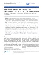
Percentage changes of bone mineral density after 12-months treatment compared between Alendronate (ALN) and pla-cebo control group (CON) (Data are in mean ± SE)Figure 1
Percentage changes of bone mineral density after 12-months
treatment compared between Alendronate (ALN) and pla-
cebo control group (CON) (Data are in mean ± SE). *: p <
0.05; **: p < 0.01.
Table 2: BMD data at baseline, 6- and 12-months. Comparison of BMD at baseline and its changes at 6- and 12-months in patients with
established osteoporosis compared between Alendronate group and placebo control group (Data: mean ± SD)
BMD measurements Placebo control group (n = 25) Alendronate group (n = 22)
Baseline 6-month 12-month % difference
+
Baseline 6-month 12-month % difference
+
DXA (g/cm2) Spine (L2-L4) 0.718 ± 0.101 0.709 ± 0.105 0.710 ± 0.102 -0.7 ± 3.3 0.719 ± 0.097 0.743 ± 0.101 * 0.756 ± 0.094 ** 5.1 ± 4.2
a
Femoral Neck 0.631 ± 0.060 0.633 ± 0.053 0.632 ± 0.055 -0.1 ± 4.1 0.653 ± 0.121 0.658 ± 0.129 0.670 ± 0.129 ** 2.5 ± 3.2
b
pQCT
(mg/cm3)
Distal radius
tBMD 138.6 ± 29.3 135.3 ± 31.5 137.3 ± 31.9 -0.6 ± 6.4 126.8 ± 46.4 130.4 ± 44.3 * 130.8 ± 44.5 * 0.9 ± 5.1
iBMD 424.5 ± 73.0 421.0 ± 75.6 429.9 ± 82.3 0.1 ± 3.2 388.1 ± 79.4 383.4 ± 79.6 385.4 ± 91.0 0.2 ± 3.8
cBMD 1124.2 ± 172.9 1070.7 ± 289.3 1113.0 ± 178.4 ** -1.4 ± 2.2 1084.3 ± 202.8 1024.7 ± 329.8 1077.6 ± 203.3 0.1 ± 2.4
b
+: percentage compared between baseline and 12-month
* p < 0.05; **p < 0.01: compared with baseline
a: p < 0.01; b: p < 0.05: compared for changes in BMD between Alendronate and placebo control group
Journal of Orthopaedic Surgery and Research 2007, 2:9 />Page 5 of 6
(page number not for citation purposes)
uncertain how long the patient should remain on drug
therapy for prevention or treatment of osteoporosis and
prevention of osteoporotic fractures. However, short- or
mid-term drug intervention for 1–2 years might be effec-
tive for not only improving BMD but also preventing cur-
rent and late fractures as there was no any acceleration in
bone loss after discontinuing the drug treatment found by
others [21,23,25,26].
In the present study, the treatment tolerability for the anti-
osteoporotic drugs was reflected partially by the dropout
rate. We recorded 13.6% and 4% dropouts in Alendronate
and control group during 12 months intervention, respec-
tively. However, none of them dropped out due to dis-
comfort of stomach after Alendronate treatment. This data
was similar to the Alendronate studies on prevention of
osteoporosis in the same reference population [8,9]. In
summery, this 12-months intervention study demon-
strated the treatment effect of Alendronate on osteoporo-
sis at both axial and appendicualr skeletons in Chinese
postmenopausal women with low-energy Colles' fracture,
with more effect on the weight-bearing spine and hip than
the non-weigh bearing distal radius. Such effects were
explained by significant suppression in bone turnover
evaluated biochemically.
Competing interests
The institution of the authors has received funding from
the Hong Kong Health Services Research Committee.
Authors do not have any potential conflicts of interests
related to this work
Acknowledgements
This study was supported with a Health Services Research Committee/
Health Care & Promotion Fund, Hong Kong SAR, China (Ref. HSRC/HCPF:
298104). Clinical Research Ethics approval was obtained from the Clinical
Research Ethics Committee of the Chinese University of Hong Kong (Ref.
No. CRE-660). Miss WY Hung provided assistance in bone mineral density
measurement.
References
1. Hung LK, Wu HT, Leung PC, Qin L: Low BMD is a risk factor for
'low energy' Colles' fracture in pre- and postmenopausal
women in Hong Kong Chinese – A DXA and pQCT study.
Clin Orthop Rel Res 2005, 435:219-25.
2. Cummings SR, Melton LJ: Epidemiology and outcomes of oste-
oporotic fractures. Lancet 2002, 359(9319):1761-7.
3. Lau EM, Lee JK, Suriwongpaisal P, Saw SM, Das De S, Khir A: The
incidence of hip fracture in four Asian countries: the Asian
Osteoporosis Study (AOS). Osteoporos Int 2001, 12(3):239-43.
4. Melton LJ 3rd, Thamer M, Ray NF, Chan JK, Chesnut CH 3rd, Einhorn
TA, Johnston CC, Raisz LG, Silverman SL, Siris ES: Fractures attrib-
utable to osteoporosis: report from the National Oste-
oporosis Foundation. J Bone Miner Res 1997, 12(1):16-23.
5. Anonymous: Osteoporosis: review of the evidence for preven-
tion, diagnosis and treatment and cost-effectiveness analysis.
Executivesummary. Osteoporos Int 1998, 8(4):S3-6.
6. Brown JP, Josse RG: Scientific Advisory Council of the Oste-
oporosis Society of Canada: Clinical practice guidelines for
the diagnosis and management of osteoporosis in Canada.
CMAJ 2002, 167(10):S1-34.
7. Christensen PM, Brixen K, Gyrd-Hansen D, Kristiansen IS: Cost-
effectiveness of alendronate in the prevention of oste-
oporotic fractures in Danish women. Basic Clin Pharmacol Toxicol
2005, 96(5):387-96.
8. Kung AWC, Yeung SS, Chu LW: The efficacy and tolerability of
alendronate in postmenopausal osteoporotic Chinese
women: A randomized placebo-controlled study. Calcif Tissue
Int 2000, 67:286-90.
9. Lau EM, Woo J, Chan YH, Griffith J: Alendronate prevents bone
loss in Chinese women with osteoporosis. Bone 2000,
27(5):677-80.
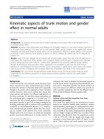
Percentage changes of Bone Alkaline Phosphate (BALP) and Deoxypyridoline (DPD) level after 12-month treatment com-pared between Alendronate (ALN) and placebo control group (CON) (Data are in mean ± SE)Figure 2
Percentage changes of Bone Alkaline Phosphate (BALP) and
Deoxypyridoline (DPD) level after 12-month treatment com-
pared between Alendronate (ALN) and placebo control
group (CON) (Data are in mean ± SE). *: p < 0.05; **: p <
0.01.
Table 3: Bone formation and resorption markers. Bone Alkaline Phosphate (BALP) and Deoxypyridinoline (DPD) level in patients with
established osteoporosis compared between Alendronate group (ALN) (n = 22) and placebo control group (CON) (n = 25) at baseline
and follow-up at 6- and 12 months (Data: mean ± SD)
Biochemical Markers Group Baseline 6-month 12-month % difference
+
BALP ALN 62.27 ± 16.86 40.33 ± 9.98 ** 34.35 ± 9.87 ** -39.9 ± 33.6
a
CON 66.17 ± 14.91 56.72 ± 18.24 ** 55.13 ± 15.76 ** -16.7 ± 14.5
DPD ALN 50.82 ± 35.10 25.11 ± 18.65 ** 23.03 ± 14.72 ** -42.6 ± 30.4
b
CON 33.70 ± 20.28 40.11 ± 36.92 27.28 ± 10.95 -9.4 ± 26.7
+: percentage difference compared between baseline and 12-month
a: p < 0.01; b: p < 0.05: significant difference in BALP found between ALN and CON group (independent t-test)
*p < 0.05; ** p < 0.01: compared with baseline (paired t-test)
Publish with BioMed Central and every
scientist can read your work free of charge
"BioMed Central will be the most significant development for
disseminating the results of biomedical research in our lifetime."
Sir Paul Nurse, Cancer Research UK
Your research papers will be:
available free of charge to the entire biomedical community
peer reviewed and published immediately upon acceptance
cited in PubMed and archived on PubMed Central
yours — you keep the copyright
Submit your manuscript here:
/>BioMedcentral
Journal of Orthopaedic Surgery and Research 2007, 2:9 />Page 6 of 6
(page number not for citation purposes)
10. Morrison LS, Tobias JH: Effect of a case-finding strategy for
osteoporosis on bisphosphonate prescribing in primary care.
Osteoporosis Int 2005, 16(1):71-7.
11. Kanis JA, Borgstrom F, Zethraeus N, Johnell O, Oden A, Jonsson B:
Intervention thresholds for osteoporosis in the UK. Bone
2005, 36(1):22-32.
12. Qin L, Au SK, Leung PC, Lau MC, Woo J, Choy WY, Hung WY, Dam-
bacher MA, Leung KS: Baseline BMD and bone loss at distal
radius measured by peripheral quantitative computed tom-
ography in peri- andpostmenopausal Hong Kong Chinese
women. Osteoporos Int 2002, 13:962-970.
13. Greenspan SL, Parker RA, Ferguson L, Rosen HN, Maitland-Ramsey
L, Karpf DB: Early changes in biochemical markers of bone
turnover predict the long-term response to alendronate
therapy in representative elderly women: a randomized clin-
ical trail. J Bone Miner Res 1998, 13:1431-8.
14. Kanis JA, Brazier JE, Stevenson M, Calvert NW, Lloyd Jones M:
Treatment of established osteoporosis: a systematic review
and cost-utility analysis. Health Technol Asse 2002, 6(29):1-146.
15. WHO: Guidelines for preclinical evaluation and clinical trials
in osteoporosis. WHO, Geneva; 1998.
16. Bouxsein ML, Parker RA, Greenspan SL: Forearm bone mineral
densitometry cannot be used to monitor response to alendr-
onate therapy in postmenopausal women. Osteoporosis Int
1999, 10:505-9.
17. Kohrt WM, Birge SJ Jr: Differential effects of estrogen treat-
ment on bone mineral density of the spine, hip, wrist and
total body in late postmenopausal women. Osteoporo Int 1995,
5(3):150-5.
18. Plotkin LI, Weinstein RS, Parfitt AM, Roberson PK, Manolagas SC,
Bellido T: Prevention of osteocyte and osteoblast apoptosis by
bisphosphonates and calcitonin. J Clin Invest 1999,
104(10):1363-74.
19. Stepan JJ, Alendeld F, Boivin G, Feyen JH, Lakatos P: Mechanisms of
action of antiresorptive therapies of postmenopausal oste-
oporosis. Endocr Regul 2003, 37(4):225-38.
20. Downs RW Jr, Bell NH, Ettinger MP, Walsh BW, Favus MJ, Mako B,
Wang L, Smith ME, Gormely GJ, Melton ME: Comparison of Alen-
dronate and Intranasal Calcitonin for treatment of oste-
oporosis in postmenopausal women. J Clin Endocrinol Metab
2000, 85(5):1783-8.
21. Harris ST, Gertz BJ, Genant HK, Eyre DR, Survill TT, Ventura JN,
DeBrock J, Ricerca E, Chesnut CH 3rd: The effect of short term
treatment with alendronate on vertebral density and bio-
chemical markers of bone remodeling in early postmeno-
pausal women. J Clin Endocrinol Metab 1993, 76(6):1399-406.
22. Chan K, Qin L, Lau M, Woo J, Au S, Choy W, Lee K, Lee S: A rand-
omized, prospective study of the effects of Tai Chi Chun
exercise on bone mineral density in postmenopausal
women. Arch Phys MedRehab 2004, 85:717-22.
23. Black DM, Thompson DE, Bauer DC, Ensrud K, Musliner T, Hochberg
MC, Nevitt MC, Suryawanshi S, Cummings SR: Fracture riskreduc-
tion with alendronate in women with osteoporosis: the Frac-
tureIntervention Trail. FIT research group. J Clin
EndocrinolMetab 2000, 85(11):4118-24.
24. Riis BJ, Hansen MA, Jensen AM, Overgaard K, Christiansen C: Low
bone mass and fast rate of bone loss at menopause: equal
risk factors for future fracture: a 15-year follow-up study.
Bone 1996, 19(1):9-12.
25. Cummings SR, Black DM, Thompson DE, Applegate WB, Barrett-
Connor E, Musliner TA, Palermo L, Prineas R, Rubin SM, Scott JC,
Vogt T, Wallace R, Yates AJ, LaCroix AZ: Effect of alendronate on
risk of fracture in women with low bone density but without
vertebral fractures: Results from the fracture intervention
trial. JAMA 1998, 280(24):2077-82.
26. Pols HA, Felsenberg D, Hanley DA, Stepan J, Munoz-Torres M, Wilkin
TJ, Qin-Sheng G, Galich AM, Vandormael K, Yates AJ, Stych B: Mul-
tinational, placebo-controlled, randomized trial of the
effects of alendronate on bone density and fracture risk in
postmenopausal women with low bone mass: results of the
FOSIT study. Fosamax International Trial Study Group.
Osteoporos Int 1999, 9(5):461-8.