báo cáo hóa học:" Varus distal femoral osteotomy in young adults with valgus knee" pot
Bạn đang xem bản rút gọn của tài liệu. Xem và tải ngay bản đầy đủ của tài liệu tại đây (924.77 KB, 5 trang )
BioMed Central
Page 1 of 5
(page number not for citation purposes)
Journal of Orthopaedic Surgery and
Research
Open Access
Research article
Varus distal femoral osteotomy in young adults with valgus knee
Farzad Omidi-Kashani*
1
, Ibrahim G Hasankhani
2
, Mahdi Mazlumi
1
and
Mohamad H Ebrahimzadeh
1
Address:
1
Department of orthopedic surgery, Qhaem hospital, Mashhad University of Medical Sciences, Mashhad, Iran and
2
Department of
orthopedic surgery, Imam Reza hospital, Mashhad University of Medical Sciences, Mashhad, Iran
Email: Farzad Omidi-Kashani* - ; Ibrahim G Hasankhani - ;
Mahdi Mazlumi - ; Mohamad H Ebrahimzadeh -
* Corresponding author
Abstract
Background: Musculoskeletal disorders specially knee osteoarthritis are the most common
causes of morbidity in old patients. Disturbance of the mechanical axis of the lower extremity is
one of the most important causes in progression of knee osteoarthritis. The purpose of the present
study was to analyze the surgical results of distal femoral varus osteotomy in patients with genu
valgum.
Methods: In this study, after recording history and physical examination, appropriate radiographs
were taken. We did varus distal femoral osteotomy by standard medial subvastus approach and 90-
angle blade plate fixation then followed the patients clinically and radiographically.
Results: This study was done on 23 knees (16 patients) age 23.3 years (range, 17 to 41 years). The
mean duration of following up was 16.3 months (range, 8 to 25 months). Based on paired T test,
there were statistically significant difference between pre- and postoperative tibiofemoral and
congruence angles (p < 0.001, t = 21.3 and p < 0.001, t = 10.1 respectively). Pearson correlation
between the amount of tibiofemoral and congruence angle correction was also statistically
significant (p = 0.02 and r = 0.46).
Conclusion: Distal femoral varus osteotomy with blade plate fixation can be a reliable procedure
for the treatment of valgus knee deformity. In this procedure, with more tibiofemoral angle
correction, more congruence angle correction can be achieved. Therefore, along with genu valgum
correction, the patella should be stabilized simultaneously.
Background
Musculoskeletal disorders specially knee osteoarthritis are
the most common causes of morbidity in old patients [1].
Disturbance of the mechanical axis of the lower extremity
is one of the most important causes in progression of knee
osteoarthritis [2,3].
Whereas high tibial osteotomy has been used successfully
to treat medial compartment disease with varus deform-
ity, the results of tibial osteotomy for valgus deformity
have varied [4-6]. Because of the inherent valgus femo-
rotibial angulation at the knee joint, tibial varus osteot-
omy could be used to correct a femorotibial angulation of
no more than 12° of valgus. Correction of a larger
deformity creates a varus or medial tilt of the joint line
Published: 13 May 2009
Journal of Orthopaedic Surgery and Research 2009, 4:15 doi:10.1186/1749-799X-4-15
Received: 12 July 2008
Accepted: 13 May 2009
This article is available from: />© 2009 Omidi-Kashani et al; licensee BioMed Central Ltd.
This is an Open Access article distributed under the terms of the Creative Commons Attribution License ( />),
which permits unrestricted use, distribution, and reproduction in any medium, provided the original work is properly cited.
Journal of Orthopaedic Surgery and Research 2009, 4:15 />Page 2 of 5
(page number not for citation purposes)
which is subjected to increased lateral shear forces, and
this tends to cause the femur to subluxate medially on the
tibia during gait [7-9].
In light of this, several authors stated that if a knee shows
an anatomic tibiofemoral angle >10–12° of valgus or if
the plane of the joint deviates from the horizontal in the
superolateral direction more than 10°, a distal femoral
varus osteotomy is the preferred method of limb realign-
ment [8,10-12]. This procedure corrects deformity in the
lower femur, which is more pronounced than in knees
with varus deformity. It also restores the orientation of the
joint line toward the horizontal and does not disturb
medial collateral ligament stability [13-15].
The purpose of the present study was to study and analyze
the surgical results of distal femoral varus osteotomy in
patients with genu valgum.
Methods
Between June 2000 and November 2006, 25 distal femo-
ral osteotomies were performed in 18 patients (14 women
and 4 men) at our institution.
Standing anteroposterior and lateral radiographs as well
as Merchant radiographs of the knee were made preoper-
atively and at the time of each follow-up. The sulcus and
congruence angles of the patellofemoral joint were meas-
ured with the use of the method described by Merchant et
al. before the osteotomy and at the time of the latest fol-
low-up [16]. Subluxation of the patella was defined as
being present if the congruence angle was >16° on the
Merchant radiograph [17].
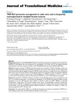
We used the method described by Stevens PM et al. who
believed that the knee can be divided into four radio-
graphic quadrants (figure 1), designating varus as negative
and valgus as positive [18]. The mechanical axis measured
on a full-length film can be readily correlated to any of
these zones with little interobserver error. Plus or minus
zone I, the central quadrants, represent physiologic
deformities. Plus or minus zone II often correlate with
symptomatic deformities that may warrant surgical inter-
vention. Plus or minus zone III are outside the confines of
the knee and usually warrant surgical intervention.
Our surgical indications were genu valgum with the
mechanical axis in plus zone III, the intermalleolar dis-
tance >5 cm, a painful deformity associated with a valgus
tibiofemoral angulation of > 12° and narrowing of the lat-
eral joint space, patellar instability, circumduction gait,
and cosmetic concerns. We tried to mainly consider the
deformity with a deviated mechanical axis as a principle
indication for surgery, even though the patient is com-
pletely asymptomatic. Without intervention, biomechan-
ically the knee most likely has an increased risk of
developing early osteoarthritis [19,20].
Our exclusion criteria included severe arthritis of the
medial compartment of the knee, severe tricompartment
osteoarthritis, and tibiofemoral subluxation greater than
one centimeter.
The Hospital for Special Surgery knee-rating system was
used for the clinical evaluation of all patients [21]. Laxity
of the medial collateral ligament was classified according
to Scuderi and Scott classification [22]. All patients were
designed to follow regularly at 6 weeks, 3 months, 6
months, 1 year, and then yearly thereafter. Two patients
were lost to follow-up and so excluded them from the
study.
Surgical Technique
The technique for distal femoral varus osteotomy was
based on the method described by McDermott et al [13].
The osteotomy was performed through a medial incision
with removal of a 5 to 10-mm medially based wedge of
bone from the distal part of the femur. A prebent 90°
The relation between the mechanical axis and the kneeFigure 1
The relation between the mechanical axis and the
knee.
Journal of Orthopaedic Surgery and Research 2009, 4:15 />Page 3 of 5
(page number not for citation purposes)
dynamic-compression blade plate was inserted into the
femoral condyle, parallel to the knee-joint line. The oste-
otomy site was closed with the plate in contact with the
medial femoral cortex. This spontaneously achieved a tibi-
ofemoral angle of approximately 0° and then final osteo-
synthesis was performed with the dynamic-compression
plate. A cortical or cancellous bone lag screw was inserted
through the hole above the bend in the blade-plate, across
the osteotomy site, to provide additional stability. A lat-
eral retinacular release was performed in 6 knees because
of lateral subluxation of the patella [23] or excessive lat-
eral retinacular tightness palpated intraoperatively and
after fixation of the osteotomy site.
Postoperatively, the affected limb was immobilized in a
hinged knee brace until healing of the osteotomy site
occurred. Patients began to use a continuous-passive-
motion machine 48 hours after the operation and contin-
ued to use it until 90° of knee flexion was achieved. Non-
weight bearing walking was commenced on the second
postoperative day and was continued until initial healing
of the osteotomy site had been confirmed radiographi-
cally, usually after six weeks of follow-up. Full weight-
bearing was only permitted after 3 months of follow-up
and after radiographs showed good healing of the osteot-
omy site.
Statistical Analysis
The preoperative and most recent knee score were com-
pared with use of the paired T test. The level of signifi-
cance was set at p < 0.05. All analyses were performed with
use of Statistical Package Science Software (SPSS version
10).
Results
We could successfully follow 23 distal femoral osteoto-
mies in 16 patients (12 women and 4 men) at our institu-
tion. All of the seven bilaterally involved patients were
women. The mean age of the patients at the time of the
index operation was 23.3 years (range, 17 to 41 years). Of
these, 6 knees had patellar subluxation. No patient with
complete patellar dislocation was present in our patients.
The mean knee score improved from 90.7 points (range,
77 to 96 points) preoperatively to 98.13 points (range, 93
to100 points) at the time of the most recent follow-up (p
> 0.05).
The laxity of the medial collateral ligament was classified
as grade 1 for 8 knees, and grade 2 for 5; the remaining 10
knees had no laxity. Knee stability was achieved in all
patients after solid union of the osteotomy site. The mean
pre- and postoperative tibiofemoral and congruence
angles were shown in table 1.
Before surgery, in 43.5% of knees, the mechanical axis
passed from zone II, and in other 56.5%, zone III was the
site of the drawn mechanical axis. Postoperatively, all of
the knees' mechanical axes were laid in zone I.
The mean duration of follow-up was 16.3 Months (range,
8 to 25 months). Union of the osteotomy site was
achieved in all but one knee, with an average time to
union of 4.1 months (range, 2 to 6 months). An autoge-
nous iliac crest bone graft was required to achieve healing
of one nonunion. Superficial wound infection occurred in
2 (8.7%) knees; treated successfully with oral antibiotics.
Implant failure occurred in 1(4.3%) patient aged 23 years
who had fallen 2 months after surgery and bent the plate,
then suffered nonunion of the osteotomy. Revision of the
fixation using a blade plate, supplemented with bone
grafting, resulted in satisfactory union.
Clinical evaluation revealed no loss of knee motion com-
pared with the preoperative examination. The distance
between the medial malleoli was reduced in all subjects;
most of the patients could approximate ankles and knees
simultaneously, indicating complete correction of genu
valgum. Circumduction gait, which was common before
surgery, was corrected in all patients. No patient had varus
laxity >5 mm. There were no patients with limb-length
inequality >1.5 cm.
Discussion
Although the mean knee score in our patients improved
with surgery, in contrast to most other studies
[10,11,14,15], this difference was not statistically signifi-
cant mostly because the operation was done in younger
healthy patients (not in osteoarthritic patients). The main
aim of our operation was to avoid late knee osteoarthritic
derangement.
In our study, based on paired T test, there were statistically
significant difference between pre- and postoperative tibi-
ofemoral and congruence angles (p < 0.001, t = 21.3 and
p < 0.001, t = 10.1 respectively). Therefore, the procedure
could sufficiently correct the deformity.
Pearson correlation between the amount of tibiofemoral
and congruence angle correction was statistically signifi-
Table 1: Mean pre- and postoperative tibiofemoral and
congruence angle
Angle
(Degree)
Preoperative
Mean (SD
❈
)
Postoperative
Mean (SD)
Difference
Mean (SD)
Tibiofemoral 20.3 (4.2) 0.13 (2.9) 20.2 (4.5)
Congruence 14.8 (7.2) 1.48 (3.8) 13.3 (6.2)
❈
SD: Standard Deviation
Journal of Orthopaedic Surgery and Research 2009, 4:15 />Page 4 of 5
(page number not for citation purposes)
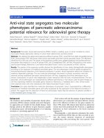
cant (p = 0.02 and r = 0.46). In respect to positive "r", one
can concluded that with more tibiofemoral angle correc-
tion, more congruence angle correction can be achieved.
In other word, along with genu valgum correction, the
patella should be stabilized simultaneously (figure 2).
The method of fixation after the osteotomy appears to
have a great influence on the results of this procedure. Use
of blade-plate for fixation at the osteotomy site has been
associated with a high healing rate and promising results
in short-term follow-up studies [10,11,13]. In a study was
done by Wang JW et al. on 30 knees that distal femoral
varus osteotomy was fixed with a 90° blade-plate,
reported that 83% had satisfactory result and only one
nonunion occurred [11].
Healy WL et al. evaluated 23 distal femoral varus osteoto-
mies at an average of 4 years postoperatively. The average
tibiofemoral angle preoperatively was 18° of valgus,
which was corrected to an average of 2° of valgus. Accord-
ing to the Hospital for Special Surgery Knee score, 19
(83%) of the 23 knees were rated as good or excellent. 15
osteotomies were performed on osteoarthritic knees and
all but one (93%) knee were rated as good or excellent
[14].
Aglietti P et al reported the results of 18 distal femoral
varus osteotomies in patients with osteoarthritis of the lat-
eral compartment of the valgus knee. The osteotomy site
was fixed with a 90° blade-plate. With an average follow-
up of 9 years, they cited 77% good or excellent results
according to the Knee Society rating system. No patients
had nonunion or infection. They advised the procedure
for the treatment of symptomatic valgus knee in both
young and older active patients [10].
In another study conducted by Mathews J and coauthors,
they described 21 patients treated with distal femoral
varus osteotomies immobilized by casting, staples and
casting, and rigid internal fixation with an AO blade plate.
They reported satisfactory results only in those patients
who had less severe degrees of osteoarthritis confined to
the lateral compartment, adequate correction of valgus
deformity (the anatomic axis within 2° from zero), and
rigid internal fixation to permit postoperative early mobi-
lization [15].
Conclusion
In conclusion, although a prospective trial is required to
investigate the optimal postoperative alignment angle,
distal femoral varus osteotomy with blade plate fixation
can be a reliable procedure for the treatment of valgus
knee deformity. In this procedure, with more tibiofemoral
angle correction, more congruence angle correction can be
achieved. Therefore, along with genu valgum correction,
the patella should be stabilized simultaneously
Competing interests
The authors declare that they have no competing interests.
Authors' contributions
FOK, the senior surgeon and has made substantial contri-
butions to conception and design of the manuscript. IGH
has been involved in drafting the manuscript, participated
in the sequence alignment. MM has made substantial con-
tributions to acquisition of data from literature. MHE has
had substantial role in preparing and revising the manu-
script.
Acknowledgements
Authors cordially appreciate the helps that provided by the personnel of
Qaem statistics department and operating room of Qaem hospital, Mash-
had, Iran. We would like to express our gratitude to Katayoun Z. Toossi
for her help in reviewing, editing and verifying this paper.
References
1. Vince KG, Cameron HU, Hungerford DS, Laskin RS, Ranawat CS,
Scuderi GR: What would you do? Case challenges in knee sur-
gery. J Arthroplasty 2005, 20(4 Suppl 2):44-50.
2. Sharma L, Song J, Felson DT, Cahue S, Shamiyeh E, Dunlop DD: The
role of knee alignment in disease progression and functional
decline in knee osteoarthritis. JAMA 2001, 286(2):188-95.
3. Sabharwal S, Zhao C: Assessment of lower limb alignment:
supine fluoroscopy compared with a standing full-length
radiograph. J Bone Joint Surg Am 2008, 90(1):43-51.
4. Habata T, Uematsu K, Hattori K, Kasanami R, Takakura Y, Fujisawa
Y: High tibial osteotomy that does not cause recurrence of
varus deformity for medial gonarthrosis. Knee Surg Sports Trau-
matol Arthrosc 2006, 14(10):962-7.
5. Amendola A, Panarella L: High tibial osteotomy for the treat-
ment of unicompartmental arthritis of the knee. Orthop Clin
North Am 2005, 36(4):497-504.
Correlation between tibiofemoral and congruence angle cor-rectionFigure 2
Correlation between tibiofemoral and congruence
angle correction.
Publish with BioMed Central and every
scientist can read your work free of charge
"BioMed Central will be the most significant development for
disseminating the results of biomedical researc h in our lifetime."
Sir Paul Nurse, Cancer Research UK
Your research papers will be:
available free of charge to the entire biomedical community
peer reviewed and published immediately upon acceptance
cited in PubMed and archived on PubMed Central
yours — you keep the copyright
Submit your manuscript here:
/>BioMedcentral
Journal of Orthopaedic Surgery and Research 2009, 4:15 />Page 5 of 5
(page number not for citation purposes)
6. Michaela G, Florian P, Michael L, Christian B: Long-term outcome
after high tibial osteotomy. Arch Orthop Trauma Surg 2008,
128(1):111-115.
7. Matokoviæ D, Haspl M: Corrective osteotomy in the treatment
of degenerative changes in the knee joint. Lijec Vjesn 2000,
122(9–10):229-33. Croatian
8. Preston CF, Fulkerson EW, Meislin R, Di Cesare PE: Osteotomy
about the knee: applications, techniques, and results. J Knee
Surg 2005, 18(4):258-72.
9. Gunther KP: Surgical approaches for osteoarthritis. Best Pract
Res Clin Rheumatol 2001, 15(4):627-43.
10. Aglietti P, Menchetti PPM: Distal femoral varus osteotomy in the
valgus osteoarthritic knee. Am J Knee Surg 2000, 13(2):89-95.
11. Wang JW, Hsu CC: Distal femoral varus osteotomy for oste-
oarthritis of the knee. J Bone Joint Surg Am 2005, 87(1):127-33.
12. Backstein D, Morag G, Hanna S, Safir O, Gross A: Long-term fol-
low-up of distal femoral varus osteotomy of the knee. J Arthro-
plasty 2007, 22(4):2-6.
13. McDermott AG, Finkle JA, Farine I, Boynton EL, MacIntosh DL, Gross
A: Distal femoral varus osteotomy for valgus deformity of
the knee. J Bone Joint Surg Am 1988, 70:110-6.
14. Healy WL, Anglen JO, Wasilewski SA, Krackow KA: Distal femoral
varus osteotomy. J Bone Joint Surg Am 1988, 70(1):102-109.
15. Mathews J, Cobb AG, Richardson S, Bentley G: Distal femoral
osteotomy for lateral compartment osteoarthritis of the
knee. Orthopedics 1998, 21(4):437-440.
16. Merchant AC: Patellofemoral imaging. Clin Orthop Relat Res 2001,
389:15-21.
17. Merchant AC, Mercer RL, Jacobsen RH, Cool CR: Roentgeno-
graphic analysis of patellofemoral congruence. J Bone Joint Surg
Am 1974, 56:1391-6.
18. Stevens PM, Maguire M, Dales MD, Robins AJ: Physeal stapling for
idiopathic genu valgum. J Ped Orthop 1999, 19(5):645-9.
19. Hunter DJ, Sharma L, Skaife T: Alignment and osteoarthritis of
the knee. J Bone Joint Surg Am 2009, 91(Suppl 1):85-9.
20. Eckstein F, Wirth W, Hudelmaier M, Stein V, Lengfelder V, Cahue S,
Marshall M, Prasad P, Sharma L: Patterns of femorotibial carti-
lage loss in knees with neutral, varus, and valgus alignment.
Arthritis Rheum 2008, 59(11):1563-70.
21. Insall JN, Ranawat CS, Aglietti P, Shine J: A comparison of four
models of total knee-replacement prosthesis. J Bone Joint Surg
AM 1976, 58:754-65.
22. Scuderi GR, Scott WN: Classification of the knee ligament inju-
ries. In Surgery of the knee Volume 1. 3rd edition. Edited by: Insall JN,
Scott WN. New York: Churchill Livingstone; 2001:585-99.
23. Arendt EA, Fithian DC, Cohen E: Current concepts of lateral
patella dislocation. Clin Sports Med 2002, 21(3):499-519.