Báo cáo hóa học: " Infectious salmon anaemia virus replication and induction of alpha interferon in Atlantic salmon erythrocytes" potx
Bạn đang xem bản rút gọn của tài liệu. Xem và tải ngay bản đầy đủ của tài liệu tại đây (340.04 KB, 12 trang )
BioMed Central
Page 1 of 12
(page number not for citation purposes)
Virology Journal
Open Access
Research
Infectious salmon anaemia virus replication and induction of alpha
interferon in Atlantic salmon erythrocytes
Samuel T Workenhe
1
, Molly JT Kibenge
1
, Glenda M Wright
2
,
Dorota W Wadowska
3
, David B Groman
4
and Frederick SB Kibenge*
1
Address:
1
Department of Pathology and Microbiology, Atlantic Veterinary College, University of Prince Edward Island, 550 University Avenue,
Charlottetown, PE. C1A 4P3, Canada,
2
Department of Biomedical Sciences, Atlantic Veterinary College, University of Prince Edward Island, 550
University Avenue, Charlottetown, PE. C1A 4P3, Canada,
3
Electron Microscopy Laboratory, Atlantic Veterinary College, University of Prince
Edward Island, 550 University Avenue, Charlottetown, PE. C1A 4P3, Canada and
4
Aquatic Diagnostic Services, Atlantic Veterinary College,
University of Prince Edward Island, 550 University Avenue, Charlottetown, PE. C1A 4P3., Canada
Email: Samuel T Workenhe - ; Molly JT Kibenge - ; Glenda M Wright - ;
Dorota W Wadowska - ; David B Groman - ; Frederick SB Kibenge* -
* Corresponding author
Abstract
Background: Infectious salmon anaemia (ISA) virus (ISAV), which causes ISA in marine-farmed
Atlantic salmon, is an orthomyxovirus belonging to the genus Isavirus, family Orthomyxoviridae. ISAV
agglutinates erythrocytes of several fish species and it is generally accepted that the ISAV receptor
destroying enzyme dissolves this haemagglutination except for Atlantic salmon erythrocytes.
Recent work indicates that ISAV isolates that are able to elute from Atlantic salmon erythrocytes
cause low mortality in challenge experiments using Atlantic salmon. Previous work on ISAV-
induced haemagglutination using the highly pathogenic ISAV strain NBISA01 and the low pathogenic
ISAV strain RPC/NB-04-0851, showed endocytosis of NBISA01 but not RPC/NB-04-0851. Real-
time RT-PCR was used to assess the viral RNA levels in the ISAV-induced haemagglutination
reaction samples, and we observed a slight increase in viral RNA transcripts by 36 hours in the
haemagglutination reaction with NBISA01 virus when the experiment was terminated. However, a
longer sampling interval was considered necessary to confirm ISAV replication in fish erythrocytes
and to determine if the infected cells mounted any innate immune response. This study examined
the possible ISAV replication and Type I interferon (IFN) system gene induction in Atlantic salmon
erythrocytes following ISAV haemagglutination.
Results: Haemagglutination assays were performed using Atlantic salmon erythrocytes and one
haemagglutination unit of the two ISAV strains, NBISA01 and RPC/NB-04-0851, of differing
genotypes and pathogenicities. Haemagglutination induced by the highly pathogenic NBISA01 but
not the low pathogenic RPC/NB-04-0851 resulted in productive infection as evidenced by
increased ISAV segment 8 transcripts and increase in the median tissue culture infectious dose
(TCID
50
) by 5 days of incubation. Moreover, reverse transcription (RT) quantitative PCR used to
compare mRNA levels of key Type I IFN system genes in erythrocyte lysates of haemagglutination
reactions with the two ISAV strains showed a higher relative fold increase of IFN-α in NBISA01
haemagglutinations compared to RPC/NB-04-085-1 haemagglutinations (33.0 – 44.26 relative fold
increase compared to 11.29). Erythrocytes exposed to heat-inactivated virus or to
polyinosinic:polycytidylic acid (polyI:C) or to L-15 medium alone (negative control assays) had
Published: 28 February 2008
Virology Journal 2008, 5:36 doi:10.1186/1743-422X-5-36
Received: 7 January 2008
Accepted: 28 February 2008
This article is available from: />© 2008 Workenhe et al; licensee BioMed Central Ltd.
This is an Open Access article distributed under the terms of the Creative Commons Attribution License ( />),
which permits unrestricted use, distribution, and reproduction in any medium, provided the original work is properly cited.
Virology Journal 2008, 5:36 />Page 2 of 12
(page number not for citation purposes)
minimal late induction (<3.5 relative fold increase) of STAT1 and/or ISG15 and Mx genes, whereas
erythrocytes exposed to UV-inactivated virus lacked any cytokine induction.
Conclusion: ISAV-induced haemagglutination by a highly pathogenic virus strain results in virus
uptake and productive infection of Atlantic salmon erythrocytes accompanied by significant
induction of IFN-α. This study also highlights the critical role of ISAV strain variation in the initial
stages of the virus-cell interaction during haemagglutination, and possibly in the pathogenesis of ISA.
Moreover, the study shows for the first time that fish erythrocytes immunologically respond to
ISAV infection.
Background
Infectious salmon anaemia (ISA) is a highly fatal viral dis-
ease affecting marine-farmed Atlantic salmon (Salmo salar
L). This fish disease is caused by ISA virus (ISAV), a fish
orthomyxovirus assigned to the genus Isavirus within the
family Orthomyxoviridae [1]. The ISAV strains are envel-
oped particles of 90–140 nm diameter with surface pro-
jections consisting of a combined haemagglutinin-
esterase (HE) protein [2] and a separate fusion (F) protein
[3]. The genome is composed of eight segments of linear,
single-stranded negative sense RNA ranging in length
from 1.0 to 2.4 kb with a total molecular size of approxi-
mately 14.3 kb [4].
The clinical disease caused by ISAV in marine-farmed
Atlantic salmon is associated with anaemia [5], which is
hypothesized to be linked with uptake of virus-coated
erythrocytes by immune cells [6]. The fish erythrocytes
would probably be coated with ISAV through interaction
of the cellular sialic acid receptors and the viral HE glyco-
protein as occurs during the haemagglutination reaction.
ISAV haemagglutination of fish erythrocytes, similar to
influenza A virus haemagglutination of avian and mam-
malian erythrocytes, involves three independent phenom-
ena: [1] adsorption of viruses at the erythrocyte
membrane, [2] subsequent elution [7-9], which is not
always complete, and [3] uptake of viruses by the erythro-
cytes [10,11]. For ISAV, elution from erythrocytes was
originally reported to occur with erythrocytes of several
fish species except Atlantic salmon [7] in which the virus
causes a natural clinical disease (as reviewed in [12]).
However, recent work indicates that ISAV isolates that are
able to elute from Atlantic salmon erythrocytes cause low
mortality in challenge experiments using Atlantic salmon
[13]. In our previous work on ISAV-induced haemaggluti-
nation using the highly pathogenic NBISA01 and the low
pathogenic RPC/NB 04-0851, only NBISA01 was taken up
by the erythrocytes from Atlantic salmon and rainbow
trout (Oncorhynchus mykiss) [11]. In contrast, the uptake of
influenza A virus by avian and mammalian erythrocytes
via pinocytosis was non-specific [10] indicating a lack of
involvement of virus strain-specific differences such as
pathogenicity level. This suggested to us that the lack of
dissolution of pathogenic ISAV-induced haemagglutina-
tion of Atlantic salmon erythrocytes favours endocytosis
of the virus particles by the erythrocytes [11] and this phe-
nomenon may contribute to the anaemia in ISA.
Members of the family Orthomyxoviridae are known to
have a nuclear replication phase [1]. Enucleated BS-C-1
cells have been shown to be non-permissive for replica-
tion of influenza A viruses [14]. By inference, mature
mammalian erythrocytes, which are enucleated, are also
non-permissive for replication of influenza A viruses. In
the case of the nucleated avian (turkey or chicken) eryth-
rocytes, haemagglutination by avian influenza A virus
resulted only in de novo synthesis of viral proteins but not
production of new infectious virus [15]. We previously
used real-time RT-PCR to assess the viral RNA levels in the
ISAV-induced haemagglutination reaction samples, and
observed a slight increase in viral RNA transcripts by 36
hours in the haemagglutination reaction with NBISA01
virus when the experiment was terminated [11]. However,
a longer sampling interval was considered necessary to
confirm ISAV replication in fish erythrocytes and to deter-
mine if the infected cells mounted any innate immune
response.
The Type I IFN system constitutes the major antiviral
defence mechanism in the innate immune system of
mammals as well as fish [16]. Most cell types are able to
detect viral replication dsRNA intermediates and respond
by secretion of IFN, which then uses the JAK/STAT signal-
ling pathway to stimulate induction of the components of
the Type I IFN system genes such as Mx, ISG15 and STAT1
[17]. Atlantic salmon organs and the macrophage-like fish
cell line TO [18] respond to ISAV infection by up-regulat-
ing the expression of key Type I IFN system genes [19].
The limited immunological studies on the nucleated fish
erythrocytes suggest that they possess a certain level of
immune functions; the mature erythrocyte populations of
rainbow trout were shown to surround macrophages
phagocytosing Candida albicans and to secrete cytokine-
like macrophage inhibitory factors [20]. However, there is
no report of IFN induction in intact erythrocytes either in
fish or avian or mammalian species.
Virology Journal 2008, 5:36 />Page 3 of 12
(page number not for citation purposes)
The goal of this study was to determine whether ISAV
uptake by fish erythrocytes results in a productive infec-
tion and, if so, whether there is any effect of differences in
the pathogenicity level of the virus on the cellular
response. This information is needed to further clarify the
pathogenesis of the clinical disease in fish. In order to
obtain information on the putative associated innate
immune response in erythrocytes, we used reverse tran-
scription (RT) quantitative PCR assays with SYBR Green
chemistry to evaluate mRNA levels of Type I IFN system
genes IFN-α, Mx, ISG15, STAT1 and PKZ at regular inter-
vals up to 5 days following virus-induced haemagglutina-
tion.
Results
ISAV replicates in Atlantic salmon erythrocytes
Haemagglutination by pathogenic ISAV is associated with
endocytosis of the virus particles, which seems to be con-
sistent with virus infection [11]. To determine whether
ISAV endocytosis by fish erythrocytes results in a produc-
tive infection, and to further analyze the differences
between virus strains of differing pathogenicity, we mon-
itored the haemagglutination assays with NBISA01 and
RPC/NB-04-085-1 strains for transcription of viral genes
on ISAV segment 8 using real-time RT-PCR. In the previ-
ous report, the haemagglutination assays were carried out
with 10
9.75
TCID
50
/ml for NBISA01 and 10
4.25
TCID
50
/ml
for RPC/NB-04-085-1 [11]. In order to use an equal
number of haemagglutination (HA) units in the present
study, the HA units of the stock virus preparations were
determined, and all haemagglutination assays used 1 HA
unit of virus preparation. For NBISA01, 1 HA unit con-
tained 10
8.45
TCID
50
and had a cycle threshold (Ct) value
of 26.57, whereas for RPC/NB-04-085-1, 1 HA unit con-
tained 10
5.75
TCID
50
and had a Ct value of 20.39. Thus, the
present study, by using the standard 1 HA unit in haemag-
glutination assays, had less NBISA01 virus but more RPC/
NB-04-085-1 virus content in each haemagglutination
reaction than in our previous study [11]. It was possible to
maintain the erythrocytes viable for up to 5 days in hae-
magglutination assays in the present study by changing
the medium of the haemagglutination assay from PBS to
L-15 growth medium. The real-time RT-PCR data for
quantification of viral transcripts are presented in Table 1.
All the Ct values were confirmed to be for positive ampli-
cons by melting curve analysis. Agarose gel electrophore-
sis of the RT-PCR products also confirmed virus-specific
amplification for NBISA01 and RPC/NB-04-085-1,
whereas Atlantic salmon erythrocytes without virus,
which were incubated in L-15 medium alongside the hae-
magglutinations as a negative control showed no virus-
specific amplification by both melting curve analysis and
agarose gel electrophoresis (data not shown). The Ct val-
ues for the highly pathogenic NBISA01 strain show a
steady decline from the 0 hour (26.57 ± 0.14) to day 5
(20.48 ± 0.29) indicating an increase in viral gene tran-
scription. To confirm if the decrease in Ct value was from
newly synthesized viral mRNA we used oligodT primers
for cDNA synthesis followed by real-time PCR amplifica-
tion. A similar decrease in Ct value from 0 hour (22.22 ±
0.05) to day 5 (19.29 ± 0.12) was observed. In this case,
the Ct value of 22.22 at time 0, at which no viral mRNA
should be present, was attributed to residual transcripts in
the viral inocula which were cell culture lysates. For a PCR
reaction with 100% efficiency, a 3.3 Ct difference between
two samples is equal to a 10-fold difference in starting
sample concentration [21]. The F5/R5 primer pair used in
the present study has an amplification efficiency of
96.842%. Thus the changes in the Ct values for NBISA01
at each sampling point beginning at day 2 (for the one-
step RT-PCR using F5/R5) or day 3 (for two-step RT-PCR
with RT primed with oligodT and PCR primed using F5/
R5) corresponded to more than 10-fold increase in ampli-
cons in the starting sample concentrations from that of
the 0 hour, suggesting that there was de novo synthesis of
viral RNA in the erythrocytes of Atlantic salmon. In con-
trast, the low pathogenic RPC/NB-04-085-1 strain showed
no significant change in the Ct values for any time point
(Table 1), indicating absence of virus replication.
Table 1: Transcript levels of viral genes on ISAV segment 8 in extended haemagglutination assays
1
Sampling points in days NBISA01 haemagglutination RPC/NB-04-085-1 haemagglutination
One-step RT-PCR with F5/R5 primer
pair
RT with oligodT primer & PCR with F5/
R5 primer pair
One-step RT-PCR with F5/R5 primer
pair
0 26.57 ± 0.14 22.22 ± 0.05 20.39 ± 0.21
1 24.43 ± 0.15 20.63 ± 0.05 21.23 ± 0.15
2 22.25 ± 0.21* 19.40 ± 0.02 20.46 ± 0.26
3 21.51 ± 0.33* 18.31 ± 0.02* 20.76 ± 0.36
4 21.04 ± 0.40* 18.67 ± 0.27* 20.27 ± 0.22
5 20.48 ± 0.29* 19.29 ± 0.12* 20.55 ± 0.29
1
Ct values are average ± SD of triplicate observation.
*denotes more than 3.3 Ct difference in the starting sample concentrations from that of the 0 hour.
Virology Journal 2008, 5:36 />Page 4 of 12
(page number not for citation purposes)
We examined if the de novo synthesis of viral RNA by
NBISA01 in the erythrocytes of Atlantic salmon was
accompanied with production of new infectious virus by
titrating the haemagglutination reactions on the TO cell
line, which is highly permissive for ISAV [18,22]. For this,
the haemagglutination reactions were sampled at 0, 3 and
5 days post-haemagglutination and 10-fold dilutions of
each sample were inoculated on TO cell monolayers in
quadruplicate. The 0 hour sample showed a TCID
50
of
10
7.0 ± 0.433
/ml and day 3 sample showed a TCID
50
of
10
6.92 ± 0.143
/ml while the day 5 sample had a TCID
50
of
10
7.75 ± 0.25
/ml, demonstrating a 0.75 log
10
increase in
virus titre by day 5. This indicated a productive infection
during ISAV-induced haemagglutination with the highly
pathogenic NBISA01 virus. The significant decrease in Ct
value in contrast to the small increase in virus titre is
attributed to differences in sensitivity between the two
assays used here to demonstrate virus replication, and to
the limited viral replication possible during the haemag-
glutination reaction.
ISAV-induced haemagglutination induces IFN-
α
in fish
erythrocytes
It is generally accepted that key proteins of the Type I IFN
system are induced during ISAV infection but they are
unable to inhibit the replication of ISAV in vitro and in vivo
[19]. Constitutive expression in CHSE-214 cells of Atlan-
tic salmon IFN-induced Mx1 protein does, however, con-
fer resistance to ISAV [23]. Moreover, recently, the ISAV
segment 7 ORF1 product was reported to be an IFN-sig-
nalling antagonist that enables the virus to counteract
IFN-induced antiviral proteins of the host, a function sim-
ilar to that of the non-structural (NS1) protein encoded by
segment 8 of influenza A virus [24]. To determine whether
fish erythrocytes mount any cytokine response to ISAV
during haemagglutination, and to show the effect of virus
replication on the quality of the response, we used quan-
titative RT-PCR assays to evaluate mRNA levels of key
Type I IFN system genes IFN-α, Mx, ISG15, STAT1, and
PKZ at regular intervals during haemagglutination reac-
tions using native virus and virus inactivated by exposure
either to UV light or to heat. The data are summarized in
Figures 1 and 2. The data show that the highly pathogenic
NBISA01 virus had a higher relative fold increase for IFN-
α transcripts than the less pathogenic RPC/NB-04-085-1
virus that did not replicate in erythrocytes. NBISA01 hae-
magglutinations showed increased IFN-α transcript levels,
with a biphasic peak at day 1 (44.26 ± 1.95) and day 3
(33.0 ± 5.4) (Fig. 1A). In contrast, the RPC/NB-04-085-1
haemagglutinations showed a moderate increase by day 2
(11.29 ± 2.59) (Fig. 1B). NBISA01 induced haemaggluti-
nations had a statistically significant (p < 0.05) mean fold
increase compared to RPC/NB-04-085-1 haemagglutina-
tions at all sampling days except day 4. The Mx transcript
levels in the NBISA01 haemagglutinations were moderate
with a maximum by day 3 (8.71 ± 1.33) (Fig. 1A). Surpris-
ingly the Mx transcript levels in the RPC/NB-04-085-1
haemagglutinations had a statistically significant (p <
0.05) mean fold increase compared to NBISA01 haemag-
glutinations at all sampling days except day 3. ISG15 tran-
scripts had a similar maximum peak for erythrocytes
haemagglutinated with either NBISA01 or RPC/NB-04-
085-1, except that the peak was by day 3 for NBISA01
where there was a statistically significant mean fold
increase compared to RPC/NB-04-085-1. For RPC/NB-04-
085-1, days 1, 4, and 5 showed statistically significant
mean fold increases of ISG15 compared to NBISA01.
STAT1 is a signal transducer and activator of transcription
involved in the JAK/STAT signalling for IFN response
(reviewed in [17]). NBISA01 haemagglutinations showed
up-regulation of STAT1 by day 3 (7.42 ± 0.98). In contrast,
RPC/NB-04-085-1 haemagglutinations showed a more
stable up-regulation from day 2 to day 5 (Figs 1A and 1B)
and statistically significant mean fold increase compared
to NBISA01 at all the days except day 3. PKR is the most
studied member of the alpha subunit of eukaryotic initia-
tion factor-2α (eIF-2α)-specific kinase subfamily. It is a
serine/threonine characterized by two kinase activities:
autophosphorylation in response to binding of dsRNA
with high affinity and ssRNA with low affinity, and phos-
phorylation of eIF-2α to impair protein synthesis during
virus infection [17]. In addition, PKR has a role in signal
transduction control through IκB/NF-κB pathway.
NBISA01 haemagglutination showed increase in PKZ
transcript levels by day 3 (18.46 ± 0.79) (Fig. 1A). RPC/
NB-04-085-1 haemagglutinations did not show specific
amplification for PKZ mRNA (data not shown). The neg-
ative control erythrocytes kept in L-15 medium had very
minimal induction with a maximum 2.34 ± 1.21 relative
fold increase of Mx transcripts by day 5 (Fig. 2). The UV-
inactivated NBISA01 and RPC/NB-04-085-1 preparations
induced very low transcript levels below the 0 hour con-
trol (Figs 3A and 3C), whereas the heat-inactivated prepa-
rations of both strains showed slight up-regulation of the
transcripts (Figs 3B and 3D). These results indicate that
fish erythrocytes are immunologically active and produce
key Type I IFN genes, particularly IFN-α, upon detection
of virus associated molecular patterns.
PolyI:C stimulated erythrocytes show minimal induction of
Type I IFN system genes
PolyI:C is a synthetic double stranded (ds)RNA that sim-
ulates viral replication nucleic acid intermediates. PolyI:C
stimulation of the TO cell line has been shown to induce
the expression of key Type I IFN system genes [19].
PolyI:C was included in this study as a direct positive con-
trol for inactivated virus preparations. To determine
whether fish erythrocytes respond to polyI:C stimulation,
Atlantic salmon erythrocytes were exposed to a large dose
of polyI:C and incubated as for the haemagglutination
Virology Journal 2008, 5:36 />Page 5 of 12
(page number not for citation purposes)
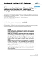
mRNA levels of key Type I IFN system genes in Atlantic salmon erythrocytes in response to native virus during haemagglutina-tion assayFigure 1
mRNA levels of key Type I IFN system genes in Atlantic salmon erythrocytes in response to native virus dur-
ing haemagglutination assay. Relative fold increase of the key Type I IFN system genes in response to NBISA01 or RPC/
NB-04-085-1 haemagglutination calibrated to the 18S rRNA housekeeping gene and the 0 hour control (values are average of a
triplicate observation ± standard deviation): (A) relative fold stimulation of Atlantic salmon IFN genes (SasaIFN-α1 and Sas-
aIFN-α2), Mx, ISG15 and STAT1 by NBISA01 virus; (B) relative fold stimulation of Atlantic salmon IFN genes (SasaIFN-α1 and
SasaIFN-α2), Mx, ISG15 and STAT1 by RPC/NB-04-085-1.
A
0
5
10
15
20
25
30
35
40
45
50
0 hour day 1 day 2 day 3 day 4 day 5
Sampling days after haemagglutination with NBISA01
Relative fold increase of the type I IFN system
genes
IFN Mx ISG15 STAT1 PKZ
B
0
2
4
6
8
10
12
14
16
18
20
day 0 day 1 day 2 day 3 day 4 day 5
Sampling days after haemagglutination with RPC/NB-04-085-1
Relative fold increase of the type I IFN genes
IFN Mx ISG15 STAT1
Virology Journal 2008, 5:36 />Page 6 of 12
(page number not for citation purposes)
reactions. As shown in Figure 4, there was only minimal
induction of the genes investigated except for ISG15 and
Mx by 72 hours after stimulation. These results show that,
unlike TO cells, Atlantic salmon erythrocytes do not effi-
ciently respond to polyI:C stimulation. However, the
response was similar to that of erythrocytes exposed to
heat-inactivated ISAV or to L-15 medium alone (negative
control assays).
Discussion
Two models of haemagglutination-infection phenotypes
have been proposed to account for the anaemia associated
with the clinical disease due to ISAV infection in fish. The
first model is that anaemia in the clinical disease is due to
uptake by immune cells of fish erythrocytes coated with
ISAV [6], and the receptor destroying enzyme (RDE) activ-
ity, which is related to the pathogenicity of the virus [13],
allows the virus to elute from fish erythrocytes except
those of Atlantic salmon [7]. An alternative model is that
failure of ISAV to elute from Atlantic salmon erythrocytes
favours virus infection of the erythrocytes, which might
result in cell death, and this combination is related to the
pathogenicity of the virus [11]. Such a haemagglutina-
tion-infection phenotype is fundamentally different from
haemagglutination by avian and mammalian orthomyxo-
viruses, and may be indicative of a different pathogenesis
for the fish orthomyxovirus.
In the present study we set up haemagglutination assays in
L-15 growth medium to compare two phenomena (elu-
tion and uptake) of the ISAV-induced haemagglutination
of Atlantic salmon erythrocytes between virus strains of
differing pathogenicities. We found remarkable differ-
ences in virus replication and quality of cytokine response
in the fish erythrocytes. Real-time quantitative RT-PCR
was used to assess the viral RNA levels (i.e., both vRNA
and viral mRNA) in the haemagglutination reaction sam-
ples. Only the Ct values for the NBISA01 haemagglutina-
tions showed any decrease from the 0 hour to day 5. This
decrease was evident even when oligodT primers were
used for cDNA synthesis, confirming that there was de
novo synthesis of virus genes in the erythrocytes. However,
there are relatively long stretches of adenosine residues in
the ISAV target gene that could allow detections of vRNA
as well. The RPC/NB-04-085-1 haemagglutinations
showed no changes in the Ct values at any sampling time
point, indicating that the low pathogenic virus did not
replicate in erythrocytes. Moreover, using virus titrations
in the TO cell line, it was shown that NBISA01 haemag-
glutinations resulted in a productive infection. The
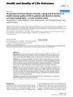
mRNA levels of key Type I IFN system genes in Atlantic salmon erythrocytes in L-15 medium (negative control for haemagglu-tination assay)Figure 2
mRNA levels of key Type I IFN system genes in Atlantic salmon erythrocytes in L-15 medium (negative con-
trol for haemagglutination assay). Relative fold stimulation of the key Type I IFN system genes in Atlantic salmon erythro-
cytes when virus is not present; i.e., in response to L-15 medium alone (negative control) calibrated to the 18S rRNA
housekeeping gene and the 0 hour control (values are average of a triplicate observation ± standard deviation): Atlantic salmon
IFN genes (SasaIFN-α1 and SasaIFN-α2), Mx, ISG15 and STAT1 in negative control erythrocytes.
0
1
2
3
4
5
0 hour 3 day 5 day
Sampling days after addition of L-15 medium
Relative fold increase of type I IFN genes
IFN Mx ISG15 Mx
Virology Journal 2008, 5:36 />Page 7 of 12
(page number not for citation purposes)
increase in virus titre between day 0 and day 5 was only
10
0.75
TCID
50
in contrast to the 10-fold increase in the
viral transcript levels detected by real-time RT-PCR within
the same samples. This may be due to three factors: [1] the
lower sensitivity of the virus titration in TO cell line com-
pared to real-time RT-PCR, [2] the fact that real-time RT-
PCR also detects non infectious or defective particles
which the TCID
50
does not, and [3] the fact that the virus
replication associated with haemagglutination involved
only a single cycle of virus replication as multiple haemag-
glutination events were unlikely. It is interesting to note
that avian erythrocytes (which have a dormant nucleus, in
contrast to the complete nucleus in fish erythrocytes) also
demonstrate virus uptake during haemagglutination by
influenza A virus and show de novo synthesis of viral pro-
teins but not production of new infectious virus particles
[5] whereas mammalian erythrocytes that do not have a
nucleus have completely lost the capacity for virus replica-
tion during influenza A virus-induced haemagglutination.
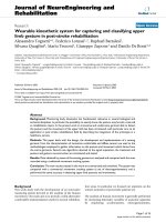
mRNA levels of key Type I IFN system genes in Atlantic salmon erythrocytes in response to inactivated virus during haemag-glutination assayFigure 3
mRNA levels of key Type I IFN system genes in Atlantic salmon erythrocytes in response to inactivated virus
during haemagglutination assay. Relative fold increase of the key Type I IFN system genes calibrated to the 18S rRNA
housekeeping gene and the 0 hour control (values are average of a triplicate observation ± standard deviation): (A) UV-inacti-
vated NBISA01; (B) heat-inactivated NBISA01; (C) UV-inactivated RPC/NB-04-085-1; and (D) heat inactivated RPC/NB-04-
085-1 haemagglutination.
0
0.5
1
1.5
2
2.5
3
3.5
4
0 hour 2 day 5 day
Sampling days after haemagglutination
Relative fold increase of the type I IFN
system genes
IFN Mx ISG15 STAT1
0
1
2
3
4
5
6
o hour 2 day 5 day
Sampling days after haemagglutination
Relative fold increase of the type I IFN
system genes
IFN Mx ISg15 STAT1
A B
0
0.5
1
1.5
2
2.5
3
3.5
4
0 hour 2 day 5 day
Sampling days after haemmaglutination
Relative fold increase of the type I IFN
system genes
IFN Mx ISG15 STAT1
0
0.5
1
1.5
2
2.5
3
3.5
4
4.5
5
0 hour 2 day 5 day
Sampling days after haemmaglutination
Relative fold increase of the type I IFN
system genes
IFN Mx ISG15 STAT1
D
C
Virology Journal 2008, 5:36 />Page 8 of 12
(page number not for citation purposes)
In addition to virus replication in haemagglutinations
induced by the highly pathogenic NBISA01 strain, we
found that there was also a higher relative fold increase of
IFN-α transcripts than with the less pathogenic RPC/NB
04-085-1 strain which did not replicate in erythrocytes.
The induction of the IFN-α gene closely followed the
increase of NBISA01 transcripts in that by day 1 the viral
transcripts started to increase simultaneously with the first
peak of IFN-α transcripts. The NBISA01 haemagglutina-
tions showed a pattern of fold increase with a peak of IFN-
α and Mx transcripts for a shorter period of time. This pat-
tern of induction is not continuous like the inductions in
TO cells infected with ISAV [19]. This may be due to dif-
ferences between the cell cycle of TO cells (which actively
multiply) and erythrocytes (which do not multiply) in
combination with the single cycle of virus replication that
probably occurs during haemagglutination in contrast to
multiple cycles of infection possible in TO cells. For the
NBISA01 haemagglutinations in the present study, the
level of IFN-α appeared to have a transient biphasic peak
at 1 and 3 days post-haemagglutination.
Both the UV- and heat-inactivated preparations of both
NBISA01 and RPC/NB-04-085-1 viruses and the L-15
medium assay (negative control) showed no haemagglu-
tination. The UV-inactivated viruses also showed no
induction of type I IFN system genes whereas the heat-
inactivated viruses and the L-15 medium assay showed
induction of the type I IFN system genes by day 5 similar
to those due to polyI:C stimulation by day 3. The absence
of haemagglutination in the UV-inactivated viruses was
unexpected since the inactivation was directed towards
the viral genome and not the surface glycoproteins
required for haemagglutination. One possible explana-
tion is that the UV lamp generated sufficient heat over the
18 hours of exposure to contribute to the denaturation of
the virus surface glycoproteins. In contrast, the heat inac-
tivation alone had no effect on the viral ssRNA genome
but probably even disrupted the structural viral proteins
so that the virus RNA was exposed and easily detected by
the erythrocyte viral pattern recognition receptors so as to
induce the observed minimal induction of the Type I IFN
system genes. Alternatively, these minimal responses were
non-specific since they were also seen with L-15 medium
alone (negative control assays).
STAT-1 expression has been studied in other fish species
including rainbow trout [25] but this is the first study to
investigate STAT1 expression in Atlantic salmon. It
appears that induction of STAT1 is not as highly respon-
sive to IFN induced by virus infection as the other compo-
nent genes of the Type I IFN system in that the fold
increase was low compared to the other genes studied.
This could be related to the multifunctional role of this
transcription factor.
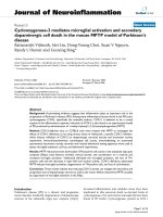
mRNA levels of key Type I IFN system genes in Atlantic salmon erythrocytes in response to PolyI:C stimulationFigure 4
mRNA levels of key Type I IFN system genes in Atlantic salmon erythrocytes in response to PolyI:C stimula-
tion. Relative fold increase of the key Type I IFN system genes in response to polyI:C stimulation calibrated to the 18S rRNA
housekeeping gene and the 0 hour control (values are average of a triplicate observation ± standard deviation).
0
2
4
6
8
10
12
14
0 hour 12 hour 24 hour 72 hour
Sampling point after polyI:C stimulation
Relative fold increase of the type I IFN
system genes
IFN Mx ISG15 STAT1
Virology Journal 2008, 5:36 />Page 9 of 12
(page number not for citation purposes)
The NBISA01 haemagglutination showed a moderate rel-
ative fold increase of PKZ transcripts. PKR gene has been
characterized in rainbow trout [26], and crucian carp [27].
PKR is one of the antiviral proteins of the IFN system [17].
For Atlantic salmon, only the sequence of a Z-DNA bind-
ing eIF-2α kinase is available in the GeneBank database.
In crucian carp cells, PKR mRNA has been shown to be up-
regulated in response to either IFN protein treatment or
virus infection. Moreover, in rainbow trout PKR has been
shown to be activated to phosphorylate eIF-2α in
response to polyI:C stimulation and virus infection. It is
very interesting that Atlantic salmon erythrocytes showed
expression of PKZ gene, albeit moderate, in response to
haemagglutination with a pathogenic ISAV strain.
In the present work the RT quantitative PCR data showed
varying levels of induction of key Type I IFN system genes
IFN-α, Mx, ISG15, STAT1, and PKZ upon haemagglutina-
tion of erythrocytes by the highly pathogenic NBISA01
virus. This virus induced significantly high relative fold
increase in IFN-α transcripts compared to the RPC/NB-04-
085-1 virus although both viruses had similar levels of
induction of Mx, ISG15 and STAT1. The slight Type I IFN
system response with RPC/NB-04-085-1 haemagglutina-
tions which involve only virus adsorption but no endocy-
tosis [11] and no replication is an interesting observation.
Various viral pathogen recognition receptors are involved
in the detection of viral pathogen associated molecular
patterns such as dsRNA, ssRNA, DNA, and viral glycopro-
teins like haemagglutinin proteins. In the case of human
cytomegalovirus [28], herpes simplex [29] and human
immunodeficiency virus [30], peripheral mononuclear
cells have been shown to induce Type I IFN independent
of virus replication, purely by the viral glycoproteins. Thus
the low level Type I IFN system gene induction detected in
the present study for the low pathogenic RPC/NB-04-085-
1 virus could possibly be associated with the detection of
the viral HE protein during haemagglutination.
PolyI:C is a synthetic dsRNA that is detected either by the
RNA helicases or the Toll-like receptor 3 (TLR3) to activate
the transcription of type I IFN system genes (reviewed in
[16]). This has been shown in the macrophage-like Atlan-
tic salmon TO cell line [19]. Stimulation of erythrocytes
with polyI:C did not, however, result in induction of Type
I IFN genes even with a polyI:C dose 10 times that used
elsewhere [19]. It was previously reported that CHSE-214
cells incubated with polyI:C show no expression of Mx
[31], probably because of inefficient response to polyI:C
stimulation. In the present study, NBISA01 endocytosis
and replication in Atlantic salmon erythrocytes resulted in
up-regulation of the IFN-α gene possibly by detection of
the viral molecular patterns by the erythrocytes. Thus the
minimal induction of the Type I IFN genes in fish erythro-
cytes by polyI:C could be due to inefficient membrane
transport activity of erythrocytes.
Conclusion
In conclusion, we report here that ISAV-induced haemag-
glutination by a pathogenic virus strain results in virus
uptake and productive infection of Atlantic salmon eryth-
rocytes accompanied by significant induction of IFN-α.
This study also highlights the critical role of ISAV strain
variation in the initial stages of the virus-cell interaction
during haemagglutination, and possibly in the pathogen-
esis of ISA. Moreover, the study shows for the first time
that fish erythrocytes immunologically respond to ISAV
infection.
Methods
Viruses
Two ISAV isolates of differing genotypes and pathogenic-
ities were used. NBISA01 is a highly pathogenic strain
belonging to the North American genotype, whereas RPC/
NB 04-085-1 is a low pathogenic strain of the European
genotype found in eastern Canada and its HE protein
places it in a unique, highly polymorphic region (HPR)
group [32]. The two isolates have variations in the amino
acid sequence of the HPR region with amino acid dele-
tions of 13 and 17 amino acids for RPC/NB-04-085-1 and
NBISA01, respectively [33]. The viruses were propagated
in the TO cell line [18] and the cell lysates were titrated on
TO cell monolayers as previously described [34] prior to
use in the subsequent studies.
Virus inactivation
The viruses were inactivated by using either UV light or
heat treatment. UV inactivation of ISAV was carried out
with a germicidal UV lamp (G30T8 with 30 Watt and 36
inch length, and a UV intensity of 125 μW/cm
2
at 1 meter
from the lamp) suspended in a biological safety cabinet
(Class II A/B3 BSC, Thermo Forma) following the proce-
dure reported by Oye and Rimstad [35], with minor mod-
ifications. Briefly, 20.0 mls of virus suspension in a 4-well
cell culture plate were placed 10 cm from the UV lamp.
The plate was left open under UV-exposure for 18 hours.
Heat inactivation of ISAV was performed by incubating
1.0 ml of the virus suspension in a 1.5-ml microfuge tube
at 56°C for 5 minutes. Complete inactivation of virus by
both methods was confirmed by titration in TO cell mon-
olayers [34] before use in the haemagglutination reac-
tions.
Haemagglutination assays
Atlantic salmon erythrocytes were collected from specific
pathogen free 100 g-Atlantic salmon using EDTA-coated
Vacutainer
®
tubes. In preliminary experiments, the
washed erythrocytes suspended in phosphate buffered
saline (PBS) were not viable beyond 48 hours. Therefore,
Virology Journal 2008, 5:36 />Page 10 of 12
(page number not for citation purposes)
common fish cell line growth media, Leibovitz's L-15
(Invitrogen) and Hanks minimum essential medium
(BioWhittaker) (HMEM), were tested to identify one that
better maintained erythrocyte viability. Using the Trypan
blue dye exclusion test, we found that erythrocytes resus-
pended in L-15 growth medium had lower cell deaths,
and those surviving maintained a normal shape in con-
trast to erythrocytes in HMEM growth medium which
were shrunken. In subsequent experiments, the erythro-
cytes were washed and then resuspended in L-15 medium
supplemented with 10% foetal bovine serum, 2 mM L-
glutamine, 100 IU/ml penicillin G, 100 μg ml
-1
strepto-
mycin, and 0.25 μg ml
-1
amphotericin B. For determining
the haemagglutination (HA) units of the stock virus prep-
arations, haemagglutination reactions were set up using
50 μl of two-fold dilutions of the two virus isolates and 50
μl of 1% erythrocytes [36]. The haemagglutination was set
using four wells for each virus dilution; 1 HA unit was
defined as the highest virus dilution that induced haemag-
glutination in four wells within 1 hr at room temperature.
Subsequent haemagglutination reactions used 1 HA unit
in 50 μl of virus preparation 50 μl of 1% erythrocytes in L-
15 growth medium. The sealed plates were kept at room
temperature for one hour, and then transferred to 16°C
for the extended incubation until sampled.
PolyI:C stimulation of Atlantic salmon erythrocytes
Washed Atlantic salmon erythrocytes were resuspended in
L-15 medium consisting of 10% FBS, 2 mM L-glutamine,
100 IU ml-1 penicillin G, 100 μg ml-1 streptomycin, and
0.25 μg ml-1 amphotericin B, and polyinosinic:polycyti-
dylic acid (polyI:C) (Amersham Biosciences) at a final
concentration of 30 μg ml-1. One hundred microliters of
1% erythrocyte suspension was added to each well of the
haemagglutination plate and incubated at 16°C. The
preparations were sampled after 12, 24, and 72 hours.
Detection of cytokine induction and virus replication using
real-time RT-PCR with SYBR Green chemistry
Total RNA from the haemagglutination samples was
extracted from 375 μl of homogeneous erythrocyte sus-
pensions using 1.25 ml of TRIZOL Reagent (Invitrogen).
RNA extraction was performed from two separate samples
at each sampling point, which were then pooled before
DNase treatment using the DNase treatment kit (Roche)
prior to RT-PCR amplification.
For quantification of the Type I IFN system genes and viral
RNA, first strand cDNA synthesis was done using the Tran-
scriptor reverse transcriptase first strand cDNA synthesis
kit (Roche). The cDNA synthesis used 125 ng of total RNA
in a master mix consisting of 4 μl of 5x RT reaction buffer,
2 μl of dNTP mix (200 μM), 2 μl of random hexamer (600
μM) or oligodT (0.8 μg/μl), 0.5 μl RNase inhibitor (40 U/
μl), 0.5 μl of Transcriptor reverse transcriptase (20 U/μl),
and nuclease free water adjusted to a final volume for 20
μl. The RT step was programmed at 25°C for 10 minutes
followed by 55°C for 30 minutes and a final enzyme
denaturation for 5 minutes at 85°C. Real-time PCR used
first strand cDNA template with LightCycler FastStart
DNA Master SYBR Green I (Roche) in the LightCycler (LC)
1.2 (Roche). The PCR primer pairs used are listed in Table
2; those for 18S rRNA, IFN-α, Mx, and ISG-15 are pub-
lished [19], and the STAT-1 primer pair was described in
Workenhe [37]. The PKZ (a Z-DNA binding orthologue of
the mammalian double stranded RNA binding PKR)
primer was designed using the coding sequence of Atlan-
tic salmon Z-DNA binding eIF-2α kinase (GenBank Acces-
sion # DQ182560
). The IFN gene primer set is designed in
the common region of the two IFN-α subtypes, α1 and α2
[38]. The 20 μl PCR reaction consisted of 2 μl of undiluted
cDNA for all genes except 18S rRNA (which was diluted
1:1000) and 18 μl of the master mix prepared using 0.5 μl
of the 10 μM of the forward and reverse primers (a final
concentration of 0.25 μM), 2 μl of the LC SYBR Green I
DNA Master mix, 1.6 μl of the stock 25 mM MgCl
2
(a final
concentration of 0.003 μM), and 13.4 μl of nuclease free
water. The real time PCR programme for amplifying PKZ
gene had a master mix consisting of 12.8 μl of water, 2.4
μl of 25 mM MgCl
2
(a final concentration of 0.004 μM),
0.4 μl of the 10 μM forward and reverse primer, and 2 μl
of SYBR Green master mix. The real-time PCR cycling con-
ditions consisted of an initial denaturation at 95°C for 10
minutes to activate the hot-start polymerase, followed by
40 cycles of 95°C for 5 s, 59°C for 10 s (60°C for the PKZ
gene), 72°C for 10 s, and detection at 80°C for 2 s. The
cycle threshold (Ct) values, the number of cycles run in
real-time RT-PCR when the fluorescence in the sample
crosses a threshold value (background) and amplification
enters a log-linear phase, were analyzed using LightCycler
software version 3.5 (Roche). Melting curve analysis with
the same software was performed from 70°C to 95°C in
0.1°C/s increments to verify the specificity of the ampli-
cons so as to interpret SYBR Green fluorescence data. For
determining amplification efficiency of each primer set
(Table 2), standard curves were generated using two-fold
dilutions of cDNA run in triplicates for six consecutive
dilutions. Each sampling point was run in triplicate and
the stability of the 18S rRNA, used as housekeeping gene,
was followed. The Ct values of positive amplicons were
then analyzed using the Pfaffl method for relative quanti-
fication in real-time RT-PCR [39] as previously used else-
where [19], to get relative fold increase of the Type I IFN
genes at each sampling point calibrated to the house keep-
ing gene and normalized with the 0 hour control. To test
if the difference in mean relative fold induction between
the two virus isolates at each sampling point was statisti-
cally significant, data were initially checked for equality of
variance using F- test in Microsoft Excel spread sheet. Then
Virology Journal 2008, 5:36 />Page 11 of 12
(page number not for citation purposes)
the t- Test was used considering the equality/inequality of
variance where applicable [40].
For quantifying the level of viral RNA, real-time RT-PCR
was done using the RNA Amplification Kit SYBR Green I
(Roche) and the primer pair designed by Devold et al. [41]
to amplify 220 bp of the ISAV segment 8, and previously
described for real time RT-PCR [42], with minor modifica-
tions. Briefly, the 20 μl reaction consisted of 50 ng of total
RNA in a master mix prepared using 0.3 μl of the 20 μM
of the forward and reverse primers (final concentration of
0.3μM), 4 μl SYBR Green, 0.2 μl LC-RT PCR enzyme mix,
3 μl of the 5x resolution solution, 1.6 μl of the 25 mM
stock MgCl
2
(a final concentration of 0.005 μM), and
nuclease free water adjusted to a final volume of 20 μl.
The cycling conditions consisted of one cycle of RT at
55°C for 30 min, initial denaturation at 95°C for 30 s fol-
lowed by 50 cycles of 95°C for 5 s, 59°C for 10 s, 72°C
for 10 s, and detection at 80°C for 2 s. The Ct values and
melting curve data were analyzed using LightCycler soft-
ware version 3.5 (Roche). Melting curve analysis was per-
formed from 70°C to 95°C in 0.1°C/s increments to
verify the specificity of the amplicons so as to interpret
SYBR Green fluorescence data. The amplicons were also
run in 1% agarose gel electrophoresis in 1x Tris acetate
EDTA buffer (40 mM Tris acetate and 1 mM EDTA) (Fisher
Scientific) and stained with ethidium bromide and photo-
graphed under 304 nm UV light.
Detection of virus replication by titration on TO cell
monolayers
Total cell lysates of the haemagglutination assays (i.e.,
total virus) were titrated to determine growth cycles of the
virus strains in Atlantic salmon erythrocytes. Virus titra-
tion utilized serial 10-fold dilutions of the samples rang-
ing from 10
-1
to 10
-8
, inoculated on 48-well cell culture
plates containing TO cell monolayers using 4 wells per
dilution, from which the median tissue culture infectious
dose (TCID
50
) was determined as previously described
[33]. Each sampling point was titrated in triplicate to
obtain a standard deviation.
Competing interests
The author(s) declare that they have no competing inter-
ests.
Authors' contributions
STW conducted all the experiments and wrote the manu-
script. MJTK helped in designing the experiments and
writing the manuscript. GMW and DWW helped in the
initial stages of conceiving the study and edited the man-
uscript. DBG helped in designing the experiments and
edited the manuscript. FSBK conceived the study, coordi-
nated the research, and helped in designing the experi-
ments, writing and editing the manuscript.
Acknowledgements
This work was supported by the Natural Sciences and Engineering
Research Council (NSERC) of Canada Discovery Grant to FSBK. We thank
the staff of Aquatic Animal Facility of the Atlantic Veterinary College, espe-
cially Vicki Leggo for the kind cooperation and provision of fish for blood
collection.
References
1. Kawaoka Y, Cox NJ, Haller O, Hongo S, Kaverin N, Klenk H-D, Lamb
RA, McCauley J, Palese P, Rimstad E, Webster RG: Infectious
Salmon Anaemia Virus. In Virus Taxonomy – Eight Report of the
International Committee on Taxonomy Viruses Edited by: Fauquet CM,
Mayo MA, Maniloff J, Desselberger U, Ball LA. Elsevier Academic
Press: New York; 2005:681-693.
2. Falk K, Asperhaug V, Vlasak R, Endresen C: Identification and
characterization of viral structural proteins of infectious
salmon anaemia virus. J Virol 2004, 78:3063-3071.
3. Aspehaug VT, Mikalsen AB, Snow M, Biering E, Villoing S: Character-
ization of the infectious salmon anaemia virus fusion protein.
J Virol 2005, 79:12544-12553.
4. Clouthier SC, Rector T, Brown NEC, Anderson ED: Genomic
organization of infectious salmon anaemia virus. J Gen Virol
2002, 83:421-428.
Table 2: The oligonucleotide primers, amplicon length and amplification efficiency of the real time RT-PCR primers amplifying IFN,
Mx, 18S, ISG15 and STAT1 genes
Primer name Primer sequence Amplicon length (bp) Amplification efficiency
As IFN Fwd
1
TGCAGTATGCAGAGCGTGTG 100 1.83
As IFN Rev
1
TCTCCTCCCATCTGGTCCAG
As Mx Fwd TGCAACCACAGAGGCTTTGAA 77 1.88
As Mx Rev GGCTTGGTCAGGATGCCTAAT
As 18S Fwd TGTGCCGCTAGAGGTGAAATT 60 1.86
As 18S Rev GCAAATGCTTTCGCTTTCG
As ISG15 Fwd CTGAAAAACGAAAAGGGCCA 100 1.83
As ISG15 Rev GCAGGGACTCCCTCCTTGTT
As STAT1 Fwd TGTCTGTTGGCTCAGTTGCG 100 1.82
As STAT1 Rev GAAATTGATGCTGTGGCGTCT
As PKZ Fwd AGATAGCGAAGGCTGTTGGA 101 1.913
As PKZ Rev TGGTTTGTCTGGTGTTGCAT
1
Primer amplifies SasaIFN-α1 and α2.
Publish with BioMed Central and every
scientist can read your work free of charge
"BioMed Central will be the most significant development for
disseminating the results of biomedical research in our lifetime."
Sir Paul Nurse, Cancer Research UK
Your research papers will be:
available free of charge to the entire biomedical community
peer reviewed and published immediately upon acceptance
cited in PubMed and archived on PubMed Central
yours — you keep the copyright
Submit your manuscript here:
/>BioMedcentral
Virology Journal 2008, 5:36 />Page 12 of 12
(page number not for citation purposes)
5. Evensen O, Thorud KE, Olsen YA: A morphological study of the
gross and microscopic lesions of infectious salmon anaemia
in Atlantic salmon (Salmo salar). Res Vet Sci 1991, 51:215-222.
6. Dale OB, Falk K, Kvellestad A: An overview of Infectious Salmon
Anemia pathology and suggested pathogenesis. Abstr. Fifth
International Symposium on Aquatic Animal Health, California USA 2006.
7. Falk K, Namork E, Rimstad E, Mjaaland S, Dannevig BH: Character-
ization of infectious salmon anemia virus, an orthomyxo-like
virus isolated from Atlantic salmon (Salmo salar L.). J Virol
1997, 71:9016-9023.
8. Hirst GK: The agglutination of red blood cells by allantoic
fluid from chick embryos infected with influenza virus. Sci-
ence 1941, 94:22-23.
9. Howe C, Lee LT: Virus erythrocyte interactions. Adv Virus Res
1972, 17:1-50.
10. Bossart W, Meyer J, Bienz K: Electron microscopic study on
influenza virus hemagglutination: pinocytosis of virions by
red cells. Virology 1973, 55:295-298.
11. Workenhe ST, Wadowska DW, Wright GM, Kibenge MJT, Kibenge
FSB: Demonstration of infectious salmon anaemia virus
(ISAV) endocytosis in erythrocytes of Atlantic salmon. Virol J
2007:4. 13
12. Kibenge FSB, Munir K, Kibenge MJT, Joseph T, Moneke E: Infectious
salmon anemia virus: causative agent, pathogenesis and
immunity. Ani Hlth Res Rev 2004, 5(1):65-78.
13. Falk K, Dale OB: Experimental evidence indicating that lack of
viral receptor destroying enzyme is a major factor for infec-
tious salmon anemia pathogenesis. Abstr. Fifth International Sym-
posium on Aquatic Animal Health, California USA 2006.
14. Follett EAC, Pringle CR, Wunner WH, Skehel JJ: Virus replication
in enucleate cells: vesicular stomatitis virus and influenza
virus. J Virol 1974, 13:394-399.
15. Cook RF, Avery J, Dimmock NJ: Infection of chicken erythro-
cytes with influenza and other viruses. Infect Immun 1979,
25:
396-402.
16. Robertsen B: The interferon system of teleost fish. Fish & Shell-
fish Immunol 2006, 20:172-191.
17. Samuel CE: Antiviral actions of interferons. Clin Microbiol Rev
2001, 14:778-809.
18. Wergeland HI, Jakobsen RA: A salmonid cell line (TO) for the
production of infectious salmon anaemia virus (ISAV). Dis
Aquat Organ 2001, 44:183-190.
19. Kileng Ø, Brundtland MI, Robertsen B: Infectious salmon anemia
virus is a powerful inducer of key genes of the type I inter-
feron system of Atlantic salmon, but is not inhibited by inter-
feron. Fish & Shellfish Immunol 2006, 23:378-389.
20. Passantino L, Altamura M, Cianciotta A, Jirillo F, Ribaud MR, Jirillo E,
Passantino GF: Maturation of fish erythrocytes coincides with
changes in their morphology, enhanced ability to interact
with Candida albicans and release of cytokine-like factors
active upon autologous macrophages. Immunopharmacology &
Immunotoxicology 2004, 26:73-585.
21. Rasmussen R: Quantification on the LightCycler. In Rapid Cycle
Real-time PCR, Methods and Applications Edited by: Meuer S, Wittwer
C, Nakagawara K. Springer, Heidelberg; 2001:21-34.
22. Kibenge FSB, Garate ON, Johnson G, Arriagada R, Kibenge MJT,
Wadowska D: Isolation and identification of infectious salmon
anaemia virus (ISAV) from Coho salmon in Chile. Dis Aquat
Organ 2001, 45:9-18.
23. Kibenge MJT, Munir K, Kibenge FSB: Constitutive expression of
Atlantic salmon Mx protein in CHSE-214 cells confers resist-
ance to infectious salmon anaemia virus. Virol J 2005, 2:75.
24. McBeath AJA, Collet B, Paley R, Duraffour S, Aspehaug V, Biering E,
Secombes CJ, Snow M: Identification of an interferon antago-
nist protein encoded by segment 7 of infectious salmon
anaemia virus. Virus Res 2006, 115:176-184.
25. Collet B, Munro SE, Gahlawat S, Acosta F, Garcia J, Roemelt C, Zou
J, Secombes CJ, Ellis AE: Infectious pancreatic necrosis virus
suppresses type I interferon signalling in rainbow trout
gonad cell line but not in Atlantic salmon macrophages.
Fish
& Shellfish Immunol 2007, 22:44-56.
26. Garner YN, Joshi B, Jagus R: Characterization of rainbow trout
and zebrafish eukaryotic initiation factor 2α and its response
to endoplasmic reticulum stress and IPNV infection. Dev &
Comp Immunol 2003, 27:217-231.
27. Hu C-Y, Zhang Y-B, Huang G-P, Zhang Q-Y, Gui J-F: Molecular
cloning and characterisation of a fish PKR-like gene from cul-
tured CAB cells induced by UV-inactivated virus. Fish & Shell
immunol 2004, 17(4):353-366.
28. Boehme KW, Singh J, Perry ST, Compton T: Human cytomegalo-
virus elicits a coordinated cellular antiviral response via
envelope glycoprotein B. J Virol 2004, 78(3):1202-1211.
29. Mossman KL, Ashkar AA: Herpesviruses and the innate
immune response. Viral Immunol 2005, 18:267-281.
30. Ankel H, Capobianchi MR, Castilletti C, Dianzani F: Interferon
induction by HIV glycoprotein 120: role of the V3 loop. Virol-
ogy 1994, 205:34-43.
31. Jensen I, Larsen R, Robertsen B: An antiviral state induced in Chi-
nook salmon embryo cells (CHSE-214) by transfection with
the double-stranded RNA poly I:C. Fish & Shellfish Immunol 2002,
13:367-378.
32. Kibenge FSB, Kibenge MJT, Groman D, McGeachy S: In vivo corre-
lates of infectious salmon anemia virus pathogenesis in fish.
J Gen Virol 2006, 87:2645-2652.
33. Kibenge FSB, Kibenge MJT, Wang Y, Qian B, Hariharan S, McGeachy
S: Mapping of putative virulence motifs on infectious salmon
anaemia virus surface glycoprotein genes. J Gen Virol 2007,
88:3100-3111.
34. Kibenge FSB, Kibenge MJT, McKenna PK, Stothard P, Marshall R,
Cusack RR, McGeachy S: Antigenic variation among isolates of
infectious salmon anaemia virus correlates with genetic var-
iation of the viral haemagglutinin gene. J Gen Virol 2001,
82:2869-2879.
35. Øye AK, Rimstad E:
Inactivation of infectious salmon anaemia
virus, viral haemorrhagic septicaemia virus and infectious
pancreatic necrosis virus in water using UVC irradiation. Dis
Aquat Organ 2001, 48:1-5.
36. Food and Agricultural Organization of the United Nations
[ />]
37. Workenhe ST: Expression of type I IFN genes: antiviral Mx and
the transcription factors STAT-1 and IRF-7. M.Sc. Thesis, Uni-
versity of Tromso, Norway 2006.
38. Bergan V, Steinsvik S, Xu H, Kileng Ø, Robertsen B: Promoters of
type I interferon genes from Atlantic salmon contain two
main regulatory regions. FEBS Journal 2006, 273:3893-3906.
39. Pfaffl MW: A new mathematical model for relative quantifica-
tion in real-time RT-PCR. Nucl Acids Res 2001, 29(9):e45.
40. Richardson BA, Overbaugh J: Basic statistical considerations in
virological experiments. J Virol 2005, 79:669-676.
41. Devold M, Krossøy B, Aspehaug V, Nylund A: Use of RT-PCR for
diagnosis of infectious salmon anaemia virus (ISAV) in car-
rier sea trout Salmo trutta after experimental infection. Dis
Aquat Organ 2000, 40(1):9-18.
42. Munir K, Kibenge FSB: Detection of infectious salmon anaemia
virus by real-time RT-PCR. J Virol Meth 2004, 117(1):37-47.