Báo cáo hóa học: " The inhibition of the Human Immunodeficiency Virus type 1 activity by crude and purified human pregnancy plug mucus and mucins in an inhibition assay" ppt
Bạn đang xem bản rút gọn của tài liệu. Xem và tải ngay bản đầy đủ của tài liệu tại đây (969.81 KB, 10 trang )
BioMed Central
Page 1 of 10
(page number not for citation purposes)
Virology Journal
Open Access
Research
The inhibition of the Human Immunodeficiency Virus type 1 activity
by crude and purified human pregnancy plug mucus and mucins in
an inhibition assay
Habtom H Habte
1
, Corena de Beer
2
, Zoë E Lotz
1
, Marilyn G Tyler
1
,
Leann Schoeman
3
, Delawir Kahn
1
and Anwar S Mall*
1
Address:
1
Department of Surgery, University of Cape Town, Cape Town, South Africa,
2
Discipline of Medical Virology, University of Stellenbosch
and National Health Laboratory Service, Tygerberg Business Unit, Stellenbosch, South Africa and
3
Obstetrics and Gynaecology, University of Cape
Town, Cape Town, South Africa
Email: Habtom H Habte - ; Corena de Beer - ; Zoë E Lotz - ;
Marilyn G Tyler - ; Leann Schoeman - ; Delawir Kahn - ;
Anwar S Mall* -
* Corresponding author
Abstract
Background: The female reproductive tract is amongst the main routes for Human
Immunodeficiency Virus (HIV) transmission. Cervical mucus however is known to protect the
female reproductive tract from bacterial invasion and fluid loss and regulates and facilitates sperm
transport to the upper reproductive tract. The purpose of this study was to purify and characterize
pregnancy plug mucins and determine their anti-HIV-1 activity in an HIV inhibition assay.
Methods: Pregnancy plug mucins were purified by caesium chloride density-gradient ultra-
centrifugation and characterized by Western blotting analysis. The anti-HIV-1 activities of the crude
pregnancy plug mucus and purified pregnancy plug mucins was determined by incubating them with
HIV-1 prior to infection of the human T lymphoblastoid cell line (CEM SS cells).
Results: The pregnancy plug mucus had MUC1, MUC2, MUC5AC and MUC5B. The HIV inhibition
assay revealed that while the purified pregnancy plug mucins inhibit HIV-1 activity by approximately
97.5%, the crude pregnancy plug mucus failed to inhibit HIV-1 activity.
Conclusion: Although it is not clear why the crude sample did not inhibit HIV-1 activity, it may be
that the amount of mucins in the crude pregnancy plug mucus (which contains water, mucins, lipids,
nucleic acids, lactoferrin, lysozyme, immunoglobulins and ions), is insufficient to cause viral
inhibition or aggregation.
Background
Cervical mucus is reported to regulate sperm penetration
and transport to the upper reproductive tract [1,2]. It also
provides lubrication to the cervix by enhancing its wetness
and thus preventing its desiccation, and retards enzymatic
degradation of the cervix and providing it with protection
from pathogenic invasion and infection [3-5]. Its secre-
tion, at a rate of 20–60 mg per day acts as a fence to sperm
and pathogen entrance [6]. Although a reduction in
mucus viscosity may allow foreign agent penetration, mil-
Published: 19 May 2008
Virology Journal 2008, 5:59 doi:10.1186/1743-422X-5-59
Received: 19 February 2008
Accepted: 19 May 2008
This article is available from: />© 2008 Habte et al; licensee BioMed Central Ltd.
This is an Open Access article distributed under the terms of the Creative Commons Attribution License ( />),
which permits unrestricted use, distribution, and reproduction in any medium, provided the original work is properly cited.
Virology Journal 2008, 5:59 />Page 2 of 10
(page number not for citation purposes)
lions of micro-organisms a day are reported to be cleared
from the reproductive tract by cervical secretions that are
the tract's most effective first line of defence [7].
Thus far six mucin genes have been reported to be
expressed by the female reproductive tract, namely
MUC1, MUC2, MUC4, MUC5AC, MUC5B and MUC6
[6]. The genes for MUC2, MUC5B, MUC5AC and MUC6,
are found on chromosome 11p15.5 and express the
secreted gel forming mucins, whereas MUC1 and MUC4
are membrane associated mucins expressed by the epithe-
lium of the ecto-cervix and vagina [7]. Of these, MUC4
and MUC5B are reported to be the major mucin genes
expressed by the endo-cervix [8]. The variation, under hor-
monal influence, of the viscoelastic and rheological prop-
erties of these mucins during the menstrual cycle is well
documented [4].
Human crude saliva is known to inhibit Human Immun-
odeficiency Virus type 1 (HIV-1) activity in an in vitro
assay [9,10]. These authors speculated that it was the
mucus component that inhibited the virus. We very
recently showed that both crude saliva and its purified
mucin components MUC5B and MUC7 inhibited HIV-1
activity [11] and so did the purified MUC1 of breast milk
[12]. The MUC1 of breast milk also showed anti-pox viral
activity [13]. Our hypothesis is that cervical mucins
should have a similarly inhibitory effect on HIV-1 activity,
an important question considering that the vagina and
cervix are significant routes for HIV transmission. The aim
of this study therefore was to extract and purify the mucins
in the pregnancy plug mucus and to determine their anti-
HIV-1 activity using an HIV inhibition assay.
We therefore extracted and purified mucins from the preg-
nancy plug mucus which occludes the cervical canal
throughout the pregnancy period [2,14]. This large mucus
plug which is more like the mucus of the luteal phase than
the mucus of the mid-cycle [2] was obtained during
labour and just prior to delivery.
Sub-Saharan Africa is reported to be home to about 25
million adults and children who are HIV positive [15]. In
Southern Africa 25.7% of the population has HIV/AIDS,
making this the most highly prevalent region of infection
compared to the Eastern and the Western regions with
11.4% and 4.3% prevalence respectively [16]. In South
Africa alone, between 4.68 and 7.03 million people were
living with HIV/AIDS in 2004 [17], of whom 55% were
female [18]. Thus this preliminary study could make a sig-
nificant contribution to the efforts being made in control-
ling this epidemic.
In this study we report the anti-HIV-1 activities of crude
and purified human pregnancy plug mucus and mucins in
an in vitro inhibition assay. We have demonstrated that
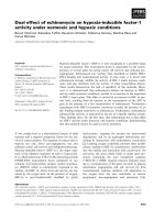
Caesium chloride density gradient purification of the pregnancy plug mucinsFigure 1
Caesium chloride density gradient purification of the pregnancy plug mucins. Samples in 4 M GuHCl were adjusted
to a density of 1.39 to 1.40 g ml
-1
with solid caesium chloride. Density gradient centrifugation was performed in a Beckman L45
ultra-centrifuge for 48 h at a 105 000 g at 4°C. Mucin positive fractions (u) at a density (s) between 1.37–1.42 and still associ-
ated with some protein (n) (a) were pooled and prepared for the second step centrifugation (b). Finally fractions (fraction
number 3, 4 and 5) were pooled, dialysed against three changes of distilled water and freeze-dried.
Virology Journal 2008, 5:59 />Page 3 of 10
(page number not for citation purposes)
the purified mucins from the pregnancy plug mucus
inhibited HIV-1 infection of the CEM SS cells. However,
the crude pregnancy plug mucus failed to inhibit HIV-1
infection of these cells.
Results
Mucin purification
Pregnancy plug mucins were purified by density gradient
centrifugation, twice in caesium chloride/4 M GuHCl
with a buoyant density between 1.39 and 1.40 g/ml to
remove proteins and nucleic acids. The purification pro-
file in Fig. 1 demonstrates a clear separation of the lower
density proteins positive for Lowry from the higher-den-
sity glycoproteins positive for PAS. The mucin-rich frac-
tions (fractions number 3, 4 and 5) (Fig. 1b) were pooled,
dialysed against three changes of distilled water and
freeze-dried.
SDS-PAGE analysis
Pregnancy plug mucus (20 μg) was dissolved in gel load-
ing buffer containing 0.2 M 2-mercaptoethanol and
loaded onto 10% SDS-PAGE (Fig. 2). Gels were stained
either with PAS for carbohydrate or Coomassie Brilliant
Blue G-250 for protein. An intense PAS positive band (M
r
>220 kDa) appeared on the top of the running gel below
which there was another band of size <220 kDa (Fig. 2a,
lane 3). Coomassie Blue staining also showed material at
the top of the running gel and a number of bands of
higher electrophoretic mobility and therefore of relatively
smaller size within the gel (Fig. 2a, lane 2).
Caesium chloride density gradient ultra-centrifugation
removed most of the contaminant protein from crude
mucus as shown clearly by subsequent gel electrophoresis
(Fig. 2b, lane 4). Bands at the top of the running gel, stain-
ing both for protein and carbohydrate confirmed the pres-
ence of the mucin and its purity (Fig. 2b, lanes 4 and 5).
Western blotting
Western blot analysis was performed to determine the
identity of the mucins present in the pregnancy plug
mucus. Samples (40 μg each) were loaded on a 1% agar-
ose gel and subjected to electrophoresis. Mucins were then
transferred from the gel to a nitrocellulose membrane and
probed with mouse anti-MUC1 monoclonal (Fig. 3 lanes
1, 2 and 3) and rabbit anti-MUC2 (lanes 4, 5 and 6), rab-
bit anti-MUC5AC (lanes 7, 8 and 9) and rabbit anti-
MUC5B (lanes 10, 11 and 12) polyclonal antibodies. The
Western blotting result confirmed the presence of MUC1,
MUC2, MUC5AC and MUC5B mucins in the pregnancy
plug mucus (Fig. 3 lanes 3, 6, 9 and 12 respectively).
While MUC5AC was strongly expressed (Fig. 3 lane 9)
MUC2 appeared in relatively smaller amounts and as a
doublet (Fig. 3 lane 6, arrows) [19]. While the positive
controls MUC1 (lane 1), colonic mucus (lane 4), pseu-
domyxoma peritonei (lanes 7 and 10) [20] reacted with
the anti-MUC1, anti-MUC2, anti-MUC5AC and anti-
MUC5B antibodies respectively, the negative controls
namely the salivary MUC5B (lane 2), tracheal sputum
(lane 5), salivary MUC7 (lane 8) and gastric mucus (lane
11) did not react with the anti-MUC1, anti-MUC2, anti-
MUC5AC and anti-MUC5B antibodies respectively.
However, due to the lack of Western blotting antibodies
against MUC4 and MUC6 the identification of these
mucins was not done in this study.
Toxicity assay
Prior to the HIV inhibition assay the toxicity of the crude
pregnancy plug mucus and purified pregnancy plug
mucins to the CEM SS cells was determined by toxicity
assay. As shown in Table 1, no toxicity of these compo-
nents or no cell death was detected.
Inhibition assay
The anti-HIV-1 activities of the crude pregnancy plug
mucus and purified pregnancy plug mucins were deter-
mined by HIV inhibition assay. When HIV-1 was incu-
bated with crude pregnancy plug mucus for an hour and
the mixture subsequently added to or incubated with the
SDS-PAGE analyses of the pregnancy plug mucinsFigure 2
SDS-PAGE analyses of the pregnancy plug mucins.
Freeze-dried pregnancy plug mucins (20 μg) before (a) and
after (b) caesium chloride density gradient purification were
separated on 10% SDS-PAGE and stained with Coomassie
Brilliant Blue (lanes 1, 2 and 4) and PAS (lanes 3 and 5). Lane
1 is molecular weight marker in kDa.
Virology Journal 2008, 5:59 />Page 4 of 10
(page number not for citation purposes)
CEM SS cells for 30 min, a 100% HIV-1 infection of the
CEM SS cells was measured by the p24 antigen assay (Fig.
4). However, when the virus was first incubated with puri-
fied mucins from the pregnancy plug for an hour and then
the mixture subsequently incubated with the CEM SS cells
for 30 min, an approximately 97.5% inhibition of the
viral activity or an approximately 2.5% infection of the
CEM SS cells was detected. This suggests that compared to
the crude pregnancy plug mucus the purified pregnancy
plug mucins reduce the infection of CEM SS cells by an
approximately 39 fold (Fig. 4).
To determine the effect of time (incubation period) on the
rate of viral infection or inhibition ability of the samples,
the mixtures of (HIV-1 plus crude pregnancy plug mucus)
and (HIV-1 plus purified pregnancy plug mucins) were
incubated with the CEM SS cells for longer time periods (1
h and 3 h). However, no difference in the rate of viral
infection or inhibition ability of the samples due to incu-
bation time difference was observed (Fig. 4). To deter-
mine the anti-HIV-1 activity of the purified pregnancy
plug mucins at the highest dilution or lowest concentra-
tion, serial tenfold fold dilutions (i.e. 10
-1
, 10
-2
, 10
-3
and
10
-4
) of the mucins were also done. Again, no difference
in the anti-HIV-1 activity of the purified pregnancy plug
mucins was detected down to10
-4
(Fig. 4a,b,c and 4d).
As shown in Fig. 4, when HIV-1 was incubated with the
media (positive control) instead of the pregnancy plug
mucins prior to addition to the CEM SS cells at all time
points (30 min, 1 h and 3 h), HIV-1 infection of the CEM
SS cells was not inhibited and 100% HIV-1 replication or
infection of the CEM SS cells was measured by the p24
antigen assay. Surprisingly the heat inactivated HIV-1
(negative control) was also shown to cause an approxi-
mately 30% infection of the CEM SS cells at all time
points (Fig. 4).
To determine or compare the efficiency of HIV-1 aggrega-
tion by the crude pregnancy plug mucus and purified
pregnancy plug mucins, at the end of the incubation
period (1 h), the mixtures of (HIV-1 plus crude pregnancy
plug mucus), (HIV-1 plus purified pregnancy plug
mucins) and the control (HIV-1 plus media) were filtered
through 0.45 μm pore size cellulose acetate filter (25 mm
diameter) and the filtrates were added to or incubated
with the CEM SS cells at different time-points (30 min, 1
h and 3 h). The result demonstrated that the filtrates from
the mixtures of (HIV-1 plus crude pregnancy plug mucus)
and (HIV-1 plus media) caused 100% HIV-1 infection of
the CEM SS cells (results not shown).
Discussion
According to various studies [9,10,21,22], salivary macro-
molecules (possibly mucins) aggregate HIV-1 prior to
host cell entry, thus preventing transmission of HIV-1
through saliva. Wiggins et al. [7] reported that mucus is
the first line of defence against pathogenic micro-organ-
isms. Studies in our laboratory have also confirmed these
findings [11]. Crude saliva (from individuals with a self-
declared risk free lifestyle and thus presumably unin-
fected), and its purified mucins MUC5B and MUC7 [11]
and purified MUC1 from breast milk [12] show anti-HIV-
1 activity in an in vitro inhibition assay.
It thus remains to be asked why other areas such as the
female reproductive tract and breast milk, so rich in
mucus and mucins quite similar in substance and confor-
mation to those in saliva, still remain major routes of
transmission of the virus. In the case of breast milk we
showed that its MUC1 component inhibited the HIV-1
from infecting CEM SS cells in an in vitro assay only after
it was dissociated from the milk fat globules and isolated
and purified by caesium chloride density gradient ultra-
centrifugation. Crude breast milk had no such inhibitory
effect on HIV-1 [12]. In the light of this we decided to
investigate whether cervical mucus and mucin display any
anti-HIV-1 properties, considering that the cervix is a sig-
nificant route of transmission in women.
The quality and quantity of cervical mucins during the dif-
ferent phases of the menstrual cycle are reported to vary
either through the influence of oestrogen (proliferative
phase) or of progesterone (luteal phase). For example the
production of MUC5B was reported to increase at the
mid-cycle and decrease during the secretory phase of the
menstrual cycle whilst MUC4 increases during the luteal
phase of the menstrual cycle [8,23]. These cyclical varia-
tions together with the fact that cervical scrapings, which
yielded very small amounts of crude material made it dif-
ficult to investigate the anti-HIV-1 activity of these mucins
per se. Therefore mucus plugs at the mouth of the cervix
rich in mucin [2,14], were obtained from women in
labour. However, a comparison of the effect of purified
plug mucin versus purified cervical mucin on HIV is being
planned.
Table 1: Toxicity of crude pregnancy plug mucus and purified pregnancy plug mucins to CEM SS cells.
Sample Con CEM SS cells % of dead cells % of live cells
Pregnancy plug mucus 0.9 mg 2.5 × 10
6
/ml 0 100
Pregnancy plug mucins 0.9 mg 2.5 × 10
6
/ml 0 100
Virology Journal 2008, 5:59 />Page 5 of 10
(page number not for citation purposes)
In this study we have demonstrated that the purified
mucins from the pregnancy plug inhibited HIV-1 infec-
tion of the CEM SS cells. However, the crude pregnancy
plug mucus and the media failed to inhibit HIV-1 infec-
tion of these cells. Though the mechanism of inhibition is
not clear, it is likely that when the HIV-1 was incubated
with the mucins, the virus was trapped by aggregation
through the sugar side-chains of the mucins, a purely
physical phenomenon [10,24-26], resulting in preventing
the virus from entering the host cells (CEM SS cells). This
was supported by our finding that salivary MUC7 inhib-
ited HIV-1 infection of the CEM SS cells when it was incu-
bated with the virus prior to addition to the CEM SS cells.
However, the mucin failed to inhibit viral infection of
these cells when it was incubated with CEM SS cells prior
to addition of the virus (unpublished data). This suggests
that the mucin inhibits HIV-1 infection by physically
aggregating the virus than by blocking putative viral bind-
ing sites or receptors on the cells.
The virus and mucins were incubated together with the
cells for different incubation periods, i.e. 30 min, 1 h and
3 h to determine the effect of time on infection or lack
thereof. Cultures were then washed three times after each
incubation period to remove free virus and cultured for
another 4 days in IL-2 rich media. This was done to deter-
mine if the virus had entered the cells during the initial
incubation step and was able to replicate inside the cells
for the extended incubation period to produce p24 anti-
gen, or if the mucins were successful in preventing viral
Western blotting analyses of the purified pregnancy plug mucinsFigure 3
Western blotting analyses of the purified pregnancy plug mucins. Lane 1, MUC1 (positive control), lane 2, salivary
MUC5B (negative control), lane 4, colonic mucus (positive control), lane 5, tracheal sputum (negative control), lane 7, pseu-
domyxoma peritonei (positive control), lane 8, salivary MUC7 (negative control), lane 10, pseudomyxoma peritonei (positive
control), lane 11, gastric mucus (negative control) and lanes 3, 6, 9 and 12 purified pregnancy plug mucins were separated by a
1% agarose gel and transferred to nitrocellulose membrane. Following overnight blocking, the membranes were incubated for
2 h with mouse anti-MUC1 monoclonal (lanes 1, 2 and 3) and rabbit anti-MUC2 (lanes 4, 5 and 6), rabbit anti-MUC5AC (lanes
7, 8 and 9) and rabbit anti-MUC5B (lanes 10, 11 and 12) polyclonal antibodies. Membranes were then incubated for 1 h with
HRPO linked goat anti-mouse and goat anti-rabbit secondary antibodies and bands that interacted with the antibodies were
detected by ECL detection. NB the two bands of MUC2 (lane 6) are indicated by the arrows.
Virology Journal 2008, 5:59 />Page 6 of 10
(page number not for citation purposes)
entry into the cells and therefore prevent the production
of p24 antigen.
To further confirm the hypothesis that mucins inhibit
HIV-1 activity by physically aggregating the virus, the
CEM SS cells were incubated with the filtrates from the
mixtures. The lower infection (2.5%) of the CEM-SS cells
by the filtrate from the mixture of HIV-1 plus purified
pregnancy plug mucins suggests the presence of insignifi-
cant amount of viruses in the filtrate or almost complete
aggregation of the virus by the mucins, leaving no free
viruses to pass through the filter paper into the filtrate to
cause viral infection. On the other hand the 100% infec-
tion of the CEM-SS cells caused by the filtrates from the
mixtures of HIV-1 plus crude pregnancy plug mucus and
HIV-1 plus media suggests the presence of higher amount
Inhibition of HIV-1 activity by crude pregnancy plug mucus and purified pregnancy plug mucins in vitro assayFigure 4
Inhibition of HIV-1 activity by crude pregnancy plug mucus and purified pregnancy plug mucins in vitro assay.
Crude pregnancy plug mucus and purified pregnancy plug mucins (0.9 mg each) were incubated with subtype D HIV-1 for 60
min and filtered through 0.45 μm pore size cellulose acetate filter. As controls HIV-1 treated with media and heat inactivated
HIV-1 were used. The unfiltered samples were then incubated with CEM SS cells at a concentration of 0.5 × 10
6
cells ml
-1
for 30
min, 1 h and 3 h. After PBS wash cells were cultured and viral replication was measured by a qualitative p24 antigen assay. Let-
ters a, b, c and d indicate the anti-HIV-1 activity of each sample in a serial tenfold dilution of 10
-1
, 10
-2
, 10
-3
and 10
-4
respec-
tively. P. plug represents pregnancy plug.
Virology Journal 2008, 5:59 />Page 7 of 10
(page number not for citation purposes)
of viruses in the filtrates or the failure of the crude preg-
nancy plug mucus and the media to aggregate the viruses.
This finding agreed with the report that HIV-1 may bind
to the high-molecular weight components which results
in macromolecular complex formation which is remova-
ble by filtration through 0.45 μm pore filter paper [10,24-
26].
The lack of inhibition by crude pregnancy plug mucus
compared to the inhibition by purified pregnancy plug
mucins is not clear. However it should be considered that
mucins constitute only about 0.5–1% of total crude
mucus [27] which is known to contain water, glycopro-
teins, lipids, nucleic acids, lactoferrin, lysozyme, immu-
noglobulins and ions [7]. It is likely therefore that the
potency of mucins would in this case be in their purified
form rather than when they are a minor part of a larger
secretion in which their concentration would be diluted.
This was quite different in the case of crude saliva, the
inhibitory effect of which was similar to that of its purified
mucins, separable by gel filtration and individually effec-
tive against the virus [11]. However, quantification of the
amount of mucins in the crude mucus prior to any assay
should be considered before drawing this conclusion.
The heat inactivated HIV-1 (negative control) caused an
approximately 30% infection of the CEM SS cells suggest-
ing that the viruses, when inactivated but not completely
killed are still infective, albeit to a lesser degree. To deter-
mine whether there is a dose/effect relationship and the
lowest possible effective concentration with anti-HIV-1
activity, ten fold serial dilutions (10
-1
to 10
-4
) of the
mucins were also done from a starting concentration of
purified mucin of 0.9 mg. The mucins showed strong anti-
HIV-1 activity down to a dilution of 10
-4
, but in this study
the lowest possible concentration which can cause inhibi-
tion of HIV-1 activity was not identified. Thus a lower
starting concentration of purified mucin than 0.9 mg
would be advisable.
There was also no effect of time (incubation period) on
the inhibitory effect of mucins or the infectivity of the
virus. This suggested that the mucins aggregated the virus
immediately and permanently. However, shorter starting
times of incubation of mucins and the virus would be nec-
essary to determine the shortest time mucins take to aggre-
gate the virus.
Although HIV-1 Subtype C is currently the most prevalent
in South Africa, the Subtype D which was used in this
study was found during the early HIV epidemic in the
country and is quite prevalent here, albeit to a lesser
degree. Even though we wished to use the Subtype C
strain, the Subtype D strain is unfortunately the only lab
adapted strain we had available to us in the vicinity of
Cape Town and it is possible that this is the only labora-
tory based HIV assay in the country. As described in the
Methods section, this virus was first isolated from an AIDS
patient by the Department of Medical Virology, Tygerberg
Hospital, University of Stellenbosch, South Africa, in Feb-
ruary 1988, and it was fully characterised and sequenced
subsequently [28]. The human T lymphoblastoid cell line
(CEM SS cells), which was used in this study, is reported
to express CD4, CXCR4, ICAM-3 and MHC class II mole-
cules [29]. These cells are capable of developing easily
quantifiable syncytia formation in four to six days upon
the addition of HIV-1 [30]. Although Subtype C predom-
inantly uses CCR5, several instances of co-receptor switch
to CXCR4 or even dual tropism have been observed in
Subtype C, especially later in infection. Therefore this
study could be relevant to in vivo situations, where trans-
mitted viruses are most often CCR5 tropic.
Extraction of mucus was in 6 M GuHCl and proteolytic
inhibitors which included 10 mM EDTA, 5 mM NEM, and
1 mM PMSF to reduce endogenous proteolysis of mucins
[2]. PMSF and EDTA inhibit serine and metallo-protease
activity respectively whilst NEM inhibits thiol proteases
and minimizes thiol-disulfide exchange [1].
Caesium chloride density gradient purification removes
all contaminants such as non-mucin proteins, lipids, pro-
teoglycans and nucleic acids from mucins [31]. Purifica-
tion of the mucins was confirmed by SDS-PAGE [32]. The
removal of these contaminants from mucins was believed
to be by dissociative conditions through the presence of
GuHCl [1], known to be a widely used denaturant [33]
which in this case could well dismantle the tertiary struc-
ture of mucins [14].
The presence of MUC1, MUC2, MUC5AC and MUC5B in
the pregnancy plug mucus was confirmed by Western
blotting with MUC2 expressed as a doublet and in small
amount compared to the other mucins. Immunohisto-
chemistry confirmed previous reports of the expression of
MUC4 and MUC6 by the endometrial tissue (data not
shown), but their presence in the mucus plug could not be
confirmed due to the lack of antibodies to these mucins
for Western blotting. This result agreed with that of Gip-
son et al. [6], Wiggins et al. [7], Gipson et al. [23] and
Wickstrom et al. [34], studies which reported the expres-
sion of MUC1, MUC2, MUC4, MUC5AC, MUC5B and
MUC6 by the female reproductive tract.
Conclusion
In summary, we have shown the in vitro inhibition of HIV-
1 activity by purified mucins from the pregnancy plug.
However, the crude pregnancy plug mucus failed to
inhibit HIV-1 activity. Although it is not clear why the
crude sample did not inhibit HIV-1 activity, it is likely that
Virology Journal 2008, 5:59 />Page 8 of 10
(page number not for citation purposes)
the amount of mucins in the crude pregnancy plug mucus
is of too low a concentration to cause viral inhibition or
aggregation. Future studies will attempt to establish the
lowest amount of purified mucin required to cause aggre-
gation of the virus. Also different HIV strains, cell lines
and samples from different donors for statistical validity
to strengthen this preliminary finding, will be carried out.
A comparison between the anti-HIV-1 activity of each cer-
vical mucin from the different stages of the menstrual
cycle has also been planned.
Materials and methods
Ethics
The University of Cape Town Research and Ethics Com-
mittee approved this study; ethics number REC REF: 283/
2004
Materials
Mouse anti-MUC1 monoclonal (NCL-MUC1, 201607)
and goat anti-mouse horse radish peroxidise (HRPO)
linked secondary antibodies (sc-2005) were from Novo-
castra (Newcastle, UK) and Santa Cruz (California, USA)
respectively. Polyclonal rabbit anti-MUC2 (LUM2-3),
anti-MUC5AC (LUM5-1), anti-MUC5B (LUM5B-2) and
goat anti-rabbit HRPO linked secondary antibodies were
kindly provided by Sara Kirkham (Manchester, UK). The
CEM SS cells were from AIDS Research and Reference Rea-
gent Programme (Germantown, USA). The p24 antigen
kit was from Vironostika HIV-1 Antigen kit Biomérieux
(France). Sepharose CL-4B and reagent solvents such as
guanidinium chloride (GuHCl) and caesium chloride
(CsCl) were from Sigma (UK). Trypan Blue Dye solution
was from Merck (Germany).
Pregnancy plug mucus collection
Pregnancy plug mucus was obtained from the Groote Sch-
uur Hospital Maternity Division at the University of Cape
Town. The pregnancy plug mucus was retrieved prior to
delivery and collected into cold 6 M GuHCl containing
proteolytic inhibitors, namely 10 mM EDTA, 5 mM NEM
and 1 mM PMSF pH 6.5 and stored at -20°C.
Mucus preparation
Crude pregnancy plug mucus was prepared according to
the method of Carlstedt et al. [2]. The pregnancy plug
mucus was collected into 0.1 M Tris-HCl, 2% (w/v) EDTA
and 5 mM PMSF pH 7.5 and prepared for the HIV inhibi-
tion assay. After gentle stirring for 15 h at 4°C, insoluble
materials were removed by high-speed centrifugation at 9
000 g for 2 h at 4°C. The supernatant was dialysed against
three changes of distilled water at 4°C and freeze-dried.
Mucin preparation
Pregnancy plug mucus was thawed and stirred gently for
15 h at 4°C in 6 M GuHCl and a cocktail of proteolytic
inhibitors as described above. Insoluble materials were
removed by high-speed centrifugation at 9 000 g for 2 h at
4°C. The soluble material was then pooled and subjected
to density gradient ultra-centrifugation, twice for 48 h at a
105 000 g at 4°C in a Beckman L45 ultra-centrifuge [31].
Briefly, samples in 4 M GuHCl containing 10 mM EDTA,
5 mM NEM and 0.05% CHAPS pH 6.5 were adjusted to a
density of 1.39 to 1.40 g/ml with caesium chloride prior
to centrifugation. Mucin rich fractions were pooled, dia-
lysed against three changes of distilled water at 4°C and
freeze-dried.
SDS-PAGE analysis
Pregnancy plug mucins (20 μg) were prepared in reducing
gel loading buffer containing 2% sodium dodecyl sulfate
(SDS), 10% glycerol, 0.01% bromophenol blue and 5%
mercaptoethanol and boiled for 2 min prior to loading.
Electrophoresis was performed by the method of Laemmli
[35] in a 10% (w/v) running gel and a 4% (w/v) stacking
gel using the Hoeffer Mighty Small mini-electrophoresis
system. After electrophoresis gels were stained for carbo-
hydrate with Periodic Acid Schiff (PAS) and for protein
with Coomassie Brilliant Blue G-250.
Agarose gel electrophoresis
Purified pregnancy plug mucins (40 μg) were prepared in
a sample loading buffer containing 40% glycerol, 0.01%
bromophenol blue and 5% mercaptoethanol in 1 × Tris-
acetate buffer (TAE) and boiled for 2 min prior to loading.
Electrophoresis was carried out according to the method
of Thornton et al. [36], in a 1% (w/v) agarose gel (15 × 15
cm) prepared in running buffer containing 40 mM TAE, 1
mM EDTA, and 0.1% SDS pH 8.0. Briefly, agarose (1.6 g
in 160 ml of running buffer) was boiled in a microwave
until completely dissolved and cooled down to approxi-
mately 50°C before pouring into the Bio-Rad DNA sub
cell gel apparatus. Upon polymerization the apparatus
was filled with running buffer and electrophoresis was
performed at 100 V for 2.5 h at room temperature.
Western blotting
After agarose gel electrophoresis the purified pregnancy
plug mucins were transferred to nitrocellulose membrane
(Nitrocellulose, 0.22 μ) by vacuum blotting for 1 h at a
suction pressure of 40 mbar, according to the method of
Thornton et al. [36]. The transfer buffer contained 4 × SSC
(0.6 M NaCl, 60 mM Tri-sodium citrate, pH 7.0). After
electro-blotting non-specific binding was blocked by
incubating the membranes overnight in 5% (m/v) low fat
milk powder in TBS, 0.05% Tween-20 (TBST) at 4°C. The
membranes were then washed with TBST 3 × 5 min and
incubated for 2 h with mouse anti-MUC1 monoclonal
and rabbit anti-MUC2, anti-MUC5AC and anti-MUC5B
polyclonal antibodies diluted in 5% (m/v) low fat milk
powder in TBST at a dilution of 1 in 100 (mouse anti-
Virology Journal 2008, 5:59 />Page 9 of 10
(page number not for citation purposes)
MUC1), 1 in 5000 (rabbit anti-MUC2 and anti-MUC5AC)
and 1 in 2000 (rabbit anti-MUC5B). The membranes were
washed 3 × 5 min with TBST and incubated for 1 h with
HRPO linked goat anti-mouse and goat anti-rabbit sec-
ondary antibodies diluted in 5% (m/v) low fat milk pow-
der in TBST at 1 in 1500 and 1 in 2000 respectively. After
another TBST wash (3 × 5 min) bands that interacted with
the antibody were detected by exposing the membranes to
ECL detection kit.
Toxicity assay
The toxicity of crude pregnancy plug mucus and purified
pregnancy plug mucins to the phytohaemagglutinin
(PHA) stimulated CEM SS cells was tested. Briefly 500 μl
of the CEM SS cells in RPMI complete containing 10%
Fetal Calf Serum, 1% Penicillin/Streptomycin antibiotic,
10 μmol Fungin and 50 μmol 2-mercaptoethanol (final
concentration 2.5 × 10
6
cells/ml) were incubated with 250
μl of IL-2 and 250 μl (0.9 mg) of crude pregnancy plug
mucus and purified pregnancy plug mucins in CO
2
incu-
bator for 24 h. As controls CEM SS cells with IL-2 only and
IL-2 without CEM SS cells (blank) were used. After spin-
ning at 100 g for 5 min cells were re-suspended in 500 μl
of RPMI and live and dead cells were counted using
Trypan blue exclusion criteria. The percentage of viable
cells was calculated as live cells/total cells × 100.
HIV inhibition assay
The anti-HIV-1 activities of the crude pregnancy plug
mucus and purified pregnancy plug mucins from HIV
negative pregnant women were tested in an inhibition
assay according to the method of Nagashunmugam et al.
[10]. Briefly the crude pregnancy plug mucus and purified
pregnancy plug mucins were dissolved in 0.25% PBS and
(500 μl or 0.9 mg each) were mixed with 4 ml of the sub-
type D HIV-1 supernatant fluid (SNF) and incubated for
60 min at 37°C separately. As controls heat inactivated
HIV-1 and HIV-1 plus media (RPMI 1640 with 10% fetal
calf serum and IL-2) were used. The virus was first isolated
from an AIDS patient by the Department of Medical Virol-
ogy, Tygerberg Hospital, in February 1988, and it was fully
characterised and sequenced subsequently [28]. At the
end of the incubation period the mixtures (HIV-1 plus
crude pregnancy plug mucus), (HIV-1 plus purified preg-
nancy plug mucins) and the control (HIV-1 plus media)
were filtered through 0.45 μm pore size cellulose acetate
filter (25 mm diameter) and both the unfiltered and fil-
tered samples were incubated with the CEM SS cells at
37°C at a concentration of 0.5 × 10
6
cells/ml for 30 min,
1 h and 3 h. Cells were then washed three times with PBS
to remove free virus and cultured. Supernatant fluid was
harvested on Day 4 and viral replication was measured by
a qualitative p24 antigen assay. Endpoints were calculated
by the Reed-Muench formula and the 50% tissue culture
infective dose (TCID
50
) was expressed as the highest dilu-
tion that produced a positive qualitative p24 antigen
result. All samples were done in triplicate and the anti-
HIV-1 activity of mucins was tested in a serial tenfold dilu-
tion (10
-1
to 10
-4
).
Analytical determinations
Glycoprotein was estimated by the PAS procedure of Man-
tle and Allen [37] and protein according to the method of
Lowry et al. [38].
Competing interests
The authors declare that have no competing interests
Authors' contributions
HHH carried out the biochemical studies and drafted the
manuscript. CdB established and carried out the HIV inhi-
bition assay. ZEL and MGT participated in the biochemi-
cal studies. LS participated in pregnancy plug mucus
collection and analysis. DK contributed ideas to the
design and coordination of the study. ASM conceived of
the study, participated in its design and coordination and
finalised the manuscript. All authors read and approved
the final manuscript.
Acknowledgements
We thank Sara Kirkham from Manchester (UK) for kindly providing anti-
bodies and the University of Cape Town Postgraduate Funding Office for
financial support. This work was supported by the South African Medical
Research Council (MRC) grant CHM504-415566 and the National
Research Foundation of South African (NRF) reference number and/or
GUN number FA2005040800007.
References
1. Carlstedt I, Sheehan J, Ulmsten U, Wingerup L: Isolation and puri-
fication of the mucin component of human cervical mucus.
Adv Exp Med Biol 1982, 144:273-275.
2. Carlstedt I, Lindgren H, Sheehan JK, Ulmsten U, Wingerup L: Isola-
tion and characterization of human cervical-mucus glyco-
proteins. Biochem J 1983, 211:13-22.
3. Gipson IK, Spurr-Michaud SJ, Tisdale AS, Kublin C, Cintron C, Keut-
mann H: Stratified squamous epithelia produce mucin-like
glycoproteins. Tissue Cell 1995, 4:397-404.
4. Idris N, Carraway KL: Sialomucin complex (MUC4) expression
in the rat female reproductive tract. Biol Reprod 1999,
61:1431-1438.
5. Venegas MF, Navas EL, Gaffney RA, Duncan JL, Anderson BE, Schaef-
fer AJ: Binding of type 1-piliated Escherichia coli to vaginal
mucus. Infect Immun 1995, 63:416-422.
6. Gipson IK, Ho SB, Spurr-Michaud SJ, Tisdale AS, Zhan Q, Torlakovic
E, Pudney J, Anderson DJ, Toribara NW, Hill JA: Mucin genes
expressed by human female reproductive tract epithelia. Biol
Reprod 1997, 56:999-1011.
7. Wiggins R, Hicks SJ, Soothill PW, Millar MR, Corfield AP: Mucinases
and sialidases: their role in the pathogenesis of sexually
transmitted infections in the female genital tract. Sex Transm
Infect 2001, 77:402-408.
8. Argüeso P, Spurr-Michaud S, Tisdale A, Gipson IK: Variation in the
amount of T antigen and N-acetyllactosamine oligosaccha-
rides in human cervical mucus secretions with the menstrual
cycle. J Clin Endocrinol Metab 2002, 87:5641-5648.
9. Bergey EJ, Cho MI, Blumberg BM, Hammarskjold ML, Rekosh D,
Epstein LG, Levine MJ: Interaction of HIV-1 and human salivary
mucins. J Acquir Immune Defic Syndr 1994, 7:995-1002.
Publish with Bio Med Central and every
scientist can read your work free of charge
"BioMed Central will be the most significant development for
disseminating the results of biomedical research in our lifetime."
Sir Paul Nurse, Cancer Research UK
Your research papers will be:
available free of charge to the entire biomedical community
peer reviewed and published immediately upon acceptance
cited in PubMed and archived on PubMed Central
yours — you keep the copyright
Submit your manuscript here:
/>BioMedcentral
Virology Journal 2008, 5:59 />Page 10 of 10
(page number not for citation purposes)
10. Nagashunmugam T, Friedman HM, Davis C, Kennedy S, Goldstein LT,
Malamud D: Human submandibular saliva specifically inhibits
HIV type 1. AIDS Res Hum Retroviruses 1997, 13:371-376.
11. Habte HH, Mall AS, de Beer C, Lotz ZE, Kahn D: The role of crude
human saliva and purified salivary MUC5B and MUC7
mucins in the inhibition of Human Immunodeficiency Virus
type 1 in an inhibition assay. Virol J 2006, 3:99.
12. Habte HH, de Beer C, Lotz ZE, Tyler MG, Kahn D, Mall AS: Inhibi-
tion of Human Immunodeficiency Virus type 1 activity by
purified human breast milk mucin (MUC1) in an inhibition
assay. Neonatology 2008, 93(3):162-170.
13. Habte HH, Lotz ZE, Tyler MG, Abrahams M, Rodriques J, Kahn D,
Kotwal GJ, Mall AS: Anti viral activity of purified human breast
milk mucin. Neonatology 2007, 92:96-104.
14. Eriksen GV, Carlstedt I, Uldbjerg N, Ernst E: Cervical mucins
affect the mobility of human spermatozoa in vitro. Fertil Steril
1998, 70:350-354.
15. Losina E, Anglaret X, Yazdanpanah Y, Wang B, Toure S, Seage GR III,
N'Dri-Yoman T, Walensky RP, Dakoury-Dogbo N, Goldie SJ, Messou
E, Weinstein MC, Deuffic-Burban S, Salamon R, Freedberg KA:
Impact of opportunistic diseases on chronic mortality in
HIV-infected adults in Côte d' Ivoire. S Afr Med J 2006,
96:526-529.
16. Shaikh N, Abdullah F, Lombard CJ, Smit L, Bradshaw D, Makubalo L:
Masking through averages-intraprovincial heterogeneity in
HIV prevalence within the Western Cape. S Afr Med J 2006,
96:538-543.
17. Strode A, Slack C, Mushariwa M: HIV vaccine research-south
Africa's ethical-legal framework and its ability to promote
the welfare of trial participants. S Afr Med J 2005, 95:598-601.
18. Kagee A, Toefy Y, Simbayi L, Kalichman S: HIV prevalence in three
predominantly Muslim residential areas in the Cape Town
metropole. S Afr Med J 2005, 95:512-516.
19. Govender U: The biochemical and molecular characterization
of respiratory mucins in TB. In MSc thesis University of Cape
Town, Surgery Department; 2006.
20. Chirwa N, Tyler M, Govender D, Kavin B, Goldberg P, Krige J, Lotz
Z, Alistair H, Kahn D, Mall A: The biochemical and immunohis-
tochemical characterisation of mucins in colonic disease. A
pilot study. S Afr J Surg 2007, 45:18-23.
21. Malamud D, Davis C, Berthold P, Roth E, Friedman H: Human sub-
mandibular saliva aggregates HIV. AIDS Res Hum Retroviruses
1993, 9:633-637.
22. Malamud D, Nagashunmugam T, Davis C, Kennedy S, Abrams WR,
Kream R, Friedman HM: Inhibition of HIV infectivity by human
saliva. Oral Dis 1997:58-63.
23. Gipson IK, Moccia R, Spurr-Michaud SJ, Argüeso P, Gargiulo AR, Hill
JA, Offner GD, Keutmann HT: The amount of MUC5B mucin in
cervical mucus peaks at midcycle. J Clin Endocrinol Metab 2001,
86:594-600.
24. Archibald DW, Cole GA: In vitro inhibition of HIV-1 infectivity
by human salivas. AIDS Res Hum Retroviruses 1990, 6:1425-1432.
25. Shugars DC, Alexander AL, Fu K, Freel SA: Endogenous salivary
inhibitors of human immunodeficiency virus. Arch Oral Biol
1999, 44:445-453.
26. Shugars DC, Wahl SM: The role of the oral environment in HIV-
1 transmission. J Am Dent Assoc 1998, 129:851-858.
27. Creeth JM: Constituents of mucus and their separation. Br Med
Bull 1978, 34:17-24.
28. Treurnicht FK, Smith TL, Engelbrecht S, Claassen M, Robson BA,
Zeier M, van Rensburg EJ: Genotypic and phenotypic analysis of
the env gene from South African HIV-1 subtype B and C iso-
lates. J Med Virol 2002, 68:141-146.
29. Lallos LB, Laal S, Hoxie JA, Zolla-Pazner S, Bandres JC: Exclusion of
HIV Coreceptors CXCR4, CCR5 and CCR3 from the HIV
envelope. AIDS Res Hum Retroviruses 1999, 15:895-897.
30. Nara PL, Hatch WC, Dunlop NM, Robey WG, Arthur LO, Gonda
MA, Fischinger PJ: Simple, rapid, quantitative, syncytium-form-
ing microassay for the detection of human immunodefi-
ciency virus neutralizing antibody. AIDS Res Hum Retroviruses
1987, 3:283-302.
31. Creeth JM, Denborough MA:
The use of equilibrium-density-
gradient methods for the preparation and characterization
of blood-group-specific glycoproteins. Biochem J 1970,
117:879-91.
32. Mall AS: Gastro-duodenal mucin isolation and structure. In
PhD thesis University of Newcastle upon Tyne, Department of Physi-
ological Sciences; 1988.
33. Francis RD, Bradford HB: Some biological and physical proper-
ties of molluscum contagiosum virus propagated in cell cul-
ture. J Virol 1976, 19:382-388.
34. Wickstrom C, Davies JR, Eriksen GV, Veerman ECI, Carlstedt I:
MUC5B is a major gel forming, oligomeric mucin from
human salivary glands, respiratory tract and endocervix:
identification of glycoforms and C-terminal cleavage. Bio-
chem J 1998, 334:685-693.
35. Laemmli UK: Cleavage of structural proteins during the
assembly of the head of bacteriophage T4. Nature 1970,
227:680-685.
36. Thornton DJ, Khan N, Mehrotra R, Howard M, Veerman E, Packer
NH, Sheehan JK: Salivary mucin MG1 is comprised almost
entirely of different glycosylated forms of the MUC5B gene
product. Glycobiology 1999, 9:293-302.
37. Mantle M, Allen A: A colorimetric assay for glycoproteins
based on the periodic acid/Schiff stain [proceedings]. Biochem
Soc Trans 1978, 6:607-609.
38. Lowry OH, Rosebrough NJ, Farr L, Randall RJ: Protein measure-
ment with the Folin phenol reagent. J Biol Chem 1951,
193:265-275.