Báo cáo hóa học: " Comparative analysis of full genomic sequences among different genotypes of dengue virus type 3" pdf
Bạn đang xem bản rút gọn của tài liệu. Xem và tải ngay bản đầy đủ của tài liệu tại đây (476.57 KB, 13 trang )
Virology Journal
BioMed Central
Open Access
Research
Comparative analysis of full genomic sequences among different
genotypes of dengue virus type 3
Chwan-Chuen King1, Day-Yu Chao*2, Li-Jung Chien3, Gwong-Jen J Chang4,
Ting-Hsiang Lin3, Yin-Chang Wu3 and Jyh-Hsiung Huang3
Address: 1Institute of Epidemiology, College of Public Health, National Taiwan University, Taipei, Taiwan(10020), PRoC, 2Institute of Veterinary
Public Health, College of Veterinary, National Chung-Shin University, Taipei, Taiwan(402), PRoC, 3Center for Disease Control, Department of
Health, Taipei, Taiwan (100), PRoC and 4Division of Vector-Borne Infectious Diseases, National Center for Infectious Diseases, Centers for Disease
Control and Prevention (CDC), Fort Collins, Colorado (80521), USA
Email: Chwan-Chuen King - ; Day-Yu Chao* - ; Li-Jung Chien - ; GwongJen J Chang - ; Ting-Hsiang Lin - ; Yin-Chang Wu - ; Jyh-Hsiung Huang -
* Corresponding author
Published: 21 May 2008
Virology Journal 2008, 5:63
doi:10.1186/1743-422X-5-63
Received: 28 January 2008
Accepted: 21 May 2008
This article is available from: />© 2008 King et al; licensee BioMed Central Ltd.
This is an Open Access article distributed under the terms of the Creative Commons Attribution License ( />which permits unrestricted use, distribution, and reproduction in any medium, provided the original work is properly cited.
Abstract
Background: Although the previous study demonstrated the envelope protein of dengue viruses
is under purifying selection pressure, little is known about the genetic differences of full-length viral
genomes of DENV-3. In our study, complete genomic sequencing of DENV-3 strains collected from
different geographical locations and isolation years were determined and the sequence diversity as
well as selection pressure sites in the DENV genome other than within the E gene were also
analyzed.
Results: Using maximum likelihood and Bayesian approaches, our phylogenetic analysis revealed
that the Taiwan's indigenous DENV-3 isolated from 1994 and 1998 dengue/DHF epidemics and one
1999 sporadic case were of the three different genotypes – I, II, and III, each associated with DENV3 circulating in Indonesia, Thailand and Sri Lanka, respectively. Sequence diversity and selection
pressure of different genomic regions among DENV-3 different genotypes was further examined
to understand the global DENV-3 evolution. The highest nucleotide sequence diversity among the
fully sequenced DENV-3 strains was found in the nonstructural protein 2A (mean ± SD: 5.84 ±
0.54) and envelope protein gene regions (mean ± SD: 5.04 ± 0.32). Further analysis found that
positive selection pressure of DENV-3 may occur in the non-structural protein 1 gene region and
the positive selection site was detected at position 178 of the NS1 gene.
Conclusion: Our study confirmed that the envelope protein is under purifying selection pressure
although it presented higher sequence diversity. The detection of positive selection pressure in the
non-structural protein along genotype II indicated that DENV-3 originated from Southeast Asia
needs to monitor the emergence of DENV strains with epidemic potential for better epidemic
prevention and vaccine development.
Background
Dengue fever (DF) and its more severe forms, dengue
hemorrhagic fever (DHF) and dengue shock syndrome
(DSS), have emerged as major public health problems in
Page 1 of 13
(page number not for citation purposes)
Virology Journal 2008, 5:63
tropical and subtropical areas [1,2]. Infection with dengue
viruses (DENV), which are maintained in a human-mosquito transmission cycle involving primarily Aedes aegypti
and Aedes albopictus, can result in various clinical manifestations ranging from asymptomatic to DF, DHF, DSS and
death [3]. The occurrences of dengue epidemics in the
past 30 years have been characterized by the rising incidence rates of infection and continuous expansion in geographic distribution of DHF epidemics [4]. Importantly,
the epidemics of DHF have become progressively larger in
the last 20 years in many dengue endemic countries [5].
The increasingly widespread distribution and the rising
incidence of DF and DHF are related to increased distribution of A. aegypti, global urbanization and rapid and frequent international travel.
Epidemiological analysis reveals that some DENV strains
are associated with mild epidemics with low occurrences
of DHF cases and inefficient virus transmission, whereas
others are more likely to cause severe epidemics with high
incidence of DHF/DSS and rapid virus transmission [6,7].
The large DHF epidemics in Indonesia in the 1970s and
Sri Lanka after 1989 provided evidence supporting this
phenomenon [8,9]. Dengue virus serotype 3 (DENV-3)
re-appeared in Latin Americain 1994 after its absence for
seventeen years. The virus was detected initially in Panama and soon dispersed throughout Central and South
America during the following years [10,11]. This introduction coincided with an increased number of DHF cases in
this region. Although the genotype originating in Southeast Asia has been postulated as the major cause of the
increased virulence, the molecular marker associated with
a difference in virulence among genotypes at the fullgenomic level is still largely unknown.
Dengue is caused by four antigenically related but genetically distinct viruses (DENV-1, -2, -3 and -4) belonging to
the genus Flavivirus, family Flaviviridae [12]. DENV is a
single stranded, positive-sense RNA virus, approximately
10,700 nucleotides in length. The genome contains a single open reading frame (ORF) that encodes a polyprotein,
which is co- and post-translationally processed to produce
three structural proteins, including capsid (C), pre-membrane (prM) and envelope (E), and seven nonstructural
(NS) proteins (NS1, NS2A, NS2B, NS3, NS4A, NS4B and
NS5) [12,13]. A considerable number of studies have
revealed that each serotype of DENV is composed of phylogenetically distinct clusters that have been classified into
"genotypes" or "subtypes," and each genotype is also
composed of phylogenetically distinct "groups" or
"clades." A previous study has classified DENV-3 strains
into four genotypes based on limited numbers of nucleic
acid sequences from the prM and E protein genes [6];
DENV-3 strains have also been re-classified into five genotypes [14]. Growing evidence suggests the existence of
/>
DENV strains with different epidemic potentials. This evidence is supported by the following observations: (1) the
differences in fitness among various genotypes of DENV2 reflect their different replication capabilities in human
monocytes and dendritic cells [15]; (2) around 1991,
clade replacement among DENV-3 genotype II containing
isolates from Thailand was associated with changing serotype prevalence and incidence of DHF epidemics [16];
and (3) sudden changes in the genotype of DENV at a single locality have been observed that appeared to originate
from the genetic bottleneck of a large viral population
[14,17]. This sudden genotype replacement has been
associated with more severe DHF epidemics in Indonesia
and Sri Lanka [9,18]. However, most of these studies
involved the E gene alone. This raises an important question: Is the introduction of different DENV genotypes in
disparate geographical locations a result of sequence differences outside of the E gene altering their epidemic
potential, or it is simply a stochastic event in viral evolution?
Dengue epidemics in Taiwan are usually initiated by
imported index cases (King et al., 2000). The re-emergence of dengue outbreaks in Taiwan started when DENV2 was re-introduced into the off-islet of Hsiao-Liu-Chiu in
1981. In 1987–1988, another large-scale DENV-1 outbreak occurred in Kaohsiung and Pingtung in southern
Taiwan [19]. Although DENV-3 was detected sporadically
from imported index cases, no DENV-3-related epidemic
occurred until 11 DHF cases were confirmed in Kaohsiung
in 1994 and 23 DHF cases in Tainan in 1998 [20]. Taiwan
neighbors many Southeast Asian countries and more than
25,000 travelers visit these adjacent countries annually.
The surveillance system implemented by the Center for
Disease Control in Taiwan (Taiwan-CDC) routinely
detects many imported dengue cases each year. Thus, Taiwan is an ideal place to study the evolution and dispersion of DENV that may have different epidemic potential,
particularly in the 1994 and 1998 DHF epidemics in Taiwan that coincided with the DHF epidemics in Southeast
Asian countries [21]. Complete genomic sequencing of
DENV-3 strains collected from different geographical
locations and isolation years offers the opportunity to
understand the genetic stasis and possible selection pressure sites in the DENV genome other than within the E
gene.
Methods
Sources of DENV-3 viruses
The blood samples of suspected dengue patients,
obtained from the sentinel hospitals/clinics located in
Tainan, Kaohsiung and Pingtung in southern Taiwan,
were sent to the Infectious Disease Epidemiology Laboratory at National Taiwan University (NTU) and TaiwanCDC for laboratory confirmation. The study protocol was
Page 2 of 13
(page number not for citation purposes)
Virology Journal 2008, 5:63
/>
approved by the College of Public Health Research
Human Subject Ethics Review Committee at NTU. A suspected and confirmed dengue case was defined as previously described and confirmed by both laboratories
[20,22]. Imported and indigenous dengue cases were
defined based on the patients' travel history to dengueendemic or -epidemic countries within 3–14 days before
the onset of the disease.
Due to few DENV-3 epidemics and limited DENV-3 isolates identified before 1998 in Taiwan, we focused our
study on comparing the sequences of different DENV-3
isolates in 1998 and considering various epidemiological
characteristics, including temporal, geographical and host
factors. Six DENV-3 isolates were selected for full-length
sequencing: (1) an isolate from the imported DENV-3
infected case in 1998; (2) an isolate from the indigenous
DF and DHF cases during the 1998 epidemic in Tainan,
Taiwan; (3) the 1998 isolate from a geographical location
in Tainan other than the 1998 epidemic area; (4) an isolate from the same geographical location as the 1998
Tainan's epidemic but in 1999; and (5) an isolate from
indoor mosquitoes during the 1998 dengue/DHF epidemic in Tainan. The epidemiological characteristics of
these six DENV-3 isolates are summarized in Table 1, and
their GeneBank accession numbers are DQ675520–
DQ675533. In addition to the 1998–99 DENV-3 strains,
four local isolates obtained from Taiwan during previous
years, kindly provided by Taiwan-CDC, were also used for
comparison, including four strains isolated from indigenous DF patients during the 1994–95 epidemic in Kaohsiung [94TWKH33 (Accession No.: DQ675534),
94TWKH65 (Accession No.: DQ675535), 94TWKH25
(Accession No.: DQ675536), 95TW466 (accession No.:
DQ675519)]. Isolate 95TW466 with low passage history
(two passages in C6/36 cells) was subjected to full-length
genomic sequencing together with the above six isolates
from 1998–99, constituting seven full-length DENV-3
Table 1: Characteristics of the full-length genome sequences of the DENV-3 isolates investigated in this study
Geographic origin
Disease Statusa
Year
Strain
Genotype Passage historyb
GenBank accession no
Philippines
Guangxi China
Thailand
Thailand
Indonesia, Jakarta
Indonesia, Jakarta
Indonesia, Jakarta
Indonesia, Jakarta
Indonesia, Jakarta
Indonesia, Jakarta
Indonesia, Jakarta
Indonesia, Jakarta
Indonesia, Jakarta
Indonesia, Jakarta
Indonesia, Jakarta
Indonesia, Jakarta
Indonesia
Singapore
Indonesia, Sumatra
Indonesia, Sumatra
Indonesia, Sumatra
Indonesia, Sumatra
Brazil
Martiniquw
Sri Lanka
Taiwan(Kaoshiung)
Taiwan Indonesia-imported
Taiwan (Pingtung)
Taiwan (Tainan)
Taiwan (Tainan)
Taiwan (Tainan)
Taiwan (Tainan)
?
?
DF
DHF
DF
DF
DF
DF
DF
DF
DF
DF
DF
DF
DF
DF
Vaccine candidate
unknown
DF
DHF
DSS
DSS
DSS
?
?
DF
DF
DF
DF
DHF
mosq
DF
1956
1980
1994
1994
2004
2004
2004
2004
2004
2004
2004
2004
2004
1998
1988
2004
H87
80-2
C0360/94
C0331/94
TB55i
TB16
PI64
PH86
KJ71
KJ46
KJ30i
FW06
FW01
den3_98
den3_88
BA51
Sleman/78
Singapore 8120/95
98902890
98901517
98901437
98901403
BR74886/02
D3/H/IMTSSA-MART/1999/1243
D3/H/IMTSSA-SRI/2000/1266
95TW466
98TW182
98TW358
98TW364
98TW368
98TWmosq
99TW628
V
V
II
II
I
I
I
I
I
I
I
I
I
I
I
I
I
II
I
I
I
I
III
III
III
I
II
II
II
II
II
III
M93130
AF317645
AY923865
AY876494
AY858048
AY858047
AY858046
AY858045
AY858044
AY858043
AY858042
AY858041
AY858040
AY858039
AY858038
AY858037
AY648961
AY766104
AB189128
AB189127
AB189126
AB189125
AY679147
AY099337
AY099336
In this study
In this study
In this study
In this study
In this study
In this study
In this study
1995
1998
1998
1998
1998
2002
1999
2000
1995
1998
1998
1998
1998
1998
1999
C6/36, SMB
?
?
?
?
?
?
?
?
?
?
?
?
?
?
?
?
?
?
?
?
?
?
?
?
AP61 2, C6/36 1
C6/36 1
C6/36 1
C6/36 1
C6/36 1
C6/36 1
C6/36 1
a. ? indicates no information available about the disease status of the patient from which the virus was isolated.
b. ? indicates no information available about the passage history of the virus strains. The C6/36 or AP61 number indicates that the virus strain was
obtained after the noted number of passages in a C6/36 or AP61 mosquito cell line infected with the original patient's plasma sample. SMB indicates
suckling mice brain inoculation.
Page 3 of 13
(page number not for citation purposes)
Virology Journal 2008, 5:63
sequences from Taiwan. The remaining three 1994 DENV3 isolates were sequenced only from the 5' NCR to the
COOH-terminus of the E gene region for phylogenetic
analysis.
Viral RNA extraction, RT-PCR and nucleotide sequencing
Acute-phase serum or plasma samples collected from the
dengue patients within seven days after the onset of fever
were used for both virus isolation and molecular diagnosis [23,24]. Molecular diagnosis by reverse transcriptase
polymerase chain reaction (RT-PCR) amplification and
subsequent nucleic acid sequencing was performed as previously described, and a complete list of the PCR and
sequencing primers utilized is available upon request
[25]. The RNA genomic 5' and 3' terminal 20 nucleotide
sequences were not confirmed independently and were
assumed to be of the same length and sequence as the prototype strain H87 in this study.
DENV-3 Viral Sequence and Phylogenetic analysis
A total of 25 complete genomic sequences of DENV-3
strains and one DENV-1 strain A88 (GenBank accession
number AB074761) were aligned using the multiple
sequences alignment ClustalX [26]. These sequences were
further combined with all available sequences of the complete E gene or the complete prM and partial E genes (to
nucleotide position 1140 of the E gene) of DENV-3
deposited in the GenBank database at the National Center
for Biotechnology Information (NCBI). Therefore, the
complete E gene (1479 nt) dataset consisting of a total of
168 sequences and the prM and partial E gene (705 nt)
dataset of a total of 195 sequences were used for phylogenetic analysis. A complete list of the sequences along with
associated epidemiological information is available upon
request.
The percentage of sequence similarities and differences
were calculated using Bioedit v3.6 program [27]. Pairwise
comparisons of both nucleotide and amino acid
sequences of DENV-3 isolates were performed using the
program MEGA v3.1 (Molecular Evolutionary Genetics
Analysis, Pennsylvania State University, PA) to determine
the mean and range of the proportional difference (p-distance) [28]. The model of nucleotide substitution that
best described DENV-3 sequence evolution was identified
using the program Modeltest 3.0 [29]. The resulting most
complex GTR+I+Γ substitution model (general time
reversible model, GTR, a proportion of sites modeled as
invariant, I, variation in rates among sites modeled using
the gamma distribution, Ã) was selected to be the best fit
to the data using the hierarchical likelihood ratio tests
(hLRTs) and Akaine information criterion (AIC). The estimated parameter values from this model were as follows:
relative substitution rates among nucleotides were A ↔ C
= 1.6120, A ↔ G = 9.5789, A ↔ T = 1.7255, C ↔ G =
/>
0.6272, C ↔ T = 29.7738, G ↔ T = 1.0; proportion of
invariable sites (I) was 0.4475; gamma distribution of
among-site rate variation (Ã) was 1.2293; and estimated
base composition of A = 0.3268, C = 0.2145, G = 0.2539,
and T = 0.2048. A maximum likelihood (ML) tree using
these parameter settings was estimated using the DNAML
in Phylip v3.6 package [30]. Bootstrap analysis with 1,000
re-samplings was used to determine confidence values for
groupings within the phylogenetic tree. In addition, a posterior probability distribution tree, generated by implementing the recently developed Bayesian hierarchical
phylogenetic model utilizing a Metropolis-coupled
Monte Carlo Markov Chains (MC)3 algorithm in the
MrBayes program (version 3.1, [31]) was compared with
the evolutionary tree of DENV-3 generated by the ML
method. Indeed, the Bayesian approaches for constructing
phylogenetics have several advantages. First, the primary
analysis often provides faster estimates of the tree and
measurements than the estimates obtained using ML
bootstrapping techniques. Secondly, Bayesian model
selection offers advantages over likelihood methods in
that the competing evolutionary hypotheses need not to
be nested, and it does not rely on standard likelihood
assumptions. In other words, the starting trees in Bayesian
method are randomly chosen, and multiple runs of the
same dataset are generally made with different starting
trees to check convergence of the process. The programs'
default settings for prior probability were used in our
analysis. Bayesian Markov Chain Monte Carlo (BMCMC)
processes, considering the heterogeneity in the evolutionary process and thus incorporating a discrete gamma distribution of four classes of substitution rates across
mutation sites, were run for 500,000 generations. Output
trees were sampled every 100 generations but the first
1,000 trees were discarded before the process reached the
convergence state. The resulting trees were rooted using a
DENV-1 strain A88 isolate as described.
To analyze the selection pressure in DENV-3, the
CODEML program from the PAML package was
employed by implementing a maximum-likelihood
method. This method presents major advantages over
simpler pairwise comparisons in considering the transition/transversion rate bias, non-uniform codon usage,
and phylogenetic relationships among the sequences
[32]. Positive selection at a small number of codons can
be detected by comparing various models of codon evolution which differ in how the rates of synonymous (dS)
and nonsynonymous (dN) substitutions (denoted as ω)
are treated among codons or within lineages using likelihood ratio tests. To analyze selection pressures at individual codons, we compared the M7 and M8 model. In the
M7 model, 10 categories were assigned and estimated
from the data, which specified only neutral evolution;
however, the M8 model allowed positive selection by add-
Page 4 of 13
(page number not for citation purposes)
Virology Journal 2008, 5:63
/>
ing an 11th codon category at which dN/dS can exceed 1.0.
To examine selection pressures along the lineages, the free
ratio model, which allows certain lineages to have ω ratios
different from the background, was implemented in the
M3 model. Additionally, parameters involving the incorporation of classes of codons where ω >1 were used by
comparing the value of the likelihood from M0, in which
the specified neutral evolution of ω is constrained to be
equal to or less than 1 at all codons among all lineages.
The comparison was again assessed using the likelihood
ratio test. If positive selection was found, the Bayesian
method was applied to identify the specific codon that
may have been subjected to positive selection pressure.
Results
Comparison of full-length nucleotide and amino acid
sequences among DENV-3 strains from Taiwan
We have determined the complete nucleotide sequences
(10,707 nucleotides in length with an ORF of 3,390
amino acids) of the seven different DENV-3 strains from
Taiwan (Table 1). The percentages of nucleotide and
amino acid identities of the entire ORF among these
strains, compared with the prototype DENV-3 strain H87
isolated in the Philippines in 1956, are shown in Table 2.
The indigenous DENV-3 isolates from the 1998 epidemic
area in Tainan City (98TW364 and 98TW368) and from
the sporadic case in Pingtung (98TW358) displayed the
highest similarity, with 99.9% sequence identity in both
nucleotide and amino acid sequences. The 1998 imported
98TW182 strain showed slightly lower nucleotide and
amino acid sequence identity (98%) relative to these 1998
indigenous Taiwanese DENV-3 isolates. The DENV-3 isolates of Taiwan from years other than 1998, including the
1995 Kaoshiung 95TW466 and the 1999 Tainan
99TW628 strains, showed higher sequence diversity compared with the 1998 DENV-3 Taiwan isolates (94% nucleotide and amino acid sequence identity), which suggested
that they might have originated from different countries.
Further phylogenetic analysis revealed that these viruses
belong to different genotypes (Genotype I and III; see the
section ''Phylogenetic analysis of DENV-3'' for details).
Compared to the prototype strain H87, several unique
amino acid substitutions that serve as unique signature
sites for each genotype were found within the full
genomic sequences of the selected DENV-3 isolates from
Taiwan or other countries and are listed by the order of the
gene in Table 3. Among those, several substitutions
changed the polarity, charges, or hydrophobicity of these
amino acids, which were present only in genotype III of
DENV-3, including the change from threonine (T) to
alanine (A) at position 112 of the C region, leucine (L) to
histidine (H) at position 55 of the prM region, L to T at
position 301 of the E region, isoleucine (I) to T at position
115 of the NS3 region, and lysine (K) to T and aspartic
acid (D) to asparagine (N) at positions 585 and 835 of the
NS5 region. Similar signature sites experiencing amino
acid property alterations in genotype II included a change
from T to A at position 57 of the prM region, L to serine
(S) at position 178 of the NS1 region, and A to T at position 133 of the NS2A region. Thus, our data suggested that
different genotypes of DENV-3 experience different amino
acid changes at both structural and non-structural genes,
and the sites of these substitutions could serve as signature
sites for genotype identification.
Phylogenetic analysis of DENV-3
The phylogenetic trees of DENV-3 were constructed from
the two different nucleic acid dataset alignments: (1) partial sequences of the prM and E gene region (prM/E) from
10 isolates obtained from Taiwan and 185 sequences
available from GenBank; (2) complete E gene sequences
including 168 isolates from both Taiwan and GenBank.
The trees derived from the maximum likelihood method
Table 2: Percentage identity within the entire genome of seven different DENV-3 obtained from Taiwan, as compared with the
prototype strain H87.
pairwise nucleotide identity (%)
Strain
1
2
3
4
5
6
7
8
1
2
3
4
5
6
7
8
H87
98TW182
98TW358
98TW364
98TW368
95TW466
99TW628
98TWmosq
95.0
95.0
95.0
95.0
95.3
94.8
94.9
95.1
98.0
98.0
97.9
94.0
93.9
97.9
95.0
98.0
100.0
99.9
93.8
94.0
99.9
95.0
98.0
99.9
99.9
93.8
94.0
99.9
95.0
98.0
99.9
99.9
93.8
94.0
99.9
95.3
94.2
94.0
94.0
94.0
93.8
93.8
94.9
94.1
94.2
94.2
94.2
93.9
94.0
95.0
98.0
99.9
99.9
99.9
94.0
94.2
-
pairwise amino acid identity (%)
The upper-right matrix corresponds to nucleotide sequences and the lower-left matrix to the amino acid sequences.
Page 5 of 13
(page number not for citation purposes)
Virology Journal 2008, 5:63
/>
Table 3: Description of amino acid differences among the selected DENV-3 from Taiwan and other countries as compared to
reference strain H87
Geno
V
V
I
I
I
I
II
II
II
II
II
III
III
III
III
Strain*
H87
80-2
Ind88
Ind04
TW95
Ind98
Tha94
Sing95
Tw182
Tw358
Tw368
TW99
Martini
SriLan
Brazil
Year
56'
80'
88'
04'
94'
98'
94'
95'
98'
98'
98'
99'
99'
00'
02'
35**
65
82
97
108
112
R
V
K
K
M
T
K
K
K
K
K
K
I
A
I
A
Q
I
A
I
A
55
57
128
H
T
L
L
L
L
L
F
F
F
F
68
81
124
132
154
160
169
231
270
301
303
383
452
479
I
I
S
H
E
A
A
R
T
L
T
K
I
A
V
V
V
V
V
L
P
V
P
Y
V
P
Y
V
P
Y
V
P
Y
D
V
H
T
T
T
N
T
N
T
N
T
N
T
N
V
N
V
N
V
V
178
188
217
339
L
V
L
N
S
S
S
S
F
F
F
F
V
V
V
V
T
T
T
T
A
A
R
A
A
A
A
A
A
E
I
I
E
I
I
E
I
I
E
I
I
I
I
I
I
Capsid
I
R
R
I
R
R
R
R
I
R
R
I
L
A
prM
L
A
L
A
L
A
L
A
P
P
D
V
V
D
V
V
P
Y
D
V
V
P
Y
D
V
V
N
N
N
N
Envelope
L
V
K
V
K
V
K
A
S
A
N
A
S
A
V
V
V
V
S
I
F
V
NS1
S
I
F
S
I
F
V
S
I
F
S
I
F
NS2A
37
55
133
153
175
L
H
A
T
I
115
255
324
350
356
452
589
I
R
D
E
V
V
K
89
99
148
115
190
I
D
V
V
L
116
191
V
L
281
K
R
R
M
M
R
T
K
D
A
T
T
T
V
K
T
V
NS3
V
V
V
M
K
D
A
K
E
F
V
F
E
E
E
A
R
V
E
A
R
NS4A
A
R
A
R
A
R
D
A
V
F
V
F
NS4B
F
F
F
F
R
R
R
NS5
Page 6 of 13
(page number not for citation purposes)
Virology Journal 2008, 5:63
/>
Table 3: Description of amino acid differences among the selected DENV-3 from Taiwan and other countries as compared to
reference strain H87 (Continued)
288
336
422
585
619
749
835
876
S
M
R
K
I
R
D
N
N
T
K
T
K
T
K
K
N
N
N
K
T
V
K
T
K
T
V
K
T
N
N
N
N
T
K
V
K
V
K
V
K
V
K
V
K
D
D
D
D
D
*The GeneBank accession numbers for the strains of DENV-3 compared are H87: M93130; 80-2: AF317645; Ind88: AY858038; Ind04: AY858040;
TW95: DQ675519; Ind98: AY858039; Tha94: AY923865. Sing95: AY766104; TW182: DQ675520; TW358: DQ675522; TW368: DQ675525; TW99:
DQ675533; Martini: AY099337; SriLan: AY099336; Brazil: AY679147.
**A blank cell indicates an amino acid identical to that of strain H87.
and the Bayesian method based on both datasets were
very similar to each other. Thus, only the posterior probability tree derived from the Bayesian method based on the
complete E gene sequences is shown (Fig 1). The DENV-3
strains isolated in Taiwan during the 1994–1995's outbreak were grouped into genotype I, together with the earlier DENV-3 strains from Southeast Asia, including those
from Indonesia, Malaysia, the Philippines and the South
Pacific islands. However, all the DENV-3 strains isolated
during the 1998 dengue/DHF epidemic in Taiwan were
classified as genotype II, which consists mainly of viruses
from Thailand. Interestingly, the only DENV-3 strain
(98TW182) examined that was imported to Taiwan from
Indonesia in 1998 did not cluster with the other Indonesia DENV-3 isolates. It is related closely to the isolate from
Myanmar from 1998, which grouped with the Thailand
isolates into genotype II. Genotype III of DENV-3 consists
of the strains from Sri Lanka, India, Africa and Samoa that
were recently introduced into Central and South America
and caused major DHF epidemics in many countries. The
99TW628 strain, isolated in 1999 from Tainan in Taiwan,
belongs to this genotype. Genotype IV, representing the
earlier American genotype, consists of the isolates from
Puerto Rico in 1963/77 and Tahiti in 1965, and viruses
belonging to this genotype have not been isolated since
the 1970s. Genotype V consists of the 80-2 strain isolated
from China in 1980, the H87 strain isolated from the
Philippines in 1956, and the Japanese isolate from an
imported case in 1977.
Sequence divergence in nucleotide and amino acid
sequence among various regions of full-length sequences of
different genotypes of DENV-3
With the lack of full-length sequences of viruses belonging
to old American genotype IV, only four DENV-3 genotypes, including representatives of genotype I (98TW366),
genotype II (98TW349), genotype III (98TW628) and
genotype V (H87) were compared. The sequence divergences in nucleotide and amino acid were calculated as
the p-distance by adjusting the lengths of different genes
[25,33]. The highest nucleotide diversity was found in the
NS2A gene (mean ± SD: 5.84 ± 0.54), followed by the E
gene (mean ± SD: 5.04 ± 0.32). Similar results were
observed for amino acid diversity, which was also the
highest in the capsid gene (mean ± SD: 3.13 ± 1.15), followed by the NS2A gene (mean ± SD: 2.57 ± 0.62) (Table
4).
Analysis of selection pressure among different viral regions
of DENV-3 full-length sequences
To determine whether higher sequence diversity in certain
genes could be the result of natural selection pressures, we
implemented the M7 and M8 selection models to determine whether positive selection pressure among all
codons from the full-length DENV-3 sequences could be
detected by using the CODEML program from PAML [32].
The results suggested that both structural and non-structural genes of DENV-3 were under neutral selection.
Although the E gene showed positive selection (ω = 2.15)
with statistical significance (p = 0.01) when using the
larger dataset with 73 sequences, no specific site with positive selection could be detected. To further examine the
selection pressure along the lineage, genotype I, II, III and
V, based on the phylogenetic tree of the full-length
sequences (Fig 2), were examined separately using the M3
model. The results are summarized in Table 5. Although
there were positive selection pressures detected in the C
and NS4B genes of genotype I, and in the E, NS1 and NS3
genes of genotype II, only the NS1 gene of genotype II
showed statistically significant positive selection pressure.
Furthermore, positive selection was detected at position
178 of the NS1 gene (substitution of S for L).
Changes at the 5' and 3' non-coding regions (NCR) and
secondary structure analysis
Changes occurring in the 5' NCR and 3' NCR were examined among the DENV-3 viruses isolated in Taiwan and
other countries. In the 5' NCR, positions 62, 90, 109 and
112 had nucleotide changes that were distinguishable for
the specific genotype. Among them, a G to A change at
position 62 was frequently seen in genotype I, a C to T
change at position 90 and a G to A change at position 109
were observed only in genotype II, and an A to G change
at position 112 was present in genotype III. Interestingly,
Page 7 of 13
(page number not for citation purposes)
Virology Journal 2008, 5:63
/>
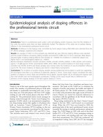
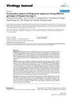
Figure 1
complete E gene sequences (1479 bp) from the 168 DENV-3 strains sampled globally
The Bayesian hierarchical consensus tree showing the phylogenetic relationships between DENV-3 genotypes is based on the
The Bayesian hierarchical consensus tree showing the phylogenetic relationships between DENV-3 genotypes is based on the complete E gene sequences
(1479 bp) from the 168 DENV-3 strains sampled globally. The names of the DENV-3 isolates refer to the year of isolation and the country of origin. In cases where there
is more than one isolate from a given country and year, a unique isolate number (or code) is also given. The abbreviations of the names of the countries are: Bangladesh (BD),
Bolivia (BL), Brazil (Br), Cambodia (Cam), Cuba (Cu), Ecuador (ECU), Indonesia (Indo), Japan (Jap), Martinique (Mart), Mexico (Mexi), Mozambique (Moza), Malaysia (Mal), Myanmar (Mya), Nicaragua (Nic), Peru (PR), Puerto Rico (PueR), Philippines (PH), Singapore (Sin), Sri Lanka (SriL), Tahiti (Tah), Thailand (TH), Taiwan (TW), Venezuela (Ven). Bootstrap values greater than 0.9 based on Bayesian posterior probabilities are shown for key nodes. The major genotypes of DENV-3 are also labeled. The tree was rooted using
DENV-1 strain A88 (GenBank accession number: AB074761) as the outgroup. Taiwan DENV-3 isolates are marked with a star.
Page 8 of 13
(page number not for citation purposes)
Virology Journal 2008, 5:63
/>
Table 4: Comparison of sequence diversity (p-distance, %) of full-length genomic sequences among different genotypes of dengue virus
type 3
Capsid
nucleotide
Amino
acid
prM
E
NS1
NS2a
NS2b
NS3
NS4a
NS4b
NS5
3.24 ± 0.54
3.13 ± 1.15
4.37 ± 0.52
1.41 ± 0.53
5.04 ± 0.32
1.60 ± 0.34
4.37 ± 0.39
1.54 ± 0.36
5.84 ± 0.54
2.57 ± 0.62
4.02 ± 0.59
0.54 ± 0.18
4.55 ± 0.30
0.99 ± 0.24
4.21 ± 0.54
1.33 ± 0.53
3.85 ± 0.41
0.85 ± 0.31
4.23 ± 0.23
1.17 ± 0.20
there was consistently an additional 11-nucleotide
sequence, AGTGAAAAAGA, inserted in the 3' NCR close
to the end of the open-reading frame (ORF) of the DENV3 strains isolated in recent years, compared to the prototype strain H87. In the 3' NCR, nucleotide changes at position 111, 129, 220 and 438 (nucleotide numbering
beginning at 5'-terminus of 3' NCR after the stop codon)
were observed from the strains circulating recently, which
differed from the strain H87. However, none of these
changes had any effect on the predicted secondary structure of the 3' NCR RNA (data not shown). The putative
genome cyclization sequence UCAAUAUG, located
between nucleotides 38 and 46 of the C gene, was conserved in all DENV-3 viruses.
Discussion and conclusion
Viral sequence comparisons among isolates from dengue
epidemics of different disease severities may provide valuable information regarding the molecular basis of the epidemic potential of the virus. DENV-3 re-appeared in 1998
in Taiwan and caused the DF/DHF epidemic in Tainan
City after its first introduction in 1994 [20]. This stimulates a great interest in understanding the molecular relationship of DENV-3 isolates in Taiwan during interepidemic periods and in comparing them with the strains
circulating globally to understand evolutionary trends
and geographical expansions. Here, we confirmed that the
Table 5: Positive selection and relevant parameter values among
different genomic regions of full-length DENV-3 sequences
Gene
dN/dS
Genotype I
Capsid
prM
E
NS1
NS2A
NS2B
NS3
NS4A
NS4B
NS5
Genotype II
Genotype III
Genotype V
5.68
0.00001
0.02
0.00001
0.00001
0.00001
0.008
0.04
19.37
0.00001
0.00001
0.14
999
18.2*
0.00001
0.00001
999
0.00001
0.00001
0.008
0.05
0.00001
0.03
0.02
0.02
0.00001
0.007
0.08
0.00001
0.018
0.00001
0.00001
0.097
0.66
0.08
0.00001
0.04
0.00001
0.1
0.00001
*p < 0.05 with statistical significance
dengue epidemics in Taiwan were strongly associated with
the globally circulating DENV-3 due to constant introduction of viruses from Southeast Asia by Taiwanese travelers.
Our data demonstrates the sequence diversity among the
full-genomic sequences of DENV-3 and the positive selection pressures exerted in different lineages (i.e. genotypes)
at sites in DENV-3 non-structural genes.
Since most Taiwan dengue epidemics were initiated by the
introduction of virus from imported cases [21], phylogenetic analysis provides essential information to understand the history and origin of all Taiwan DENV-3 isolates
originating in other countries (Fig. 1). The high nucleotide sequence identity (> 99.8%) among the strains isolated in 1998 indicates that they were from a single origin
and further spread to different townships, such as Pingtung (ID#98TW358). The only 1998 imported DENV-3
isolated from a traveler who had recently visited Indonesia was more closely associated with the genotype II isolates from Myanmar and older isolates from Thailand.
This virus differed from the virus isolated during the 1998
Tainan outbreak, which might suggest that multiple genotypes of DENV-3 circulated in Indonesia. This observation is consistent with a previous study indicating that at
least two subtypes of DENV-3 were present in Indonesia
[18]. The phylogenetic analysis also suggested that a single 1999 isolate (ID#99TW628) from the same location as
the 1998 epidemic was grouped together with the genotype III Sri Lanka isolates. Additional DENV-3 isolates
from the first DENV-3-caused DHF outbreak in Taiwan
(1994–1995) were grouped into genotype I. All these
results implicated that repeated introductions of different
genotypes of DENV-3 into Taiwan since 1994 were important causes of dengue epidemics, and that DENV-3 was
not endemic in Taiwan. This situation may be similar in
the subtropical region of China. Our country initiated airport fever screening during the severe acute respiratory
syndrome (SARS) outbreak in 2003–04, and it successfully identified 40 confirmed, imported dengue cases
[22]. Airport fever screening can thus quickly identify
imported dengue cases, and may prevent a significant
number of dengue outbreaks that would have been initiated by imported index cases. However, its cost-effectiveness in preventing any dengue epidemics in Taiwan will
need to be evaluated in the future.
Page 9 of 13
(page number not for citation purposes)
Virology Journal 2008, 5:63
/>
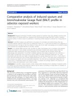
Figure
available2from likelihood
The Maximum GenBank phylogenetic tree shown here is based on the complete genomic sequences of 25 DENV-3 strains
The Maximum likelihood phylogenetic tree shown here is based on the complete genomic sequences of 25
DENV-3 strains available from GenBank. The tree was rooted using DENV-1 strain A88 (GenBank accession number:
AB074761) as the outgroup. The major amino acid changes along lineages within genotype I and II are also labeled. Taiwan
DENV-3 isolates are marked with a star.
With different DENV-3 genotypes imported into Taiwan
from Southeast Asia and other parts of the world, this
virus collection provides an excellent opportunity to
examine the sequence diversity of different genes of the
full-length DENV-3 viral RNA genome for genotypes other
than genotype IV. The highest p-distance of nucleotide
diversity of the full-length genomes occurred for the NS2A
gene (5.84% ± 0.54%), followed by the E gene (5.04% ±
0.32%). In contrast, the highest p-distance of amino acid
diversity of the full-length genomes occurred for the cap-
Page 10 of 13
(page number not for citation purposes)
Virology Journal 2008, 5:63
sid gene (3.13% ± 0.96%), followed by the NS2A gene
(2.57% ± 0.62%). This observation is consistent with the
previous analysis in DENV-1, DENV-3 and DENV-4
[16,34], although the precise cause of the increased rate of
amino acid change in the NS2A gene is unknown. A similar observation could also be made while analyzing the
full genomic sequences of West Nile virus (WNV) isolated
from different animal species [35]. The flavivirus NS2A, a
protein important for viral replication and particle formation [12], is cleaved by viral serine protease. A mutation at
the basic P1 cleavage site residue in NS2A blocks this
processing event and is lethal for virus production while
still allowing RNA replication [36,37]. Furthermore, this
basic residue in NS2A and an acidic residue in NS3 are
important determinants for virus assembly and/or release
[38]. Although the relative high sequence diversity of the
NS2A gene of DENV-3 may be due to the lesser structural
constraint required for NS2A, it is possible that positive
selection pressures may be exerted on this gene. Especially
in light of recent studies, NS2A together with NS4B and
NS4A were identified as dengue virus-encoded proteins
that could antagonize the interferon (IFN) response during viral infection [39,40]. Our analysis didn't detect any
selection pressure exerted on the NS2A gene probably due
to the small sample size; future studies will be needed to
focus the selection pressure analysis on non-structural
proteins and DENV evolution.
Several evolutionally conserved amino acid changes are
preserved, which are unique in different DENV-3 genotypes (Table 3). These substitutions resulted in changes of
its polarity, hydrophobicity or charge. Especially notable
was the change from L to S at position 178 of the NS1
region, which is an amino acid substitution unique to
genotype II. This might be the result of positive selection
within the lineage of genotype II but not other genotypes.
All DENV-3 isolates from Thailand belong to genotype II,
and interestingly, based on a previous publication [16],
strains of DENV-3 isolated prior to 1992 in Thailand may
have been replaced by two new locally evolving strains.
This could be a sign of a new genotype evolving in Thailand; however, most of the mutations or substitutions
occurring were deleterious and a purifying selection of
DENV-3 was suggested [16]. It is very likely that the previous analysis focused on only the E protein gene. Determining the possibility of a positive natural selection site
in the non-structural genes of the new Thailand lineage
will require further study. A number of T- and B-cell
epitopes are present on the non-structural proteins, especially the NS1 gene [41-43]. Even though the biological
significance of the L to S change at position 178 of the
NS1 region is unclear, growing evidence supported by in
vitro and in vivo studies suggest that there are certain evolutionary forces acting on the NS1 gene shaping the gene
flow of the dengue viral population, which might differ
/>
during viral replication in mammalian and mosquito cells
[44,45]. This is the first time that a positive selection pressure site was detected in a non-structural protein in
DENV-3 and its importance together with its functional
relevance to epidemic severity will need to be examined
with a larger sample size.
The global distribution of different genotypes of DENV-3
indicates that they originated in Southeast Asia; these genotypes demonstrated higher epidemic potential with
regards to severe DHF epidemics in Sri Lanka, Central and
South America [46,47]. Genotype III, once its transmission cycle was established locally, soon resulted in DHF
epidemics regardless of an increase in virus transmission
or a change in circulating serotypes [7,14], supporting the
hypothesis that virus strain is an important risk factor for
DHF [48,49]. Two sub-lineages (isolated before and after
1989) existed within the DENV-3 genotype III strains
from Sri Lanka, and the viruses isolated after 1989 were
associated with the DHF epidemic [7]. We found that the
strain isolated in 1999 from a indigenous dengue patient
(99TW628) that did not lead to a large-scale epidemic of
DF or DHF was more closely related to the lineage of the
DENV-3 genotype III Sri Lankan strain isolated before
1989. Similarly, in Indonesia two sub-lineages of DENV3 were present (isolated before and after 1998), and a
greater DHF epidemic, especially in adult cases, was
caused by the DENV-3 strains isolated after 1998 [50]. The
DENV-3 strain isolated in Taiwan during the DHF outbreak in 1994 was actually more closely related to the old
Indonesian strain of genotype I from 1976–78. While it is
currently unknown how the different sub-lineages within
each genotype are associated with different DHF epidemic
potential, a recent publication suggested that changing
serotype prevalence could lead to differential susceptibility to cross-reactive immune responses [16]. Furthermore,
Wearing et al suggested that both vector and short-termed
host cross-immunity are two factors responsible for dengue epidemics [51]. It would be necessary to strengthen
comprehensive dengue virological surveillance, especially
in those endemic and hyper-endemic areas/countries, to
monitor the emergence of DENV strains with epidemic
potential for better epidemic prevention and vaccine
development.
Competing interests
The authors declare that they have no competing interests.
Authors' contributions
DYC and CCK designed and performed all the experiments and drafted this manuscript together. DYC participated in the sequence alignment and statistical analysis.
JHH and YCW helped with collecting field human isolates
and LJC helped with sequencing experiments, together.
THL helped for the field mosquito collection and GJC for-
Page 11 of 13
(page number not for citation purposes)
Virology Journal 2008, 5:63
mulated the idea for this study and also provided critical
comments regarding this manuscript. All authors read and
approved the final manuscript.
/>
19.
20.
Acknowledgements
We sincerely thank Shih-Ting Ho at the Sin-Lau Christian Hospital, ChienMing Li at the Chi-Mei Foundation Medical Center and Shih-Chung Lin at
the Kuo General Hospital for cooperation in kindly providing the clinical
samples. The study was supported by the grants from the National Health
Research Institute (NHRI), Taipei, Taiwan (grant number: NHRI#DD01861X-CR-501P and NHRI#CN-CL8903P) and the National Science Council (NSC#90-2320-B-002-200, NSC#91-2320-B-002-081) in Taiwan.
23.
References
24.
1.
2.
3.
4.
5.
6.
7.
8.
9.
10.
11.
12.
13.
14.
15.
16.
17.
18.
Gubler DJ: Dengue and dengue hemorrhagic fever. Clin Microbiol Rev 1998, 11:480-496.
Pinheiro FP, Corber SJ: Global situation of dengue and dengue
haemorrhagic fever and its emergence in the Americas. Wld
Hlth Statist Quart 1997, 50:161-169.
McBride WJH, Bielefeldr-Ohmann H: Dengue viral infections;
pathogenesis and epidemiology.
Microbes Infect 2000,
2:1041-1050.
Gubler DJ, Clark GG: Dengue/dengue hemorrhagic fever: The
emergence of a global health problem. Emerg Infect Dis 1995,
1:55-57.
Lucas GN, Amerasinghe A, Sriranganathan S: Dengue haemorrhagic fever in Sri Lanka. Indian J Pediatr 2000, 67:503-504.
Lanciotti RS, Lewis JG, Gubler DJ, Trent DW: Molecular evolution
and epidemiology of dengue-3 viruses. J Gen Virol 1994,
75:65-75.
Messer WB, Gubler D, Harris E, Sivananthan K, de Silva AM: Emergence and global spread of a dengue serotype 3, subtype III
virus. Emerging Infectious Diseases 2003, 9:800-809.
Gubler DJ, Suharyono W, Lubis I, Eram S, Saroso JS: Epidemic dengue hemorrhagic fever in rural Indonesia. I. Virological and
epidemiological studies. Am J Trop Med Hyg 1979, 28:701-710.
Messer WB, Vitarana T, Sivananthan K, Elvtigala J, Preethimala LD,
Ramesh R, Withana N, Gubler DJ, Silva AM: Epidemiology of dengue in Sri Lank before and after the emergence of epidemic
dengue hemorrhagic fever. Am J Trop Med Hyg 2002, 66:765-773.
Gubler DJ, Meltzer M: Impact of dengue/dengue hemorrhagic
fever on the developing world. Advances in virus research 1999,
53:35-70.
Anonymous: Dengue-3 in Central America. Dengue Surveillance
Summary, CDC 1995, 70:1-4.
Chambers TJ, Hahn CS, Galler R, Rice CM: Flavivirus genome:
organization, expression and replication. Annu Rev Microbiol
1990, 44:649-688.
Gubler DJ, Kuno G: Dengue and dengue hemorrhagic fever.
Cab international, New York 1997:1-23.
Wittke V, Robb TE, Thu HM, Nisalak A, Nimmannitya S, Kalayanrooj
S, Vaughn DW, Endy TP, Holmes EC, Aaskov J: Extinction and
rapid emergence of strains of dengue 3 virus during an
interepidemic period. Virology 2002, 301:148-156.
Cologna R, Rico-Hesse R: American genotype structures
decrease dengue virus output from human monocytes and
dendritic cells. J Virol 2003, 77:3929-3938.
Zhang C, Mammen MPJ, Chinnawirotpisan P, Klungthong C, Rodpradit P, Monkongdee P, Nimmannitya S, Kalayanarooj S, Holmes EC:
Clade replacement in dengue virus serotypes 1 and 3 are
associated with changing serotype prevalence. J Virol 2005,
79:15123-15130.
Sittisombut N, Sistayanarain A, Cardosa MJ, Salminen M, Damrongdachakul S, Kalayanarooj S, Rojanasuphot S, Supawadee J, Maneekarn
N: Possible occurrence of a genetic bottleneck in dengue
serotype 2 viruses between the 1980 and 1987 epidemic seasons in Bangkok, Thailand. Am J Trop Med Hyg 1997, 57:100-108.
Raekiansyah M, Pramesyanti A, Bela B, Kosasih H, Ma'roef CN, Tobing
SY, Rudiman PI, Alisjahbana B, Endi TP, Green S, et al.: Genetic variations and relationship among dengue virus type 3 strains
isolated from patients with mild or severe form of dengue
21.
22.
25.
26.
27.
28.
29.
30.
31.
32.
33.
34.
35.
36.
37.
38.
39.
40.
41.
42.
disease in Indonesia and Thailand. Southeast Asian J Trop Med
Public Health 2005, 36:1187-1197.
Hwang KP: Dengue fever and dengue hemorrhagic fever. J Formosan Med 1997, 1:48-55.
Chao DY, Lin TH, Hwang KP, Huang JH, Liu CC, King CC: 1998 dengue hemorrhagic fever epidemic in Taiwan. Emerg Infect Dis
2004, 10:552-554.
King CC, Wu YC, Chao D-Y, Lin TH, Chow L, Wang H-T, Ku C-C,
Kao C-L, Chien LJ, Chang H-J, et al.: Major epidemics of dengue
in Taiwan in 1981–2000: Related to intensive virus activities
in Asia. Dengue Bulletin 2000, 24:1-10.
Shu P, Chien L, Chang S, Su C, Kuo Y, Liao T, Ho M, Lin T, Huang J:
Fever screening at airports and imported dengue. Emerg
Infect Dis 2005, 11:460-462.
Gubler DJ, Kuno G, Sather GE, Velez M, Oliver A: Mosquito cell
cultures and specific monoclonal antibodies in surveillance
for dengue viruses. Am J Trop Med Hyg 1984, 33:158-165.
Kao C, Wu M, Chiu Y, Lin J, Wu Y, Yueh Y, Chen L, Shaio M, King C:
Flow cytometry compared with indirect immunofluorescence for rapid detection of dengue virus type 1 after amplification in tissue culture. J Clin Microbiol 2001, 39:3672-3677.
Chao DY, King CC, Wang WK, Chen WJ, Wu HL, Chang GJ: Strategically Examining the Full-Genome of Dengue Virus Type
3 in Clinical Isolates Reveals Its Mutation Spectra. Virology
Journal 2005, 24:72.
Higgins DG, Sharp PM: CLUSTAL: a package for performing
multiple sequence alignment on a microcomputer. Gene
1988, 73:237-244.
Hall T: BioEdit: Biological sequence aligment editor for
WinXP. Ibis Thearpeutics, Carlsbad, CA92008 2005.
Kumar S, Tamura K, Nei N: MEGA3: Integrated software for
Molecular Evolutionary Genetics Analysis and sequence
alignment. Brief Bioinform 2004, 5:150-163.
Posada D, Crandall KA: Modeltest: testing the model of DNA
substitution. Bioinformatics 1998, 14:817-818.
Felsenstein J: PHYLIP: Phylogeny Inference Package (Univ. of
Wahsington, Seattle), Version 3.5c. 1993.
Huelsenbeck JP, Ronquist F: MRBAYES: Bayesian inference of
phylogenetic trees. Bioinformatics 2001, 17:754-755.
Yang Z: PAML: a program package for phylogenetic analysis
by maximum likelihood. Comput Appl Biosci 1997, 13:555-556.
Wang W-K, Lin S-R, Lee C-M, King C-C, Chang S-C: Dengue type
3 virus in plasma is a population of closely related
genomes:quasispecies. J Virol 2002, 76:4662-4665.
Klungthong C, Zhang C, Mammen MPJ, Ubol S, Holmes EC: The
molecular epidemiology of dengue virus serotype 4 in Bangkok, Thailand. Virol 2004, 329:168-179.
Lanciotti R, Ebel G, Deubel V, Kerst A, Murri S, Meyer R, Bowen M,
McKinney N, Morrill W, Crabtree M, et al.: Complete genome
sequences and phylogenetic analysis of West Nile virus
strains isolated from the United States, Europe, and the Middle East. Virol 2002, 298:96-105.
Lindenbach BD, Rice CM: Flaviviridae: The viruses and their replication, p.991–1110. In Fields' Virology 4th edition. Edited by: Knipe
DM, Howley PM. Lippincott Williams & Wilkins, Philadelphia, PA;
2001.
Hori H, Lai CJ: Cleavage of dengue virus NS1-NS2A requires
an octapeptide sequence at the C terminus of NS1. J Virol
1990, 64:4573-4577.
Preugschat F, Yao CW, Strauss JH: In vitro processing of dengue
virus type 2 nonstructural proteins NS2A, NS2B, and NS3. J
Virol 1990, 64:4364-4374.
Munoz-Jordan JL, Sanchez-Burgos GG, Laurent-Rolle M, Garcia-Sastre A: Inhibition of interferon signaling by dengue virus. Proc
Natl Acad Sci 2003, 100:14333-14338.
Munoz-Jordan JL, Laurent-Rolle M, Ashour J, Martinez-Sobrido L,
Ashok M, Lipkin WI, Garcia-Sastre A: Inhibition of alpha/beta
interferon signaling by the NS4B protein of flaviviruses. J Virol
2005, 79:8004-8013.
Falconar AKI, Young PR, Miles MA: Precise location of sequential
dengue virus subcomplex and complex B cell epitopes on the
nonstructural-1 glycoprotein. Arch Virol 1994, 137:315-326.
Garcia G, Vaughn DW, del Angel RM: Recognition of snthetic oligopeptides from nonstructural proteins NS1 and NS3 of
dengue-4 virus by sera from dengue virus-infected children.
Am J Trop Med Hyg 1997, 56:466-470.
Page 12 of 13
(page number not for citation purposes)
Virology Journal 2008, 5:63
43.
44.
45.
46.
47.
48.
49.
50.
51.
/>
Callahan JD, Wu S-JL, Dion-Schultz A, Mangold BE, Peruski LF, Watts
DM, Porter KR, Murphy GR, Suharyono W, King C-C, et al.: Development and evaluation of serotype- and group-specific fluorogenic reverse transcriptase PCR (TaqMan) assays for
dengue virus. J Clin Microbiol 2001, 39:4119-4124.
Blaney JJ, Manipon GG, Murphy BR, Whitehead SS: Temperature
sensitive mutations in the genes encoding the NS1, NS2A,
NS3, and NS5 nonstructural proteins of dengue virus type 4
restrict replication in the brains of mice. Arch Virol 2003,
148:999-1006.
Chen WJ, Wu HR, Chiou SS: E/NS1 modifications of dengue 2
virus after serial passages in mammalian and/or mosquito
cells. Intervirology 2003, 46:289-295.
Usuku S, Castillo L, Sugimoto C, Noguchi Y, Yogo Y, Kobayashi N:
Phylogenetic analysis of dengue-3 viruses prevalent in Guatemala during 1996–1998. Arch Virol 2001, 146:1381-1390.
Palacio M, Amadoor JJ, Acevedo F, de los Reyes J, Ramirez A,
Gonzalez A, Guelva G: Internaltional notes dengue type 3 infection – Nicaragua and Panama, October-November 1994.
Morb Mortal Wkly Rep (MMWR) 1995, 44:21-24.
Gubler DJ: Cities spawn epidemic dengue viruses. Nat Med
2004, 10:129-130.
Rosen L: Disease exacerbation caused by sequential dengue
infections: myth or reality? Rev Infect Dis 1989, 11:S840-842.
Mcardle JL, Listyaningsih E, Suharyono W, Porter KR: Evolution of
the dengue-3 virus in Indonesia and throughout SE Asia may
contribute to recent changes in outbreak dynamics. International Conference on Emerging Infectious Diseases, July 16–19, 2000
Atlanta, Georgia, USA 2000. conference abstract, board 68
Wearing HJ, Rohani P: Ecological and immunological determinants of dengue epidemics.
Proc Natl Acad Sci 2006,
103:11802-11807.
Publish with Bio Med Central and every
scientist can read your work free of charge
"BioMed Central will be the most significant development for
disseminating the results of biomedical researc h in our lifetime."
Sir Paul Nurse, Cancer Research UK
Your research papers will be:
available free of charge to the entire biomedical community
peer reviewed and published immediately upon acceptance
cited in PubMed and archived on PubMed Central
yours — you keep the copyright
BioMedcentral
Submit your manuscript here:
/>
Page 13 of 13
(page number not for citation purposes)