báo cáo hóa học:" Ilizarov treatment of humeral shaft nonunion in an antiepileptic drug patient with uncontrolled generalized tonic-clonic seizure activity" ppt
Bạn đang xem bản rút gọn của tài liệu. Xem và tải ngay bản đầy đủ của tài liệu tại đây (1.82 MB, 7 trang )
CAS E REP O R T Open Access
Ilizarov treatment of humeral shaft nonunion in
an antiepileptic drug patient with uncontrolled
generalized tonic-clonic seizure activity
Vasileios S Sioros
†
, Marios G Lykissas
*†
, Dimitrios Pafilas
†
, Panayiotis Koulouvaris
†
, Alexandros N Mavrodontidis
†
Abstract
Nonunion of the humeral shaft in patients with antiepileptic drug associated metabolic bone disorder constitute a
challenging surgical problem difficult to treat due to seizure activity, osteoporosis, and poor stabilization options.
We report a case of nonunion of the humeral shaft in an antiepileptic drug patient with uncontrolled generalized
tonic-clonic seizure activity successfully treated with Ilizarov external fixator and a follow-up of 4 years.
Background
Humeral shaft fractures acco unt for approximately 1.3%
of all fractures [1]. Approximately 1-15% of these frac-
tures progress to nonunion [2-7]. Nonunion of the hum-
eral shaft in patients with antiepileptic drug associated
metabolic bone disorder constitute a challenging surgi-
cal problem difficult to treat due to seizure activity,
osteoporosis, and poor stabilization options. Treatment
options include internal fixation supplemented with can-
cellous bone graft, intramedullary nailing, free vascular-
ized fibular graft, and Ilizarov circular frame fixation. At
the hands of an expert surgeon, Ilizarov external thin-
wire fixator can be a viable surgical option for the treat-
ment of humeral shaft nonunion. We report a case of
nonunion of the humeral shaft in an antiepileptic drug
patient with uncontrolled generalized tonic-clonic sei-
zure activity successfully treated with Ilizarov external
fixator and a follow-up of 4 years.
Case presentation
A 43-year-old man was admitted to the emergency
department after a fall during a generalized tonic-cl onic
seizure attack (grand mal). He sustained a closed trans-
verse diaphyseal fracture of his right humerus (Figs. 1
&2). The pati ent suffered from epilepsy for the last 15
years and he was on carbamazepine (Tegretol CR 400
mg, Novartis, Greece) since then. Although well
compliant with his treatment regimen, generalized
tonic-clonic attacks occur almost once a week.
The fracture was initially managed by open reduction
and internal fixation with plate and screws through an
anterolateral longitudinal incision. Fixation was augmen-
ted with autologous bone graft obtained from the con-
tralateral iliac crest. Eighteen months after surgery,
radiographic evaluation revealed pseudarthrosis of the
shaft of the humerus (Figs. 3 &4).
Exploration of the nonunion was performed under
gen eral anesthesia and using the prior incision. Prophy-
lactic second generation cephalosporin antibiotic therapy
was administered for 72 hours after surgery. The frac-
ture site was opened and hardware materials were
removed. Fibrous scar tissue and soft avascular bone
was excised to expose fresh bleeding bone ends. The
intramedullary canals were opened at the proximal and
distal fragment. Following debridement, approximately a
1-cm segmental defect was measured. Specimens were
sent for gram stain and microbiological analysis.
A 3-ring f rame connected with 5 threaded rods was
prefabricated using the left normal humerus as a tem-
plate (Smith and Nephew plc, Memphis, Tennessee, U.S.
A.). The fixator consisted of a 2-ring frame (full ring
proximal and 5/8 ring distal) placed distally and a 5/8 1-
ring frame placed proximally to the fracture site (Figs. 5
&6). The proximal and distal rings were not circular to
facilitate active shoulder and elbow range of motion.
Four thin w ires (1.8 mm) with olives for both the distal
frames and 2 thin wires (1.8 mm) with olives for the
proximal frame were used, while 2 half pins (6.0 mm)
* Correspondence:
† Contributed equally
Department of Orthopaedic Surgery, University of Ioannina School of
Medicine, Ioannina, Greece
Sioros et al. Journal of Orthopaedic Surgery and Research 2010, 5:48
/>© 2010 Sioros et al; licensee BioMed Central Ltd. This is an Open Access article distributed under the terms of the Creative Commons
Attribution License ( licenses/by/2.0), which permits unre stricted use, distribution, and rep roduction in
any medium, provided the original work is properly cited.
were placed proximally in the mid-shaft of the humerus.
Acute shortening of 1.0 cm via the Ilizarov fixator with
immediate bone-to-bone contact at the nonunion site
was then performed. The procedure was accomplished
under fluoroscopic guidance. The radial nerve was
explored in order to avoid nerve injury during wire
insertion. Autologous cortico-cancellous bone graft har-
vested from the contralateral ilium was applied to the
nonunion. The total operating time was 120 minutes.
Immediately after surgery the arm was placed in a
sling for 6 weeks. From the first morning after surgery,
joint mobilization of the shoulder and elbow was started
as tolerated. In order to better control seizure activity,
levetiracetam (Keppra 1000 mg, UCB Pharma S.A., Bel-
gium) was added in the anticonvulsant therapy. The
patient was instructed in pin care cleaning and hygie ne
and discharged f rom the hospital 5 days after surgery.
Pin-tract infection was noticed in two skin/pin contacts
which were treated with oral antibiotics (second genera-
tion cephalosporin) for one week.
Antero-posterior and lateral radiographs demonstrated
uncomplicated fracture healing at 18 weeks. The Ilizarov
frame was removed at 24 weeks without anesthesia in
the outpatient department. No protective immobilization
was used after frame removal. At the most recent fol-
low-up, 4 years postoperatively, the alignment of the
humerus was anatomic and full range of motion was
obtained at both the shoulder and elbow joint (Figs. 7
&8). The p atient was very satisfied with his treatment
and had returned to his previous activities.
Discussion
Decreased bone density has been well documented in
patients with epilepsy [8]. The occurrence of fractures
in these patients is increased two fold to sixfold
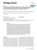
Figure 1 Anteroposteriorradiograph of the right humerus of a
43-year-old man sustained a transverse diaphyseal fracture
after a fall during a generalized tonic-clonic attack.
Figure 2 Lateral view of the right humerus.
Sioros et al. Journal of Orthopaedic Surgery and Research 2010, 5:48
/>Page 2 of 7
compared with than that expected in nonepileptic popu-
lation [9]. In a comparative study of 202 institutiona-
lized patients with epilepsy the frequency of fractures of
the humerus was increased fourfold compared with a
normal population [10]. The relative r isk for humeral
fractures is most increased in patients more than 45
years of age [11]. Seizure activity may cause fractures,
usually vertebral c ompression fractures, as a result of
spine hyperflexion during extreme muscular contrac-
tions [12]. Bilateral posterior fracture dislocation of the
shoulder is highly indicative of seizure [13]. Trauma or
fall during tonic-clonic, tonic, or atonic attack is also
associated with fracture of the humerus along with frac-
ture of the hip, ankle, and wrist [10,11]. Repetitive,
uncontrolled seizure activity, especially tonic-clonic
attacks, as in our case, may also adversely affect the pro-
cess of fracture healing, making the management of
such fractures a challenging surgical problem.
Antiepileptic drugs have been categorized as indepen-
dent risk factors for decrease of bone mineral density
regardless of patient’ s age, gender, and period of treat-
ment [14]. Their role in bone l oss is thought to be mul-
tifactorial. Conventional antiepileptic drugs, such as
carbamazepine, phenytoin, and phenobarbital, are potent
hepatic mixed-function oxidase (CYP450) inducers [15].
Valproic acid is a CYP450 inhibitor. Pregnabe × recep-
tor (PXR), a transcriptional regulator of CYP450, med-
iates the adverse effect on bone metabolism of both
CYP450 inducers and inhib itors through stimulation of
vitamin D catabolism and inhibition of 25-hydroxylation
of vitamin D [16]. The effect of antiepileptic drugs on
bone mineral density is also mediated by Vitamin D
receptor (VDR) gene, an important regulator of osteo-
clastic activity [17]. In turns, vitamin D catabolism
results in decreased calcium absorption across the small
intestine, hypocalcemia, and secondary hyperparathyr-
oidism [18]. It has also been demonstrated that CYP450
exhibits antiproliferative and antidifferentiation effects
on osteoblasts [19].
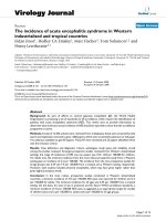
Figure 3 Anteroposteriorr adiograph of the right humerus
showing atrophic nonunion of the humeral shaft 18 months
after treatment with open reduction and internal fixation.
Figure 4 Lateral view of the right humerus 18 months
postoperatively.
Sioros et al. Journal of Orthopaedic Surgery and Research 2010, 5:48
/>Page 3 of 7
The deterioration of bone metabolism caused by con-
ventional antiepileptic drugs highlights the role of these
agents both in pathogenesis of special type of fractures
and the need of vitamin D and calcium supplementation
in this patient population [20]. The induction o f bone
loss by conventional antiepileptic drugs also emphasizes
the need of special techniques to treat difficult cases,
such as fracture nonunion.
The incidence of nonunion of humeral shaft fractures
after both conservative and surgical management is
reported to be as high as 1-15% [2-7]. Failure to unite
after surgical management of diaphyseal fractures of the
humerus could be multifactorial. Factors that may play a
role in nonunion include inadequate fracture fixation,
osteopenia/osteoporosis, infection, devitalization of
bone, and poor contact between the fracture segments.
Most nonunions of the humerus are associated with
angulation, displacement, over-riding, limb shortening,
and osteopenia. Treatment options include internal fixa-
tion supplemented w ith cancellous bone graft, intrame-
dullary nailing, free vascularized fibular graft, and
Ilizarov circular frame fixation. Locking plates and dual
plating have also been proposed as alternatives in cases
of nonunion of the humerus with poor bone stock.
Rigid internal fixation with plating is considered as the
“gold standard” for the management of humeral shaft
nonunion with union rates approaching 100% [21]. Aug-
mentation with autologous bone graft is recommended,
especially in atrophic type of nonunions, representing
the 70-90% of all cases [21]. Atrophic asept ic nonunion
of the humeral shaft after failure of surgical manage-
ment, as in our patient, is characterized by poor bone
quality. Further decrease in the bone mineral density,
secondary to anticonvulsant bone disease, makes i nter-
nal fixation less stable than in normal bone. Further
comp lications after open reduction and internal fixat ion
in a previous surgically treated hume rus include difficult
dissection in a scarry tissue environment with risk for
radialnerveiatrogenicinjuryapproaching4%[22].
Superficial or deep infection following convent ional
methods of internal fixation is reported as high as 6.7%
[22].
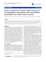
Figure 5 Radiograph of the3-ring frame.
Figure 6 Photograph of the same Ilizarov circular fram e.Note
the proximal and distal 5/8 rings that facilitate active shoulder and
elbow range of motion.
Sioros et al. Journal of Orthopaedic Surgery and Research 2010, 5:48
/>Page 4 of 7
If intramedullary nailing is selected for the manage-
ment of diaphyseal fractures o f the humerus , nonunion
is reported in a higher rate than plating, ranging from 0
to 33% [23,24]. Exchange nailing in cases of nonunion
of the diaphysis femur or tibia is a viable method for
achieving union. However, humeral shaft fractures com-
plicated by nonunion cannot achieve union after ream-
ing and exchange nailing [24]. This can easily be
explained biomechanically by the absence of axial load-
ing in the humerus and the presence of greater torsional
and d istractive forces than in tibia or femur [25].
Further drawbacks following intramedullary nailing
include shoulder or elbow stiffness, depending on the
point of insertion, radial nerve palsy, disruption of
the endosteal blood supply, and fracture instability if the
nail remains unlocked [26]. According to some authors,
higher union rates can be achieved if the intramedullary
nail is locked [21].
Ilizarov technique has been successfully used for the
management of nonunion of the humeral shaft [27,28]. It
is a very promising method because it is minimally invasive
with low intraoperative blood loss, and minimal patient
discomfort. It provides stable fixation, prompt postopera-
tive elbow and shoulder mobilization, and has no major
complications. It gives postoperative capability for mala-
lignment correction and, at the hands of an expert, Ilizarov
external fixation is not time consuming [28]. It appears
that the Ilizarov apparatus is superior to conventional fixa-
tion methods, especially in patients with severe bony d efor-
mity, limb shortening, and bone loss [29]. In such cases,
callus f ormation can be stimulated by controlled oscillating
compression and distraction [5,25]. Long-lasting nonunion
may lead to local o steoporosis wh ich is different from
osteoporosis due to old age. When severely compromised
local bone due to disuse is associated with metabolic bone
disorder, internal fixation is technically demanding and
plate loosening often occurs. In our patient, severe osteo-
porosis due to local and systemic factors was accompanied
by mechanical instability of the osteosynthesis because of
the frequent tonic-clonic seizure activity. T he Ilizarov
external fixator was the only system that could simulta-
neously provide stable fixation in an osteoporotic bone,
externally controlled compression, and interfere dynami-
cally with repetitive seizures. Ilizarov does not support the
use of bone grafting for the management of nonunions.
However, autologous bone graft obtained from the iliac
crest was used in our patient with atrophic nonunion in
Figure 8 Lateral v iew of the humeral fracture 4 years
postoperatively.
Figure 7 Anteroposteriorradiograph of the humeral fracture 4
years after surgery. Union was achieved 4.5 months after initial
application of the frame.
Sioros et al. Journal of Orthopaedic Surgery and Research 2010, 5:48
/>Page 5 of 7
order to stimulate the biology of the nonunion site, speed
the bone healing, and minimize t he fixation time.
Ilizarov technique may involve the risk of pin-tract
infections most of which can be treated by administra-
tion of antibiotics, as in our case. Other disadvantages
include re-fracture following frame removal, limb short-
ening, radial nerve palsy, and patient discomfort because
of the weight of the device and impingement of the
frame on the chest. Re-fracture can be prevented with
the use of a plastic brace after frame removal. Limb dis -
crepancy of 3 to 4 cm is generally well tolerated and
further shortening of the upper extremity can be mana-
ged by lengthening the humerus with a new Ilizarov
frame in a later stage. Nerve injury during placement of
the transosseous wires can be avoided by reducing the
amount of paralytic agents given and looking for motor
flickers to the wrist, hand o r fingers. In order to allow
early shoulder and elbow mobility and minimize the
frame interference with daily activities, a semicircular
proximal and distal ring should be used.
Conclusions
The management o f humeral shaft non union in antiepi-
leptic drug patients offers a different challenge. In such
cases, Ilizarov external fixator is an a dequ ate treat ment
option that surgeo n should always have in mind. It pro-
vides stable fixation, prompt postoperative mobilization,
and has no major complications. It gives postoperative
capability for malalignment correction and, at the hands
of an expert, Ilizarov external fixation is not time con-
suming. When conventional antiep ileptic drugs are used,
vitamin D and calcium supplementation are recom-
mended for prophylaxis and treatment of bone loss.
Consent
Written informed consent was obtained from the patient
for publication of this case report and any accompany-
ing images. A copy of the written consent is availabl e
for review by the Editor-in-Chief of this journal.
Authors’ contributions
All authors contributed equally to this work. MGL and VSS participated in
the design of the study and drafted the manuscript. ANM, DP, and PK
conceived of the study and participated in its design and coordination.
Marios G. Lykissas has had the main responsibility for the study and
manuscript preparation. All authors read and approved the final manuscript.
Competing interests
There are no competing interests; this is a basic academic research initiative.
Received: 29 January 2010 Accepted: 28 July 2010
Published: 28 July 2010
References
1. Brinker MR, O’Connor DP: The incidence of fractures and dislocations
referred for orthopaedic services in a capitated population. J Bone Joint
Surg Am 2004, 86:290-7.
2. Borus TA, Yian EH, Karunakar MA: A case series and review of salvage
surgery for refractory humeral shaft nonunion following two or more
prior surgical procedures. Iowa Orthop J 2005, 25:194-9.
3. Durbin R, Gottesman MJ, Saunders KC: Hackthal stacking nailing of
humeral shaft fractures. Experience with 30 patients. Clin Orthop Relat Res
1983, 179:168-74.
4. Healy WL, White GM, Mick CA, Brooker AF Jr, Weiland AJ: Nonunion of the
humeral shaft. Clin Orthop Rel Res 1987, 219:206-13.
5. Jupiter JB, von Deck M: Ununited humeral diaphyses. J Shoulder Elbow
Surg 1998, 7:644-53.
6. Marti RK, Verheyen CC, Besselaar PP: Humeral shaft nonunion: evaluation
of uniform surgical repair in fifty-one patients. J Orthop Trauma 2002,
16:108-15.
7. Ring D, Kloen P, Kadzielski J, Helfet D, Jupiter JB: Locking compression
plates for osteoporotic nonunions of the diaphyseal humerus. Clin
Orthop Relat Res 2004, 425:50-4.
8. Khanna S, Pillai KK, Vohora D: Insights into liaison between antiepileptic
drugs and bone. Drug Discov Today 2009, 14:428-35.
9. Mattson RH, Gidal BE: Fractures, epilepsy, and antiepileptic drugs. Epilepsy
Behav 2004, 5(Suppl 2):S36-40.
10. Desai KB, Ribbans WJ, Taylor GT: Incidence of five common fracture types
in an institutional epileptic population. Injury 1996, 27:97-100.
11. Persson HBI, Alberts KA, Farahmand BY, Tomson T: Risk of extremity
fractures in adult outpatients with epilepsy. Epilepsia 2002, 43:768-72.
12. Vasconcelos D: Compression fractures of the vertebrae during major
epileptic seizures. Epilepsia 1973, 14:323-8.
13. Elsberger ST, Brody G: Bilateral posterior shoulder dislocations. Am J
Emerg Med 1995, 13:331-2.
14. Khanna S, Pillai KK, Vohora D: Insights into liaison between antiepileptic
drugs and bone. Drug Discov Today 2009, 14
:428-35.
15. Patsalos PN, Fröscher W, Pisani F, van Rijn CM: The importance of drug
interactions in epilepsy therapy. Epilepsia 2002, 43:365-85.
16. Collins N, Maher J, Cole M, Baker M, Callaghan N: A prospective study to
evaluate the dose of vitamin D required to correct 25-hydroxyvitamin D
levels, calcium and alkaline phosphatase in patients at risk of
developing antiepileptic drug-induced osteomalacia. QJMed1991,
286:113-22.
17. Takasu H, Sugita A, Uchiyama Y, Katagiri N, Okazaki M, Ogata E, Ikeda K: c-
Fos protein as a target of anti-osteoclastogenic action of vitamin D, and
synthesis of new analogs. J Clin Invest 2006, 116:528-35.
18. Foxa SW, Lovibond AC: Current insights into the role of transforming
growth factor-b in bone resorption. Mol Cell Endocrinol 2005, 243:19-26.
19. Feldkamp J, Becker A, Witte OW, Scharff D, Scherbaum WA: Long-term
anticonvulsant therapy leads to low bone mineral density-evidence for
direct drug effects of phenytoin and carbamazepine on human
osteoblast-like cells. Exp Clin Endocrinol Diabetes 2000, 108:37-43.
20. Pack AM: The association between antiepileptic drugs and bone disease.
Epilepsy Curr 2003, 3:91-5.
21. Tomić S, Bumbasirević M, Lesić A, Mitković M, Atkinson HD: Ilizarov frame
fixation without bone graft for atrophic humeral shaft nonunion: 28
patients with a minimum 2-year follow-up. J Orthop Trauma 2007,
21:549-56.
22. Hsu TL, Chiu FY, Chen CM, Chen TH: Treatment of nonunion of humeral
shaft fracture with dynamic compression plate and cancellous bone
graft. J Chin Med Assoc 2005, 68:73-6.
23. Hems TE, Bhullar TP: Interlocking nailing of humeral shaft fractures: the
Oxfo experience 1991 to 1994. Injury 1996, 27:485-9.
24. Lin J, Hou SM: Antegrade locked nailing for humeral shaft fractures. Clin
Orthop Relat Res 1999, 365:201-10.
25. Lammens J, Bauduin G, Driesen R, Moens P, Stuyck J, De Smet L, Fabry G:
Treatment of nonunion of the humerus using the Ilizarov external
fixator. Clin Orthop Relat Res 1998, 353:223-30.
26. Cierny G III, Mader JT: Approach to adult osteomyelitis. Orthop Rev 1987,
16:259-72.
27. Patel VR, Menon DK, Pool RD, Simonis RB: Nonunion of the humerus after
failure of surgical treatment. Management using the Ilizarov circular
fixator. J Bone Joint Surg Br 2000, 82:977-83.
28. Beris AE, Lykissas MG, Sioros V, Mavrodontidis AN, Korompilias AV: Femoral
periprosthetic fracture in osteoporotic bone after a total knee
replacement. Treatment with Ilizarov external fixation. J Arthroplasty 2010.
Sioros et al. Journal of Orthopaedic Surgery and Research 2010, 5:48
/>Page 6 of 7
29. Kocaoğlu M, Eralp L, Tomak Y: Treatment of humeral shaft non-unions by
the Ilizarov method. Int Orthop 2001, 25:396-400.
doi:10.1186/1749-799X-5-48
Cite this article as: Sioros et al.: Ilizarov treatment of humeral shaft
nonunion in an antiepileptic drug patient with uncontrolled
generalized tonic-clonic seizure activity. Journal of Orthopaedic Surgery
and Research 2010 5:48.
Submit your next manuscript to BioMed Central
and take full advantage of:
• Convenient online submission
• Thorough peer review
• No space constraints or color figure charges
• Immediate publication on acceptance
• Inclusion in PubMed, CAS, Scopus and Google Scholar
• Research which is freely available for redistribution
Submit your manuscript at
www.biomedcentral.com/submit
Sioros et al. Journal of Orthopaedic Surgery and Research 2010, 5:48
/>Page 7 of 7