báo cáo hóa học:" Ovarian cancer: emerging concept on cancer stem cells" docx
Bạn đang xem bản rút gọn của tài liệu. Xem và tải ngay bản đầy đủ của tài liệu tại đây (415.64 KB, 9 trang )
BioMed Central
Page 1 of 9
(page number not for citation purposes)
Journal of Ovarian Research
Open Access
Review
Ovarian cancer: emerging concept on cancer stem cells
Moorthy P Ponnusamy
1
and Surinder K Batra*
1,2
Address:
1
Department of Biochemistry and Molecular Biology, University of Nebraska Medical Center, Omaha, NE 68198-5870, USA and
2
Eppley
Institute for Research in Cancer and Allied Diseases, University of Nebraska Medical Center, Omaha, NE 68198-5870, USA
Email: Moorthy P Ponnusamy - ; Surinder K Batra* -
* Corresponding author
Abstract
Emerging evidence suggests that the capacity of a tumor to grow and propagate is dependent on a
small subset of cells within a tumor, termed cancer stem cells. In fact, cancer cells, like stem cells,
can proliferate indefinitely through a dysregulated cellular self-renewal capacity. Cancer stem cells
may originate due to the distribution into self-renewal and differentiation pathways occurring in
multi-potential stem cells, tissue-specific stem cells, progenitor cells and cancer cells. Recent
studies have shown that ovarian cancer also contains stem cells or tumor-initiating cells. Moreover,
ovarian serous adenocarcinomas were disaggregated and subjected to growth conditions to select
for self-renewing, non-adherent spheroids previously shown to be derived from tissue stem cells.
A recent study showed that epithelial ovarian cancer was derived from a sub population of CD44
+
,
CD117
+
and CD133
+
cells. The existence of cancer stem cells would explain why only a small
minority of cancer cells is capable of extensive proliferation of the tumor. In this review, we have
discussed the studies on ovarian cancer stem cells along with the molecular pathways that could
be involved in these cancer stem cells.
Introduction
Ovarian cancer is the fifth leading cause of cancer deaths
and has the highest mortality rate among gynecologic can-
cers. It is the most lethal malignancy of the female repro-
ductive system, at the initial stage the five-year survival
rate is nearly 45%, which declines to 30% for patients
with an advanced disease [1,2]. Greater than 90% of ovar-
ian cancers arise from the surface epithelium [3], and tum-
origenesis has been associated with ovulation-associated
wound repair and/or inflammation, possibly leading to
abnormal stem cell expansion [3,4]. Over the last several
years, it has been increasingly evident that a small popu-
lation (less than 5%) of cancer cells, referred to as "cancer
stem cells (CSCs)", is responsible for the aggressiveness
of the disease, metastasis and resistance to therapy [5-7].
Cancer stem cells, like somatic stem cells, are thought to
be capable of self-renewal or unlimited proliferation [7].
The recent discovery that CSCs express certain 'stem cell-
specific' markers has renewed interest and provided a rise
in the idea that CSCs may arise from somatic stem/pro-
genitor cells. Considerable research efforts have been
directed toward the identification of cancer stem cell
markers in ovarian cancer.
Stem cells, as classically defined, are cells with a capacity
for self-renewal and generation of daughter cells that can
differentiate into all the way down different cell lineages
found in the mature tissue [8]. Stem cells always undergo
asymmetric cell divisions, with each cell generating two
cells; one that is identical to itself in stemness and another
which is committed to a certain lineage. The daughter cell
with stem cell like properties maintains its own compart-
Published: 12 October 2008
Journal of Ovarian Research 2008, 1:4 doi:10.1186/1757-2215-1-4
Received: 23 August 2008
Accepted: 12 October 2008
This article is available from: />© 2008 Ponnusamy and Batra; licensee BioMed Central Ltd.
This is an Open Access article distributed under the terms of the Creative Commons Attribution License ( />),
which permits unrestricted use, distribution, and reproduction in any medium, provided the original work is properly cited.
Journal of Ovarian Research 2008, 1:4 />Page 2 of 9
(page number not for citation purposes)
ment over time, while its sister cell undergoes a series of
cell divisions [9]. Self-renewal allows stem cells to persist
during the entire the lifetime of the organism, while their
differentiation potential allows them to perform func-
tions like tissue genesis, tissue maintenance, and regener-
ation following stress or injury [9].
Of all the types of stem cell, hematopoietic stem cells
(HSCs) are the best characterized adult stem cell [10].
HSCs can differentiate to form mature blood cells but can
also reproduce themselves, which is known as self-
renewal [10]. It is reside in distinct stem-cell niches that
vary in location depending on the developmental stages
of organism [11]. The human HSCs express high level of
CD34 and low or absent level of CD33, CD38, thy-1, and
CD71, appears to be enriched for primitive progenitor
and HSC activity, while more mature progenitors express
one or more of these markers [12]. Furthermore, in thera-
peutic target hematopoietic stem cells are the only stem
cells developed up to therapy for the cancer and other dis-
orders for the blood [11] and following HSC study for
other stem cells will lead to improve therapy for other can-
cers.
Cancer stem cells may arise following transforming muta-
tions that occur in untransformed stem cells, progenitor
cells, mature cells, and cancer cells. The genetic program
controlling self-renewal and differentiation plays a key
role in the genesis of cancer stem cells (Figure 1). Cancer
stem cells (CSCs) have been demonstrated to have roles in
several cancers, including cancers of the ovaries, breast,
brain, prostate, pancreatic, hepatocellular, head and neck
cancers and hematological malignancies [5-7,13-27].
According to the CSC model, only a specific subset of the
cancer cell population (i.e., the long-lived CSC subset)
should be able to sustain in vivo tumor growth, whereas all
other subsets (i.e., the tumor counterparts of short-lived
differentiated cells) should not. Indeed, this assumption
has now been repeatedly confirmed in several tumor sys-
tems. Three key observations classically define the exist-
ence of a CSC population: (i) Only the minority of cancer
cells within each tumor are usually endowed with tumor-
igenic potential when transplanted into immunodeficient
mice; (ii) Tumorigenic cancer cells are characterized by a
distinctive profile of surface markers and can be differen-
tially and reproducibly isolated from non-tumorigenic
ones by flow cytometry or other immunoselection proce-
dures; and (iii) Tumors grown from tumorigenic cells con-
tain mixed populations of tumorigenic and non-
tumorigenic cancer cells, thus recreating the full pheno-
typic heterogeneity of the parent tumor [28]. Further-
more, recent studies have been shown the functions of
normal and malignant stem/progenitor cells in tissue
regeneration, cancer progression and targeting therapies
[29,30]. In this review we aim to provide insight into the
evaluation of the evidence that supports the existence of
cancer stem cells and the characterization studies that
have tried to identify ovarian cancer stem cells. We also
discuss how taking this subpopulation of cells into
account may affect the way we treat ovarian cancers in the
future.
Cancer stem cells
The identification of a reservoir of stem cells within many
adult tissues raises the interesting possibility that all adult
tissues have stem cells. Stem cell populations within nor-
mal tissues are defined by certain common characteristics:
self-renewal to maintain the stem cell pool over time; reg-
ulation of stem cell number through a strict balance
between cell proliferation, cell differentiation and cell
death; and the ability to give rise to a broad range of dif-
ferentiated cells [31,32]. It is observed that like stem cells,
cancer cells are widely thought to be able to proliferate
indefinitely through a deregulated self-renewal capacity.
In fact, cancer stem cells can thus only be defined experi-
mentally by their ability to generate continuously growing
tumors. CSCs have the capacity to self-renew, undergoing
divisions that allow the generation of more CSCs and ulti-
mately some of them differentiate into the various cell
types that compose the tumor mass. To date, the practical
translation of this definition, and the gold standard to
define the 'stemness' of cancer cells, has been their ability
to generate a phenocopy of the original malignancy in
immuno-compromised mice [7].
Evidence for the existence of cancer stem cells
To assay the cancer stem cells, a xenograft model for breast
cancer was developed that allowed specific cancer tumors
isolated directly from a patient to be passaged reliably in
vivo. In this model, only a subset of cancer cells had the
ability to form new tumors [5]. The cancer stem cells iso-
lated from tumors are mostly isolated by flow cytometry
as the CD44+ CD24-/
low
lineage cell population [5]. Fur-
thermore, dilution assays demonstrated that as few as 100
tumorigenic cancer cells were able to form tumors, while
tens of thousands of the other (non-CSCs) populations of
cancer cells failed to form tumors in nude mice. These
tumorigenic cells have been serially generated in new
tumors containing additional CD44+ CD24-/low lineage
tumorigenic cells as well as the phenotypically mixed
population of non-tumorigenic cancer cells [5,7]. In addi-
tion, when cultured cells were isolated based on the
expression of CD133, a marker expressed by normal CNS
stem cells [33], only the CD133+ fraction of cells was
capable of forming spheres. These studies suggest that
CNS tumors of neural origin contain a stem cell popula-
tion. Li et al. reported that a highly tumorigenic subpopu-
lation of pancreatic cancer cells expresses the cell surface
markers CD44, CD24 and epithelial-specific antigen
(ESA) [18]. Table 1 summarizes the studies which have
Journal of Ovarian Research 2008, 1:4 />Page 3 of 9
(page number not for citation purposes)
described the direct isolation of populations containing
cancer stem cells in various malignancies. Another pheno-
type used to distinguish these cells is their presence within
the Side Population fraction as determined by their ability
to exclude the Hoechst dye [34].
Therapeutic targets for cancer stem cells
The field of stem cell research has given new hope for the
treatment and even a cure for incurable diseases in
human. Particularly, the identification of a rare popula-
tion of adult stem cells in most tissues/organs in humans
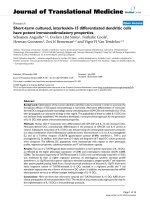
Origin of cancer stem cells. Self-renewal and differentiation potentials are the features of stem cellsFigure 1
Origin of cancer stem cells. Self-renewal and differentiation potentials are the features of stem cells. Progenitor
cells, the product of stem cells that lose the activity of self-renewal, could differentiate into mature cells, which have the fea-
ture of differentiation. The hypothesis is that cancer stem cells are caused by transforming mutations occurring in multi-poten-
tial stem cells, tissue-specific stem cells, progenitor cells, mature cells, and cancer cells.
6WHPFHOOV
3URJHQLWRUFHOOV
7HUPLQDOO\GLIIHUHQWLDWHGFHOOV
0XWDWLRQV
0XWDWHGSURJHQLWRUFHOOV
0XWDWHGVWHPFHOOV
&DQFHUFHOOV
0XWDWLRQV
0XWDWLRQV
6HOIUHQHZDO
6HOIUHQHZDO
6HOIUHQHZDO
6HOIUHQHZDO
Journal of Ovarian Research 2008, 1:4 />Page 4 of 9
(page number not for citation purposes)
has emerged as an attractive source of multiple stem/pro-
genitor cells for cell replacement-based therapies and tis-
sue engineering in regenerative medicine. Our recent
review discussed that cancer stem/progenitor cell research
also offers the possibility of targeting these undifferenti-
ated and malignant cells that provide critical function in
cancer initiation and relapse for treating patients diag-
nosed with advanced and metastatic cancer [30,35,36].
Various strategies consisting of molecular targeting of dis-
tinct oncogenic signaling elements activated in the cancer
progenitor cells and their local microenvironment during
cancer progression can be explored [37]. Furthermore,
overcoming the intrinsic and acquired resistance of cancer
stem/progenitor cells to current clinical treatments repre-
sents a major challenge in treating and curing the most
aggressive and metastatic cancers [38]. In addition,
hematopoitic stem cells are the most characterized stem
cells and it has been used for the therapy to cure cancer
[11]. In this review we also described that the molecular
mechanisms involved in the intrinsic and acquired resist-
ance of cancer cells to current cancer therapies [38].
Pathways of self-renewal and carcinogenesis
Since the cancer stem cells share common properties with
normal stem cells, it is reasonable to think that they have
overlapping regulatory mechanisms. Indeed, one of the
most outstanding questions concerning the biology of
stem cells is: how do multi-potent stem cells select a par-
ticular differentiation pathway and start to differentiate?
Another question is how do stem cells decide to maintain
self-renewal properties and continue to proliferate?
Recent studies demonstrate that the presence of various
genes and signaling pathways are involved in the regula-
tion of the aforementioned processes. Among these, the
Sonic Hedgehog (Shh), Notch and Wnt signaling trans-
duction pathways play a major role in the self-renewal of
stem cells [39-41]. Recent advances in the understanding
of the role of Wnt, Hedgehog, Shh, and Notch signaling
pathways in regulating stem cell self-renewal have shed
new light on carcinogenesis (Figure 2) [7,42,43]. The next
obvious question is the possible connection between
tumors and the (Hedgehog) Hh and Wnt pathways and
how the activation of these pathways leads, in some cases,
to such highly efficient tumorigenesis. Recent genetic evi-
dence suggests that somatic stem cells are the producers of
CSCs; that the Wnt and Hh pathways function in the nor-
mal regulation of stem-cell number in at least some tis-
sues; and that expansion of the somatic stem-cell
population may be the first step in the formation of at
least some types of cancers [44-46]. Numerous arguments
support a stem-cell origin for human cancer. Foremost is
the observation that stem cells possess many of the fea-
tures that characterize the malignant phenotype, includ-
ing self-renewal and unlimited replicative potential [47].
Also, the mutations that initiate tumor formation seem to
accumulate in cells that persist throughout life, as sug-
gested by the exponential increase of cancer incidence
with age. This is thought to reflect a requirement for four
to seven mutations in a single cell to effect malignant
transformation [47]. Although similar signaling pathways
may regulate self-renewal in normal stem cells and cancer
stem cells, there are mechanistic differences in some can-
cers. Interestingly, the mechanistic differences in self-
renewal between normal stem cells and cancer stem cells
can thus be targeted to deplete cancer stem cells without
damaging normal stem cells.
Ovarian tumors
The ovaries contain three main types of cells germ cells,
stromal cells and epithelial cells which give rise to germ
cell, stromal and epithelial ovarian tumors, respectively.
Epithelial ovarian cancers (EOC) were the most common
type of ovarian cancers. Comprising nearly 90% of all
ovarian cancers EOCs are derived from relatively pluripo-
tent cells of the celomic epithelium or "modified mes-
othelium". These cells originate from the primitive
mesoderm and can undergo metaplasia. Approximately
10% to 20% of epithelial ovarian neoplasms are border-
line or low malignant potential tumors and are character-
ized by a high degree of cellular proliferation in the
absence of stromal invasion. Of the invasive epithelial
ovarian cancers, about 55–60% are serous, 15% endome-
Table 1: Cancer type and specific marker for cancer stem cell populations
S. No Cancer type Markers for CSC population References
1. Brain Tumors CD133
+
[23]
2. Breast Cancer CD24
-/low
/CD44
+
/ESA
+
[5]
3. Ovarian Cancer CD133
+
/Side population (SP)/CD44
+
, CD117
+
[25,27,59]
4. Lung Cancer CD133
+
[15]
5. Prostate Cancer CD44
+
/α2β1
high
/CD133
+
[14]
6. Pancreatic Cancer CD44
+
/CD24
+
/ESA/CD133
+
[16,18]
7. Hepatocellular Cancer CD133
+
[24,26]
8. Hematological Malignancies CD34
+
/CD38
-
[17]
9. Colon Cancer CD133
+
/CD44
+
/Lin
-
/ESA
+
[22,28,44]
10. Head and Neck Cancer CD44
+
[21]
Journal of Ovarian Research 2008, 1:4 />Page 5 of 9
(page number not for citation purposes)
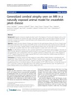
trioid, 5–10% clear cell and <5% mucinous [48] (Figure
3). The various histological subtypes of ovarian carci-
noma have identifiable precursor lesions and multiple
early genetic alterations. Figure 3 explains the various his-
tological subtypes of ovarian cancer and their associated
specific mutations. Mutations may be one of the major
factors contributing to the origin of ovarian cancer stem
cells. Many of the histological subtypes resemble the epi-
thelial component of the lower genital tract, including
papillary serous tumors that have an appearance resem-
bling the glandular epithelium lining the fallopian tube.
Mucinous tumors, on the other hand, contain cells resem-
bling endocervical glands, and endometrioid tumors con-
tain cells resembling the endometrium. Non-epithelial
types of ovarian cancer include sex cord-stromal tumors
(6% of ovarian cancers) and germ cell tumors (3% of all
malignant ovarian neoplasms) [49-51]. The histological
subtypes of ovarian carcinoma have identifiable precursor
lesions and early genetic alterations. Figure 3 explains the
histological subtypes and its specific mutations in ovarian
carcinoma. Mutations are one of the major alteration fac-
tors for the origin of cancer ovarian stem cells.
Markers and their roles in ovarian tumors
In general, tumor markers can be used for one of four pur-
poses: (i) screening a healthy population or a high risk
population for the presence of cancer; (ii) making a diag-
nosis of cancer or of a specific type of cancer; (iii) deter-
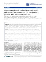
Schematic diagram of signaling pathways that are involved in normal and cancer stem cell biologyFigure 2
Schematic diagram of signaling pathways that are involved in normal and cancer stem cell biology. Wnt, Shh and
Notch1 pathways have been shown to contribute to the self-renewal of stem cells and/or progenitors in a variety of organs,
including the ovarian system. When deregulated, these pathways can contribute to oncogenesis. Mutations of these pathways
have been associated with a number of carcinomas.
:QW
*6.
ȕ
ȕFDW
ȕFDW
/35
6PR
6KK
*OL
X
3WF
*OL
7)V
7)V
7)V
&OHDYHG1RWFK
1RWFK
&%)
6(/)5(1(:$/
Journal of Ovarian Research 2008, 1:4 />Page 6 of 9
(page number not for citation purposes)
mining the prognosis of a patient; and (iv) monitoring the
course in a patient in remission or while receiving surgery,
radiation, or chemotherapy.
Furthermore, recent studies have identified different prog-
nostic and diagnostic surface markers for ovarian cancer
[52] and these markers need to be analyzed for their role
in ovarian cancer. One of the well-known tumor antigens
is the epithelial cell mucin MUC1, a transmembrane glyc-
oprotein that is differentially expressed on tumor cells
compared with normal epithelial cells [53,54]. MUC1 is
expressed either not at all or in small amounts on various
normal epithelia but aberrantly or neoexpressed at high
levels on the majority of adenocarcinomas. Tumor-associ-
ated alterations of MUC1 are characterized by hypoglyco-
sylation, increased sialylation, and altered carbohydrate
core-type expression [53]. Engelmann et al reported that
MUC1 molecule is not only expressed on mature cancer
cells, but also on tumor cells that have multiple character-
istics of stem and progenitor cells [55]. This study demon-
strates MUC1 expressed breast cancer cell line MCF7 as a
source of a minor population of cells with characteristics
of tumor stem/progenitor cells to show for the first time
that these cells also express the hypoglycosylated (tumor)
form of MUC1, previously described only on mature
MCF7 cells and other tumors and tumor cell lines. More-
over, these cells give rise to MUC1
+
tumors in vivo and that
these tumors maintain a small population of MUC1
+
cells
with the stem/progenitor characteristics [55]. Our recent
finding demonstrated the tumor-specific expression of
Tumor Associated Glycoprotein-72 (TAG-72) in ovarian
cancer and its association with disease stage may serve as
a potential marker for effective disease management [56].
In addition, surface marker mucins are overexpressed in
many epithelial malignancies including ovarian cancer,
suggesting a possible role in the pathogenesis of these can-
cers. Other studies from our laboratory have provided
experimental evidence that the MUC4 mucin interacts
with HER2 potentiates its downstream signaling and
enhances the motility of ovarian cancer cells. Our findings
provide experimental support for the hypothesis that
MUC4 mucin expression is associated with a higher met-
astatic potential and thereby a poor prognosis in ovarian
cancer [57]. The future direction of these studies will be to
explore the roles of MUC4 and TAG-72 in ovarian cancer
stem/progenitor cells.
Ovarian cancer stem cells
A recent study describes that ovarian cancer cell lines were
shown to possess "side population" (SP) cells that have
been described as cancer stem cells due to their stem-like
characteristics including the ability to differentiate into
tumors with different histologies. These putative cancer
stem cells reflect the various histological subtypes
observed in ovarian carcinoma. They also provide a
model of cancer metastasis in which these cells are able to
Schematic diagram representing the histological types and its specific mutations in ovarian carcinomaFigure 3
Schematic diagram representing the histological types and its specific mutations in ovarian carcinoma.
6(5286
(1'20(75,2,'
&/($5&(//
08&,1286
0XWDWLRQV
S%5&$.UDV SȕFDWHQLQ37(1
S S
+,672/2*,&$/7<3(62)29$5,$1&$5&,120$$1',76087$7,216
Journal of Ovarian Research 2008, 1:4 />Page 7 of 9
(page number not for citation purposes)
colonize, expand, and differentiate into heterogeneous
tumor phenotypes similar to primary tumors. In such a
model, both the primary tumors and metastasis would
display similar genetic and expression profiles because
both populations are supposedly derived from the same
lineage of cancer stem cells [58]. Ovarian cancer stem
cells, like somatic stem cells, are shown to be capable of
unlimited self-renewal and proliferation. In general,
multi-potent cancer stem cells may account for the histo-
logical heterogeneity often found in tumors [25,27,59].
Moreover, ovarian somatic stem cells would be expected
to divide asymmetrically, yielding both a daughter cell
that proceeds to terminal differentiation, and an undiffer-
entiated copy capable of self-renewal. Repeated asymmet-
ric self-renewal sets of somatic stem cells or their
immediate progenitor's stem cells lead to the accrual of
mutations over time, which might ultimately lead to their
transformation into cancer stem cells and malignant pro-
gression.
Furthermore, another study describes that two mouse
ovarian cancer cell lines such as MOVCAR7 and 4306 con-
tain candidate cancer stem cells [25]. These two murine
ovarian cancer cells have large SP, making them suitable
to study ovarian cancer stem cell biology. A similar, albeit
very small, SP was also identified in the human ovarian
cancer stem cell lines IGROV-1, SKOV-3 and OVCAR-3
and also in cells claimed from patient ascetic fluid [25].
Further, a study proved that isolated and characterized
ovarian cancer-initiating cells (OCICs) are fully capable of
reestablishing their original tumor hierarchy in vivo. These
cells are very organized self-renewing, anchorage-inde-
pendent spheres and were reproducibly dividable using
antibodies against both CD44 and CD117 [27]. These
OCICs were capable of intraperitoneal tumorigenesis and
could serially propagate tumors in animals. Conse-
quently, this study fulfills all currently accepted criteria for
the existence of a subpopulation of tumor-initiating cells
[27], and their specific detection and targeting could be
highly valuable for therapy of recurrent, chemo-resistant
disease. Whereas advanced ovarian cancer is generally ini-
tially responsive to standard chemotherapies (ciaplatin
and paclitaxel), that responsive almost inevitably fol-
lowed by drug resistant phenotype [2,60]. One accepted
hypothesis about chemoresistance is standard therapies
failed to target tumor progenitors, which are have like
normal stem cells because of expression of membrane
efflux transporters. Zhang et al showed that OCICs, under
stem cell-selective conditions, over express ABCG2 and
are more resistant to cisplatin and paclitaxel, suggesting a
possible role for these cells in ovarian cancer chemoresist-
ance [27].
Conclusion and perspective
The aforementioned studies showed that a so-called ovar-
ian cancer stem cell, with high-proliferative capacity, self-
renewal properties and multi-lineage potential, could be
responsible for tumor development and the differentia-
tion of more mature epithelial ovarian cells contributing
to tumorigenesis. There are important consequences for
cancer treatment if the growth of tumors is at least in part,
dependent on a cancer stem cell population. The cancer
stem cell hypothesis posits that cancer stem cells are a
minor population of self-renewing cancer cells that fuel
tumor growth and remain in patients after conventional
therapy has been completed. The hypothesis predicts that
effective tumor eradication will require obtaining agents
that can target cancer stem cells while sparing normal
stem cells. Experimental evidence suggests that ovarian
cancer stem cells are relatively resistant to conventional
chemotherapeutic agents. Current cancer therapies often
engender severe toxicity because of their general effects on
all rapidly dividing cells. Identification of candidate tar-
gets for more specific mechanism-based cancer therapy
using techniques such as gene chips could reveal signature
patterns of transcriptional output which are characteristic
of activated self-renewal pathways.
Emerging evidence suggests that these pathways also con-
trol patterning and growth in self-renewing adult tissues
by regulating the stem-cell compartment. Thus, pharma-
cological inhibition of these pathways in the worst case
might result in severe toxicity due to a loss of normal
stem-cell compartments. Further research will be needed
to determine whether continuous pathway activity is
required in normal and tumor tissues, and whether these
requirements differ sufficiently as to allow therapeutic
intervention. Even if pathway inhibition is prohibited by
normal physiological requirements, other mechanism-
based approaches that exploit aberrant pathway activa-
tion might be feasible. It has been proposed that malig-
nancy is determined in all tissues by mis-regulation of a
common set of genes that control growth by affecting cell
proliferation, apoptosis, invasion and angiogenesis. This
hypothesis is supported by the demonstration that multi-
ple types of normal human cells can be made tumorigenic
by the expression of a defined set of viral and cellular pro-
teins. Therapeutic agents for the treatment of such tumors
might target not only self-renewal pathway components,
but also other critical transcriptional targets of the self-
renewal pathways, or proteins that co-operate with them
to deregulate growth.
It is important that agents directed against cancer stem
cells discriminate between cancer stem cells and normal
stem cells. This will require the identification of realistic
drug targets unique to cancer stem cells. The identification
of such targets and the development of anti-cancer agents
Journal of Ovarian Research 2008, 1:4 />Page 8 of 9
(page number not for citation purposes)
will require a deeper understanding of normal stem cell
biology as well as cancer biology. More importantly, iden-
tification of the ovarian cancer stem cell would provide a
critical step in advancing the development of novel thera-
peutic strategies in the management of ovarian cancer.
Furthermore, characterizations of such progenitor or can-
cer stem cells in drug resistant (Ciaplatin, Paclitaxel and
etc) manner for ovarian cancer will likely lead to a greater
understanding of early events leading to the genesis of this
elusive disease, in addition to providing new therapeutics
targets aimed at the cells directly responsible for its prop-
agation.
Abbreviations
CSC: Cancer Stem Cell; CNS: Central Nervous System;
ESA: Epithelial-Specific Antigen; Shh: Sonic Hedgehog;
Hh: Hedgehog; EOC: Epithelial ovarian Cancer; OCIC:
Ovarian Cancer Initiating Cells; SP: Side Population
Competing interests
The authors declare that they have no competing interests.
Authors' contributions
PPM participated in drafting the full manuscript and cre-
ating figures. SKB participated in substantial contribution
to conception and revising it critically for important intel-
lectual content.
Acknowledgements
The authors thank Kristi L.W. Berger (Eppley Institute) for editorial assist-
ance. The authors on this article were supported by grants from the U.S.
Department of Defense (OC04110) and National Institutes of Health (RO1
CA78590, CA 131944 and CA133774).
References
1. Aletti GD, Gallenberg MM, Cliby WA, Jatoi A, Hartmann LC: Cur-
rent management strategies for ovarian cancer. Mayo Clin
Proc 2007, 82:751-770.
2. Pecorelli S, Favalli G, Zigliani L, Odicino F: Cancer in women. Int J
Gynaecol Obstet 2003, 82:369-379.
3. Godwin AK, Testa JR, Hamilton TC: The biology of ovarian can-
cer development. Cancer 1993, 71:530-536.
4. Ness RB, Cottreau C: Possible role of ovarian epithelial inflam-
mation in ovarian cancer. J Natl Cancer Inst 1999, 91:1459-1467.
5. Al-Hajj M, Wicha MS, ito-Hernandez A, Morrison SJ, Clarke MF: Pro-
spective identification of tumorigenic breast cancer cells.
Proc Natl Acad Sci USA 2003, 100:3983-3988.
6. Matsui W, Huff CA, Wang Q, Malehorn MT, Barber J, Tanhehco Y,
Smith BD, Civin CI, Jones RJ: Characterization of clonogenic
multiple myeloma cells. Blood 2004, 103:2332-2336.
7. Reya T, Morrison SJ, Clarke MF, Weissman IL: Stem cells, cancer,
and cancer stem cells. Nature 2001, 414:105-111.
8. Seaberg RM, van der KD: Stem and progenitor cells: the prema-
ture desertion of rigorous definitions. Trends Neurosci 2003,
26:125-131.
9. Potten CS, Loeffler M: Stem cells: attributes, cycles, spirals, pit-
falls and uncertainties. Lessons for and from the crypt. Devel-
opment 1990, 110:1001-1020.
10. Orkin SH, Zon LI: Hematopoiesis: an evolving paradigm for
stem cell biology. Cell 2008, 132:631-644.
11. Zon LI: Intrinsic and extrinsic control of haematopoietic
stem-cell self-renewal. Nature 2008, 453:306-313.
12. McNiece I: The CD34+Thy1+ cell population: are they all
stem cells? Exp Hematol 2000, 28:
1312-1314.
13. Brown MD, Gilmore PE, Hart CA, Samuel JD, Ramani VA, George NJ,
Clarke NW: Characterization of benign and malignant pros-
tate epithelial Hoechst 33342 side populations. Prostate 2007,
67:1384-1396.
14. Collins AT, Berry PA, Hyde C, Stower MJ, Maitland NJ: Prospective
identification of tumorigenic prostate cancer stem cells. Can-
cer Res 2005, 65:10946-10951.
15. Eramo A, Lotti F, Sette G, Pilozzi E, Biffoni M, Di VA, Conticello C,
Ruco L, Peschle C, De MR: Identification and expansion of the
tumorigenic lung cancer stem cell population. Cell Death Differ
2008, 15:504-514.
16. Hermann PC, Huber SL, Herrler T, Aicher A, Ellwart JW, Guba M,
Bruns CJ, Heeschen C: Distinct populations of cancer stem cells
determine tumor growth and metastatic activity in human
pancreatic cancer. Cell Stem Cell 2007, 1:313-323.
17. Lapidot T, Sirard C, Vormoor J, Murdoch B, Hoang T, Caceres-
Cortes J, Minden M, Paterson B, Caligiuri MA, Dick JE: A cell initiat-
ing human acute myeloid leukaemia after transplantation
into SCID mice. Nature 1994, 367:645-648.
18. Li C, Heidt DG, Dalerba P, Burant CF, Zhang L, Adsay V, Wicha M,
Clarke MF, Simeone DM: Identification of pancreatic cancer
stem cells. Cancer Res 2007, 67:1030-1037.
19. Ma S, Chan KW, Hu L, Lee TK, Wo JY, Ng IO, Zheng BJ, Guan XY:
Identification and characterization of tumorigenic liver can-
cer stem/progenitor cells. Gastroenterology 2007, 132:2542-2556.
20. Pardal R, Clarke MF, Morrison SJ: Applying the principles of
stem-cell biology to cancer. Nat Rev Cancer 2003, 3:895-902.
21. Prince ME, Sivanandan R, Kaczorowski A, Wolf GT, Kaplan MJ,
Dalerba P, Weissman IL, Clarke MF, Ailles LE: Identification of a
subpopulation of cells with cancer stem cell properties in
head and neck squamous cell carcinoma. Proc Natl Acad Sci USA
2007, 104:973-978.
22. Ricci-Vitiani L, Lombardi DG, Pilozzi E, Biffoni M, Todaro M, Peschle
C, De MR: Identification and expansion of human colon-can-
cer-initiating cells. Nature
2007, 445:111-115.
23. Singh SK, Hawkins C, Clarke ID, Squire JA, Bayani J, Hide T, Henkel-
man RM, Cusimano MD, Dirks PB: Identification of human brain
tumour initiating cells. Nature 2004, 432:396-401.
24. Suetsugu A, Nagaki M, Aoki H, Motohashi T, Kunisada T, Moriwaki H:
Characterization of CD133+ hepatocellular carcinoma cells
as cancer stem/progenitor cells. Biochem Biophys Res Commun
2006, 351:820-824.
25. Szotek PP, Pieretti-Vanmarcke R, Masiakos PT, Dinulescu DM, Con-
nolly D, Foster R, Dombkowski D, Preffer F, Maclaughlin DT, Dona-
hoe PK: Ovarian cancer side population defines cells with
stem cell-like characteristics and Mullerian Inhibiting Sub-
stance responsiveness. Proc Natl Acad Sci USA 2006,
103:11154-11159.
26. Yin S, Li J, Hu C, Chen X, Yao M, Yan M, Jiang G, Ge C, Xie H, Wan
D, Yang S, Zheng S, Gu J: CD133 positive hepatocellular carci-
noma cells possess high capacity for tumorigenicity. Int J Can-
cer 2007, 120:1444-1450.
27. Zhang S, Balch C, Chan MW, Lai HC, Matei D, Schilder JM, Yan PS,
Huang TH, Nephew KP: Identification and characterization of
ovarian cancer-initiating cells from primary human tumors.
Cancer Res 2008, 68:4311-4320.
28. Dalerba P, Dylla SJ, Park IK, Liu R, Wang X, Cho RW, Hoey T, Gurney
A, Huang EH, Simeone DM, Shelton AA, Parmiani G, Castelli C,
Clarke MF: Phenotypic characterization of human colorectal
cancer stem cells. Proc Natl Acad Sci USA 2007, 104:10158-10163.
29. Mimeault M, Batra SK: Functions of tumorigenic and migrating
cancer progenitor cells in cancer progression and metastasis
and their therapeutic implications. Cancer Metastasis Rev 2007,
26:203-214.
30. Mimeault M, Batra SK: Recent progress on tissue-resident adult
stem cell biology and their therapeutic implications. Stem Cell
Rev 2008, 4:27-49.
31. Gage FH: Mammalian neural stem cells. Science 2000,
287:1433-1438.
32. Morrison SJ, Shah NM, Anderson DJ: Regulatory mechanisms in
stem cell biology.
Cell 1997, 88:287-298.
33. Uchida N, Buck DW, He D, Reitsma MJ, Masek M, Phan TV, Tsu-
kamoto AS, Gage FH, Weissman IL: Direct isolation of human
central nervous system stem cells. Proc Natl Acad Sci USA 2000,
%19(97):14720-14725.
Publish with Bio Med Central and every
scientist can read your work free of charge
"BioMed Central will be the most significant development for
disseminating the results of biomedical researc h in our lifetime."
Sir Paul Nurse, Cancer Research UK
Your research papers will be:
available free of charge to the entire biomedical community
peer reviewed and published immediately upon acceptance
cited in PubMed and archived on PubMed Central
yours — you keep the copyright
Submit your manuscript here:
/>BioMedcentral
Journal of Ovarian Research 2008, 1:4 />Page 9 of 9
(page number not for citation purposes)
34. Goodell MA, Brose K, Paradis G, Conner AS, Mulligan RC: Isolation
and functional properties of murine hematopoietic stem
cells that are replicating in vivo. J Exp Med 1996, 183:1797-1806.
35. Mimeault M, Batra SK: Concise review: recent advances on the
significance of stem cells in tissue regeneration and cancer
therapies. Stem Cells 2006, 24:2319-2345.
36. Mimeault M, Hauke R, Batra SK: Recent advances on the molec-
ular mechanisms involved in the drug resistance of cancer
cells and novel targeting therapies. Clin Pharmacol Ther 2008,
83:673-691.
37. Mimeault M, Hauke R, Mehta PP, Batra SK: Recent advances in
cancer stem/progenitor cell research: therapeutic implica-
tions for overcoming resistance to the most aggressive can-
cers. J Cell Mol Med 2007, 11:981-1011.
38. Mimeault M, Hauke R, Batra SK: Stem cells: a revolution in ther-
apeutics-recent advances in stem cell biology and their ther-
apeutic applications in regenerative medicine and cancer
therapies. Clin Pharmacol Ther 2007, 82:252-264.
39. Bhardwaj G, Murdoch B, Wu D, Baker DP, Williams KP, Chadwick K,
Ling LE, Karanu FN, Bhatia M: Sonic hedgehog induces the pro-
liferation of primitive human hematopoietic cells via BMP
regulation. Nat Immunol 2001, 2:172-180.
40. Lee HY, Kleber M, Hari L, Brault V, Suter U, Taketo MM, Kemler R,
Sommer L: Instructive role of Wnt/beta-catenin in sensory
fate specification in neural crest stem cells. Science 2004,
303:1020-1023.
41. Varnum-Finney B, Xu L, Brashem-Stein C, Nourigat C, Flowers D,
Bakkour S, Pear WS, Bernstein ID: Pluripotent, cytokine-depend-
ent, hematopoietic stem cells are immortalized by constitu-
tive Notch1 signaling. Nat Med 2000, 6:1278-1281.
42. Reguart N, He B, Taron M, You L, Jablons DM, Rosell R: The role of
Wnt signaling in cancer and stem cells. Future Oncol 2005,
1:787-797.
43. Taipale J, Beachy PA: The Hedgehog and Wnt signalling path-
ways in cancer.
Nature 2001, 411:349-354.
44. Bienz M, Clevers H: Linking colorectal cancer to Wnt signaling.
Cell 2000, 103:311-320.
45. Polakis P: Wnt signaling and cancer. Genes Dev 2000,
14:1837-1851.
46. Wodarz A, Nusse R: Mechanisms of Wnt signaling in develop-
ment. Annu Rev Cell Dev Biol 1998, 14:59-88.
47. Hanahan D, Weinberg RA: The hallmarks of cancer. Cell 2000,
100:57-70.
48. Bell DA: Origins and molecular pathology of ovarian cancer.
Mod Pathol 2005, 18(Suppl 2):S19-S32.
49. Breedlove G, Busenhart C: Screening and detection of ovarian
cancer. J Midwifery Womens Health 2005, 50:51-54.
50. Hightower RD, Nguyen HN, Averette HE, Hoskins W, Harrison T,
Steren A: National survey of ovarian carcinoma. IV: Patterns
of care and related survival for older patients. Cancer 1994,
73:377-383.
51. Jemal A, Siegel R, Ward E, Murray T, Xu J, Thun MJ: Cancer statis-
tics, 2007. CA Cancer J Clin 2007, 57:43-66.
52. Chauhan SC, Singh AP, Ruiz F, Johansson SL, Jain M, Smith LM, Moni-
aux N, Batra SK: Aberrant expression of MUC4 in ovarian car-
cinoma: diagnostic significance alone and in combination
with MUC1 and MUC16 (CA125). Mod Pathol 2006,
19:1386-1394.
53. Gendler SJ, Lancaster CA, Taylor-Papadimitriou J, Duhig T, Peat N,
Burchell J, Pemberton L, Lalani EN, Wilson D: Molecular cloning
and expression of human tumor-associated polymorphic epi-
thelial mucin. J Biol Chem 1990, 265:15286-15293.
54. Vlad AM, Kettel JC, Alajez NM, Carlos CA, Finn OJ: MUC1 immu-
nobiology: from discovery to clinical applications. Adv Immunol
2004, 82:249-293.
55. Engelmann K, Shen H, Finn OJ:
MCF7 side population cells with
characteristics of cancer stem/progenitor cells express the
tumor antigen MUC1. Cancer Res 2008, 68:2419-2426.
56. Ponnusamy MP, Venkatraman G, Singh AP, Chauhan SC, Johansson
SL, Jain M, Smith L, Davis JS, Remmenga SW, Batra SK: Expression
of TAG-72 in ovarian cancer and its correlation with tumor
stage and patient prognosis. Cancer Lett 2007, 251:247-257.
57. Ponnusamy MP, Singh AP, Jain M, Chakraborty S, Moniaux N, Batra
SK: MUC4 activates HER2 signalling and enhances the motil-
ity of human ovarian cancer cells. Br J Cancer 2008, 99:520-526.
58. Chien JR, Aletti G, Bell DA, Keeney GL, Shridhar V, Hartmann LC:
Molecular pathogenesis and therapeutic targets in epithelial
ovarian cancer. J Cell Biochem 2007, 102:1117-1129.
59. Ferrandina G, Bonanno G, Pierelli L, Perillo A, Procoli A, Mariotti A,
Corallo M, Martinelli E, Rutella S, Paglia A, Zannoni G, Mancuso S,
Scambia G: Expression of CD133-1 and CD133-2 in ovarian
cancer. Int J Gynecol Cancer 2008, 18:506-514.
60. Cannistra SA: Cancer of the ovary. N Engl J Med 2004,
351:2519-2529.