báo cáo hóa học:" Microarray analysis of Foxl2 mediated gene regulation in the mouse ovary derived KK1 granulosa cell line: Over-expression of Foxl2 leads to activation of the gonadotropin releasing hormone receptor gene promoter" pptx
Bạn đang xem bản rút gọn của tài liệu. Xem và tải ngay bản đầy đủ của tài liệu tại đây (556.28 KB, 12 trang )
Escudero et al. Journal of Ovarian Research 2010, 3:4
/>
RESEARCH
Open Access
Microarray analysis of Foxl2 mediated gene
regulation in the mouse ovary derived KK1
granulosa cell line: Over-expression of Foxl2 leads
to activation of the gonadotropin releasing
hormone receptor gene promoter
Jean M Escudero1†, Jodi L Haller2, Colin M Clay3, Kenneth W Escudero1*†
Abstract
Background: The Foxl2 transcription factor is required for ovarian function during follicular development. The
mechanism of Foxl2 regulation of this process has not been elucidated. Our approach to begin to understand
Foxl2 function is through the identification of Foxl2 regulated genes in the ovary.
Methods: Transiently transfected KK1 mouse granulosa cells were used to identify genes that are potentially
regulated by Foxl2. KK1 cells were transfected in three groups (mock, activated, and repressed) and twenty-four
hours later RNA was isolated and submitted for Affymetrix microarray analysis. Genesifter software was used to carry
out analysis of microarray data. One identified target, the gonadotropin releasing hormone receptor (GnRHR) gene,
was chosen for further study and validation of Foxl2 responsiveness. Transient transfection analyses were carried
out to study the effect of Foxl2 over-expression on GnRHR gene promoter-luciferase fusion activity. Data generated
was analyzed with GraphPad Prism software.
Results: Microarray analysis identified 996 genes of known function that are potentially regulated by Foxl2 in
mouse KK1 granulosa cells. The steroidogenic acute regulatory protein (StAR) gene that has been identified as
Foxl2 responsive by others was identified in this study also, thereby supporting the effectiveness of our strategy.
The GnRHR gene was chosen for further study because it is known to be expressed in the ovary and the results of
previous work has indicated that Foxl2 may regulate GnRHR gene expression. Cellular levels of Foxl2 were
increased via transient co-transfection of KK1 cells using a Foxl2 expression vector and a GnRHR promoterluciferase fusion reporter vector. The results of these analyses indicate that over-expression of Foxl2 resulted in a
significant increase in GnRHR promoter activity. Therefore, these transfection data validate the microarray data
which suggest that Foxl2 regulates GnRHR and demonstrate that Foxl2 acts as an activator of the GnRHR gene.
Conclusions: Potential Foxl2 regulated ovarian genes have been identified through microarray analysis and
comparison of these data to other microarray studies. The Foxl2 responsiveness of the GnRHR gene has been
validated and provided evidence of Foxl2 transcriptional activation of the GnRHR gene promoter in the mouse
ovary derived KK1 granulosa cell line.
* Correspondence:
† Contributed equally
1
Department of Biological and Health Sciences, Texas A&M UniversityKingsville, Kingsville, TX, USA
© 2010 Escudero et al; licensee BioMed Central Ltd. This is an Open Access article distributed under the terms of the Creative
Commons Attribution License ( which permits unrestricted use, distribution, and
reproduction in any medium, provided the original work is properly cited.
Escudero et al. Journal of Ovarian Research 2010, 3:4
/>
Background
The transcription factor Foxl2 is vital to ovarian function as evidenced by the identification of mutations in
the gene encoding FoxL2 that result in the condition
known as blepharophimosis/ptosis/epicanthus inversus
syndrome (BPES) and in some cases, premature ovarian
failure (POF) [1]. Type I BPES is characterized by eyelid
malformation and POF suggesting that the expression of
FoxL2 is critical in the developing eyelid as well as in
the maintenance of ovarian function. Foxl2 has also
been implicated in the process of sex determination in
mice [2] as well as in humans [3].
Foxl2 knockout studies in the mouse provide compelling evidence of the critical role that Foxl2 plays in ovarian function. In a study in which a portion of the Foxl2
coding region (amino acids N-61) was fused to the bgalactosidase gene (LacZ), homozygous Foxl2-lacZ mice
exhibited ovarian failure resulting from the absence of
granulosa cell differentiation at an early stage of follicular development [4]. These investigators observed that
follicles were activated and underwent apoptosis, leading
to progressive follicular depletion and ovarian atresia. A
second study in which both copies of Foxl2 were completely knocked out determined that the development of
granulosa cells was blocked at the point of primordial
follicle formation [5].
In order to understand the mechanism through which
Foxl2 functions in follicular development, ovarian specific target genes must be identified. Microarray analysis
has been performed previously by other groups using
strategies that differ from this present study. The first
study involved over-expression of FoxL2 in the human
KGN granulosa cell line and Nimblegen gene chips [6].
A more recent study used Foxl2 knockout mice and
whole ovary preparations of RNA for their analyses.
Both Affymetrix and Agilent gene chips were used by
these investigators [7].
In this present study, mouse Foxl2 target genes were
identified using Affymetrix microarray analysis. The
effect of Foxl2 mediated gene regulation was examined
through the use of vectors expressing Foxl2 fusion proteins designed to either activate or repress gene expression (fusions are described in Methods section below).
The KK1 mouse granulosa cell line is an excellent system for these studies in that these cells exhibit granulosa cell characteristics such as gonadotropin
responsiveness and inhibin expression [8]. In addition,
KK1 cells are easy to maintain in culture and can be
transfected with high efficiency. The data generated
using the Affymetrix microarray analysis have been compared to those found in the above mentioned studies
and many of the target genes identified are common to
those studies [6,7].
Page 2 of 12
In addition, a candidate gene identified in the microarray analysis was chosen for further study. The GnRHR
gene was chosen based on the results of our previous
study which implicated Foxl2 in the regulation of
GnRHR in the pituitary derived aT3-1 cell line [9]. This
present study provides the first evidence that Foxl2
affects the expression of GnRHR in the ovary. Foxl2 regulation of the GnRHR gene has been examined in KK1
granulosa cells through the use of transient co-transfection studies of a GnRHR promoter-luciferase fusion
construct and a wild type Foxl2 expression vector. The
results of these analyses suggest that Foxl2 is a positive
regulator of the murine GnRHR gene promoter.
Methods
Cell culture
The KK1 granulosa cell line was a gift from Dr. Ilpo
Huhtaniemi whose laboratory developed the cell line
and from Dr. Deborah Segaloff who sent us the cell line.
The cells are grown in DMEM/F12 (50/50) containing
10% heat inactivated FBS (Gemini Bioproducts; West
Sacramento, CA), 100 μg/ml penicillin-streptomycin,
and 0.25 μg/ml amphotericin B. All cell culture reagents
other than FBS were purchased from Mediatech; Manassas, VA.
Affymetrix microarray analysis
The KK1 granulosa cell line was chosen as a model system for this study as a result of numerous published
studies as well as our own observations suggesting that
the site of action of Foxl2 during follicular development
is the granulosa cell. However, due to endogenous
expression of Foxl2 in KK1 cells, simple over-expression
resulting from transfection of a Foxl2 expression vector
may not affect levels of gene expression sufficiently to
be detected efficiently by microarray analysis. Therefore,
in order to increase the potential of Foxl2 to alter gene
expression levels of putative target genes, two fusions
were constructed consisting of Foxl2 fused to the activation domain of the Herpes simplex virus VP16 transcription factor (Foxl2-VP16) and Foxl2 fused to the
repression domain of the murine MAD transcription
factor (Foxl2-MAD). Levels of gene expression were
compared between mock transfected cells and those
that were transfected with Foxl2-VP16 and Foxl2-MAD,
respectively.
Foxl2 fusion protein derivatives
The construction of Foxl2-VP16 was described previously and was shown to function as a specific activator
of transcription [9]. Foxl2-MAD consists of the 40
amino acid N-terminal mSin3 interaction domain (SID)
of the Mad transcription factor fused to Foxl2 in the
expression vector pcDNA 3.1 (Invitrogen Corporation;
Escudero et al. Journal of Ovarian Research 2010, 3:4
/>
Carlsbad, CA). The repression activity of the Mad-SID is
mediated through interactions of the Mad N-terminus
with the mammalian Sin3 co-repressor protein [10,11].
The effectiveness of the Sin3 binding domain of Mad in
silencing gene expression when fused to DNA binding
proteins has been demonstrated in two studies. One
group fused the Mad SID to the c-Jun DNA binding
domain and demonstrated specific inhibition of transcription mediated by binding to AP-1 binding site elements [12]. Another group of investigators fused Mad
SID to the tetracycline receptor and demonstrated tetracycline mediated gene repression via binding to the tetracycline operator sequence [11]. The ability of Foxl2Mad and Foxl2-VP16 fusion constructs was tested utilizing the Foxl2-VP16 responsive luciferase reporter 3XGRAS-Luc [9]. In transient transfections of KK1 cells,
Foxl2-VP16 resulted in a greater than 50-fold increase
in luciferase expression. The co-transfection of Foxl2Mad attenuated the ability of Foxl2-VP16 to activate
luciferase expression by 95% (data not shown). Therefore, these fusion vectors are appropriate for activation
and repression studies of Foxl2 specific genes.
Microarray transfection
KK1 cells were grown in 150 mm tissue culture plates
to 50% confluence. Three plates were mock transfected
with 30 μl of Fugene 6 transfection reagent (Roche
Applied Science; Indianapolis, IN) and 10 μg of empty
vector (pcDNA 3.1). Three plates were transfected with
10 μg of Foxl2-VP16 expression vector in 30 μl Fugene
reagent and 3 plates were transfected with 10 μg of
Foxl2-MAD expression vector in 30 μl Fugene reagent.
After 24 hours incubation at 37°C the cells were harvested and polyA+ RNA was isolated using RNeasy (Qiagen Inc.; Valencia, CA) according to manufacturer’s
procedures. Ten μg of RNA isolated from each plate
were diluted to a concentration of 1 μg/μl and a total of
9 samples were submitted to the Colorado State University Affymetrix core facility for analysis.
Microarray and data analysis
Nine Affymetrix mouse genome 430 2.0 chips were used
(one for each plate of KK1 cells). Our microarray data
can be downloaded from the web site, i.
nlm.nih.gov/geo, via the accession number GSE18891.
Data obtained from the chips was analyzed using the
Genesifter Program (VizX Labs LLC; Seattle, WA).
Two independent analyses were performed. In the
first, our data was analyzed using pairwise analysis,
mean normalization, and T-test statistical analysis (P <
0.05). In the second, we compared our data to that of
Garcia-Ortiz et al. [7]. Eighteen Affymetrix CEL files
representing the developmental stage embryonic days 13
and 16, and birth were downloaded from http://www.
ncbi.nlm.nih.gov/geo, via the accession number
GSE12989 [7]. These files were uploaded along with our
Page 3 of 12
9 Affymetrix CEL files together into Genesifter for analysis using the MAS5 advanced upload method. A project
analysis in was set up in Genesifter with the following
parameters: the cutoff threshold was set at 1.8 fold with
T-test statistical analysis (mouse) or anova (KK1), and P
< 0.05.
Transient co-transfection and luciferase assays
Expression vectors
The original GnRHR promoter luciferase fusion reporter
vector (pMGR-600 Luc) was described previously [13].
In this study, the plasmid -600 Luc was created by subcloning the 600 base pair fragment of the mouse
GnRHR promoter into the expression vector pGL3 basic
(Promega Corporation; Madison, WI). The Foxl2
expression vector (pFoxl2) consists of the mouse Foxl2
open reading frame inserted in pCDNA 3.1 (Invitrogen
Corporation; Carlsbad, CA). The renilla luciferase control vector phRL-CMV is part of the dual luciferase
assay system (Promega Corporation; Madison, WI).
Transfection
KK1 cells were plated at a density of 2.5 × 10⁵ cells per
well of 24 well tissue culture plates the day the transfections were carried out. Transfections included 0.3 μg
-600 Luc reporter plasmid, 3 ng of phRLCMV renilla
luciferase vector to normalize for transfection efficiency,
and either 0.3 μg pcDNA 3.1 (as a control) or 0.3 μg
pFoxl2. DNA was mixed with DMEM/F12 medium containing Fugene 6 reagent (Roche Applied Science; Indianapolis, IN) at a ratio of 4:1 Fugene to total DNA and
added to cells.
Luciferase assays
Transfected cells were incubated for 24 hours at 37°C
and washed 3 times with phosphate buffered saline.
Cells were lysed and the Dual-luciferase assay was performed according to manufacturer’s instructions (Promega Corporation; Madison, WI) using a 20/20 n
luminometer (Turner Biosystems; Sunnyvale, CA). Each
transfection was carried out in triplicate and experiments were carried out a total of 4 times. Data was analyzed with the paired T-test using GraphPad Prism
software (GraphPad Software, Inc.; La Jolla, CA).
Results
Affymetrix microarray analysis
In order to increase the effectiveness of this analysis,
Foxl2 derivatives consisting of the entire open reading
frame of Foxl2 fused to either the strong transcriptional
activation domain of Herpes simplex virus VP16 [9] or
the Mad protein-SID domain, a repressor of transcription [10,14] were used. These Foxl2 fusion proteins can
be used as tools for gene discovery as the DNA binding
domain of Foxl2 will guide either the activation or
repression domain to the promoter regions of genes
Escudero et al. Journal of Ovarian Research 2010, 3:4
/>
that are normally regulated by Foxl2 via specific DNA
binding. Theoretically, Foxl2-VP16 should stimulate
genes that are normally regulated by Foxl2, particularly
those that are normally repressed in KK1 cells. Conversely, Foxl2-MAD should function to repress genes normally regulated by Foxl2, especially those that are
normally active in KK1 cells. In this study, levels of gene
expression in murine KK1 granulosa cells transfected
with the fusion protein expression vectors were compared to mock transfected cells. A pairwise analysis was
performed with the following groupings: mock vs. VP16
transfected cells (group NA); mock vs. Mad transfected
cells (group NR); and Mad vs. VP16 transfected cells
(group RA). The cumulative listings of genes for these
groups are found in Additional Files 1, 2, and 3 respectively. The results of the pairwise analysis are summarized in the comprehensive listing of 636 potential
targets of the Foxl2 transcription factor (Additional File
4). Only genes of known function are listed and all
exhibited at least a 1.75 fold change in expression levels.
In order to validate these findings, we carried out a
comparison of our data to data derived from two other
microarray studies [6,7]. One group used the human
KGN granulosa cell line and over-expression of FoxL2
to identify potential targets and qPCR to confirm
numerous FoxL2 regulated genes [6]. A second group
used mouse Foxl2 knockouts and whole ovarian preparations for microarray studies as well as analysis of
other investigator’s microarray data [7].
Garcia-Ortiz et al. [7] used Affymetrix gene chips
identical to those used in our study to compare developmental stage specific gene expression levels in ovary
preparations from wild type and Foxl2 knockout mice at
embryonic day 13 (E13), embryonic day 16 (E16), and
birth (P0). This allowed us to compare our raw data
directly to theirs and determine the similarity between
differentially expressed genes in our KK1 study and
their stage specific study. In this analysis, we compared
differentially expressed genes from mock, VP16, and
Mad transfected KK1 cell groups combined (Additional
file 5) to mouse samples [7] that compared wild type to
Foxl2 knockout for stages E13 (Additional file 6); E16
(Additional file 7), and P0 (Additional file 8).
The “Intersector” subroutine in Genesifter allowed us
to find commonalities between the groups of genes in
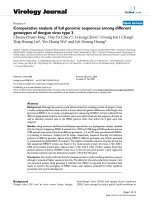
Additional files 5, 6, 7, 8. The comparisons are the following: KK1 vs. E16, KK1 vs. E13, and KK1 vs. P0 (Figure 1 and Additional file 9). This analysis resulted in the
discovery of 360 new genes of known function that were
shared between the KK1 and mouse ovary studies
increasing the total number of potential Foxl2 target
genes to 996 (Additional file 4).
We then turned our attention to the human KGN cell
line study of Batista et al. [6]. In comparing their
Page 4 of 12
confirmed human gene list to our mouse gene list, 3
common genes were identified. These were Mrgpre,
Maff, and Rspo3 (Additional file 10). Comparison of
their comprehensive listing with our study’s list of genes
resulted in finding a total of 42 genes common to both
(Additional file 10).
In order to begin to understand the significance of the
genes common to the three microarray studies in the
ovary, we then searched the Ovarian Kaleidoscope Database (OKdb) to determine if other investigators had
published evidence of ovarian expression of these genes.
A total of 71 of the genes were found to be expressed in
the ovary and have been categorized according to function: Gene Regulation (Table 1), Signaling (Table 2), and
Metabolism, Cell Adhesion, Cytoskeletal, and Structural
(Table 3).
Three of the genes that we have included in Table 2
were not found in the OKdb: Mrgpre, Ctla4 and Eda.
These are potentially important due to the fact that they
are common to all three studies. The StAR gene was not
found in either of the other group’s data sets but
included in Table 3 because it has been shown to be a
human Foxl2 target gene [15]. Our finding that the
mouse StAR gene is a Foxl2 target validates the
approach chosen for this study. The GnRHR gene was
not found in either of the other group’s data sets and is
noted in Table 2 because further evidence of Foxl2 regulation of this gene is provided in the section that
follows.
Transient co-transfection and luciferase assays
Transient co-transfection studies to determine the Foxl2
regulation of the GnRHr promoter were carried out
using KK1 cells transfected with various combinations
of luciferase reporter vectors and pcDNA 3.1 expression
vectors. The effect of Foxl2 over-expression on the
activity of the GnRHR promoter was determined by
comparing the luciferase activity of the promoter in the
presence of pFoxl2 (over-expression) to the activity of
the promoter in the absence of Foxl2 over-expression
(pcDNA 3.1). Foxl2 over-expression activated the
GnRHR promoter 5.8 fold (Figure 2).
Discussion
This study has resulted in an increased awareness of
Foxl2 function in the ovary. First, through the use of
microarray analysis we have added to the growing list of
genes that appear to be regulated by Foxl2 and thus
may play a role in follicular development in the ovary.
Second, we have demonstrated the GnRHR gene promoter is regulated in a positive manner by the transcription
factor Foxl2 in the KK1 granulosa cell line.
Escudero et al. Journal of Ovarian Research 2010, 3:4
/>
Page 5 of 12
Figure 1 Comparison of differential gene expression in KK1 cells to mouse ovary developmental stages. Genesifter software was used to
determine differentially expressed genes in KK1 cells and mouse in vivo samples [7]. The KK1 analysis for differentially expressed genes
generated a cumulative listing of 2520 differentially expressed genes among the mock, VP16, and mad transfected cell groups combined
(Additional file 5). The mouse samples compared wild type to knockout for each stage as follows: E13, 3289 genes (Additional file 6); E16, 2995
genes (Additional file 7); and P0, 4330 genes (Additional file 8). Comparisons between groups of genes were performed with Genesifter
Intersector. The comparisons are represented by circles and are as follows: KK1 vs. E16; KK1 vs. E13; and KK1 vs. P0. The number of genes in each
grouping corresponding to color codes is indicated on the right. A listing of all genes may be found in Additional file 9.
Affymetrix microarray analysis
In an effort to increase the level of differential gene
expression that could be induced by Foxl2 and thereby
efficiently detected by microarray analysis, we have used
Foxl2 derivatives in this study. This approach appears to
have succeeded in that the mouse StAR gene was
detected and had been previously demonstrated to be
Foxl2 responsive in a human system [15]. Based on our
experimental design, we would have predicted that in
comparing mock to Foxl2-VP16 transfected cells (NA)
all values would be positive due to VP16 transactivation.
However, in looking through the genes beginning with
the letter “A” in our comprehensive alphabetical listing
(Additional file 4), 16 out of 22 in the NA category were
negative. The simplest explanations for repression of
gene expression by VP16 are provided by the authors of
a study that also used a VP16 fusion for microarray analysis and also noted unexpected negative regulation [16].
These investigators speculated that VP16 caused the
Escudero et al. Journal of Ovarian Research 2010, 3:4
/>
Page 6 of 12
Table 1 Gene Regulation
Gene
Symbol
Name
Entrez
Gene ID
Group
Bcl11a**
B cell CLL/lymphoma 11a (zinc finger protein)
14025
MA -4.5
Bub3
Budding uninhibited by benzimidazoles 3 homolog
12237
E16
Cdc25a
Dazl
Cell division cycle 25 homolog A (S. pombe)
deleted in azoospermia-like
12530
13164
P0
MA -3.1
Etv1
Ets variant gene 1
14009
RA+6.5, E16
Gabpa
GA repeat binding protein, alpha
14390
P0
Greb1
Growth regulation by estrogen in breast cancer 1
268527
E13, E16
Hoxb5**
Homeo box B5
15413
RA-5.1, E16
Id4
Inhibitor of DNA binding 4
15904
MA -5.6
Mcm7
Minichromosome maintenance deficient 7
17220
E16, P0
Nr4a2
Plagl1
Nuclear receptor subfamily 4, group A, member 2
pleiomorphic adenoma gene-like 1
18227
22634
E16
MA +5.3
Serpine2
serine (or cysteine) peptidase inhibitor, clade E, member 2
20720
MR +2.4
Snw1
SNW domain containing 1
66354
E16, P0
Sox21*
SRY-box containing gene 21
223227
MR +2.5
Zfp106
zinc finger protein 106
20402
MR +5.1
All genes listed that do not have an asterisk after the gene symbol are common to our study and the in vivo in mouse ovary study only [7]. Genes denoted by a
single asterisk* are common to our study and the human KGN cell line only [6]. Those indicated by ** are common to all three studies. The (-) symbol indicates
fold decreased expression. The (+) symbol indicates fold increased expression. MA compares mock (M) transfected cells to activated (A) cells (Foxl2-VP16
transfected). MR compares mock (M) transfected to repressed (R) cells (Foxl2-Mad transfected). RA compares repressed to activated cells. E16 and E13 are mouse
embryonic stages day 16 and 13, respectively. P0 is mouse birth stage.
Table 2 Signaling
Gene Symbol
Name
Entrez
Gene ID
Group
Akt1
Thymoma viral proto-oncogene 1
11651
E16
Akt2
Thymoma viral proto-oncogene 2
11652
P0
Ccr2
chemokine (C-C motif) receptor 2
12772
RA +4
Ctla4**
cytotoxic T-lymphocyte-associated protein 4
12477
RA -5.2
Dlg5
Discs, large homolog 5
71228
MA -3.7, RA -3.6
Eda**
ectodysplasin-A
13607
RA -3.9
Fgf8
Gnrhr***
fibroblast growth factor 8
Gonadotropin releasing hormone receptor
14179
14715
MR -4.8
MA -2.5, MR -3.3
Gucy1b3
Guanylate cyclase 1, soluble, beta 3
54195
MR -2.7
Irak1
Interleukin-1 receptor associated kinase 1
16179
E16
Ltk
Leukocyte tyrosine kinase
17005
MA -2.9
Mrgpre**
MAS-related GPR, member E
244238
NA-1.91
Npy1r*
Neuropeptide Y receptor Y1
18166
RA-1.79
Pard3
Par-3(partitioning defective 3)homolog(C. elegans)
263803
E13
Ppp1r1b
Prlr
Protein phosphatase1, regulatory(inhibitor)subunit 1
Prolactin receptor
19049
19116
P0
MA -7.3, MR -7.3
Ptpn6**
Protein tyrosine phosphatase, nonreceptor type 6
15170
MR-2.5, RA+2.1
Reln*
Reelin
19699
MR +7.7
Slit2
Slit homolog 2 (drosophila)
20563
MA -6, MR -11.6
Stc1
Stanniocalcin 1
20855
P0
Stk3
Serine/threonine kinase 3(STE20 homolog, yeast)
56274
E16
Thbd*
Thrombomodulin
21824
NA-2.3, NR-2.5
Tmsb10
Wnt9a
Thymosin, beta 10
Wingless related MMTV integration site 9a
19240
216795
RA +3.4
P0
All genes listed that do not have an asterisk after the gene symbol are common to our study and the in vivo in mouse ovary study only [7]. Genes denoted by a
single asterisk* are common to our study and the human KGN cell line only [6]. Those indicated by ** are common to all three studies. The (-) symbol indicates
fold decreased expression. The (+) symbol indicates fold increased expression. MA compares mock (M) transfected cells to activated (A) cells (Foxl2-VP16
transfected). MR compares mock (M) transfected to repressed (R) cells (Foxl2-Mad transfected). RA compares repressed to activated cells. E16 and E13 are mouse
embryonic stages day 16 and 13, respectively. P0 is mouse birth stage.
Escudero et al. Journal of Ovarian Research 2010, 3:4
/>
Page 7 of 12
Table 3 Metabolism/Cell Adhesion/Cytoskeletal/Structural
Gene Symbol
Name
Entrez Gene ID
Group
Acta2
Actin, alpha 2, smooth muscle, aorta
11475
E16
Bpgm
Casq1
2,3-bisphophoglycerate mutase
Calsequestrin 1
12183
12372
E16, P0
RA +4.4
Dlg5
Discs, large homolog 5
71228
NA-3.7, RA-3.6, E13
Erp29
Endoplasmic reticulum protein 29
67397
E16, P0
Hspg2
Heparin sulfate proteoglycan 2
15530
E16, P0
Itih5
Inter-alpha (globulin) inhibitor H5
209378
E16
Klk13*
Kallikrein related-peptidase 13
626834
RA +3.3
Odc1
Ornithine decarboxylase, structural 1
18263
E16
Slc12a2
StAR***
Solute carrier family 12, member 2
Steroidogenic acute regulatory protein
20496
20845
E16, P0
MA+2.7, MR+3.9
Thbs2
Thrombospondin 2
21826
E13, E16
Usp9x
Ubiquitin specific peptidase 9, X chromosome
22284
E13, P0
Vldlr
Very low density lipoprotein receptor
22359
MR +2.1
All genes listed that do not have an asterisk after the gene symbol are common to our study and the in vivo in mouse ovary study only [7]. Genes denoted by a
single asterisk* are common to our study and the human KGN cell line only [6]. Those indicated by ** are common to all three studies. The (-) symbol indicates
fold decreased expression. The (+) symbol indicates fold increased expression. MA compares mock (M) transfected cells to activated (A) cells (Foxl2-VP16
transfected). MR compares mock (M) transfected to repressed (R) cells (Foxl2-Mad transfected). RA compares repressed to activated cells. E16 and E13 are mouse
embryonic stages day 16 and 13, respectively. P0 is mouse birth stage.
induction of repressors or squelching of coactivator
activity [16].
The repressor induction mechanism for VP16 repression is a distinct possibility in light of recent studies
that have explored the mechanisms involved in the control of Foxl2 transactivation activity. These investigators
found that deacetylation of the Foxl2 protein by the
SIRT1 deacetylase causes a decrease in Foxl2 transactivation [17]. Sirt1 was also identified as a Foxl2 regulated
gene that is activated by Foxl2 [18]. In addition, these
investigators demonstrated that the Foxl2 promoter is
repressed by Sirt1 expression as part of a feedback
mechanism of regulation in response to stress [18].
Therefore, Sirt1 induction could alter the activity of
Foxl2-VP16, as well as repress other genes, resulting in
down regulation of genes in Foxl2-VP16 transfected
cells. A specific example is a study demonstrating that
Sirt1 deacetylation of AP1 modulates its function and
causes repression of the Cox2 gene [19].
The comparison of mock to Foxl2-Mad transfected
cells (NR) of genes beginning with the letter “A” reveals
10 out of 12 are negatively regulated as expected (Additional file 4). The finding that only 2 out of the 12 were
activated by Mad domain repression suggests that
Foxl2-Mad functions more reliably as a repressor in
comparison to Foxl2-VP16 as an activator in our study.
Perhaps this is due to the Mad repression domain interacting specifically with the Sin3 complex deacetylase
leading to chromatin remodeling [10]. On the other
hand, the VP16 activation domain mechanism of transactivation is the result of interactions with a variety of
factors including histone acetylases, basal transcription
factors, and the coactivators CBP and Mediator to name
a few [20,21]. Therefore, over-expression of VP16 fusion
proteins may lead to repression due to competition for
the factors needed for endogenous gene expression [22].
With this in mind, the use of an alternative activation
domain with greater specificity in its interactions would
have resulted in fewer false repression events. However,
we should reiterate that this microarray study was
intended to provide a listing of potential Foxl2 target
genes and does not have the potential to discern Foxl2
regulatory mechanisms.
This study has compared microarray data from two in
vitro studies that utilized the human KGN [6] and
mouse KK1 (this study) cell lines respectively, and an in
vivo study that used mouse Foxl2 knockouts [7]. The
KK1 cell line was derived from a transgenic female
mouse in which SV40 T antigen expression was driven
by a 6 Kb inhibin alpha promoter fragment [8]. The
mouse developed a large ovarian tumor that was collected after 5 months. The tumor cells had the morphological characteristics of granulosa cells. Subcultures
were tested for their cAMP and steroidogenic response
to chorionic gonadotropin and the culture with the
strongest response (KK1) was characterized further. The
KK1 cells were shown to be immortalized luteinizing
granulosa cells that expressed LH and FSH receptors,
steroidogenic enzymes, and inhibin alpha [8]. The KGN
cell line was derived from a 73 year old woman in
which granulosa cell carcinoma had recurred [23]. The
KGN cells had steroidogenic activities similar to those
of normal human granulosa cells and expressed functional FSH receptor [23]. Therefore, in comparisons of
Escudero et al. Journal of Ovarian Research 2010, 3:4
/>
Figure 2 Foxl2 over-expression causes activation of the GnRHR
promoter. The GnRHR promoter-firefly luciferase vector (-600 Luc)
was co-transfected with either pFoxl2 or pcDNA 3.1. All transfections
included the control vector phRLCMV that expresses renilla
luciferase to correct for differences in transfection efficiency
between samples. Firefly luciferase values were divided by renilla
luciferase values to normalize for transfection efficiency. As an
additional control, the promoter-less luciferase vector (pGL3 basic)
was transfected with either the empty vector pCDNA3 or Foxl2
expression vector (pFoxl2) in order to show that Foxl2 did not affect
the luciferase control vector (Data not shown). Data from four
independent transfection experiments was combined to generate
the graph in Figure 1. Each of the four experiments was performed
in triplicate for a total of 12 data points represented in each
column. Each of the experiments used different KK1 cell cultures
and DNA preparations. Statistical analysis using GraphPad Prism
software (paired T-test; p = 0.0067**) allowed us to determine that
Foxl2 over-expression caused a 5.8 fold increase in promoter
activity.
these studies, we would assume that the microarray data
derived from the KGN and KK1 cell lines is representative of well differentiated granulosa cells while the
mouse microarray data (E13, E16, and P0) represents
less differentiated granulosa cells from embryonic stages
and birth [7].
As seen in Figure 1, we do find evidence that this
assumption is correct when we compare the number of
genes shared between KK1 cells and the in vivo mouse
data of Garcia-Ortiz et al. [7]. The number of shared
genes increases from the embryonic stages (211&225
genes respectively) to 325 genes at birth (P0), indicating
that KK1 cells have transcriptional profile more like that
of a mature granulosa cell in vivo. Further similarities as
well as differences in shared genes among the three
comparison groups represented by the different colors
in Figure 1 can be found in individual sheets in Additional file 9. Of the five genes that are shared by all
groups (Figure 1-white), three have known functions:
Pa2g4, Rab28, and Thbs2. Pa2g4 stands for proliferation-associated 2G4, a transcription factor involved in
cell growth and signalling [24]. Rab28 is a Ras oncogene
family member involved in the regulation of membrane
Page 8 of 12
trafficking [25]. Thbs2 is also found in Table 3, and
encodes thrombospondin 2, an antiangiogenic protein
involved in follicle development [26]. The two genes of
unknown function are RIKEN cDNA 5033428C03 which
encodes the hypothetical protein LOC74728 (entrez
gene ID 74728) and Ta0871 that encodes a hypothetical
protein from Thermoplasma acidophilum (entrez gene
ID 1456410).
Comparison of our microarray data and comprehensive listing of potential Foxl2 target genes (Additional
file 4) to those generated by two other groups of investigators [6,7] has allowed us to generate a subset of Foxl2
targets that have greater potential of being Foxl2 regulated (Additional file 10). Of particular interest are the
genes that are common to all three studies: Bcl11a,
Hoxb5, Mrgpre, and Ptpn6 (Tables 1 and 2). The functions of these genes are described below.
Mrgpre, along with Maff and Rspo3, also appear on
the qPCR confirmed gene listing of Batista et al. [6].
The Mrgpre gene product is a Mas1 related G protein
coupled receptor that may be involved in the sensation
or modulation of pain in a subset of sensory neurons
[27]. Maff stands for v-maf musculoaponeurotic fibrosarcoma oncogene homolog f (avian). The protein
encoded by the gene is a basic-leucine zipper (bZIP)
transcription factor that is up-regulated by pro-inflammatory cytokines in myometrial cells [28]. Rspo3
encodes R-spondin 3, a secreted protein that mediates
Wnt signaling and is involved in angiogenesis during
mouse development [29].
Finally, the OKdb was utilized to identify genes from
this study that were demonstrated to be expressed in
the ovary in previous studies. These genes have been
divided into three tables based on known functions: 1.
gene regulation; 2. signaling; and 3. metabolism, cell
adhesion, cytoskeletal, and structural. Our focus now
turns to a discussion regarding the functions of genes
listed in Tables 1, 2, 3 that are known to be expressed
in granulosa cells (OKdb).
In the area of gene regulation (Table 1), Bcl11a and
Serpine2 are expressed in granulosa cells although their
function in the ovary is unknown. The zinc finger transcription factor Bcl11a was shown to be up-regulated in
human granulosa cells treated with FSH [30]. In human
erythroid cells where much more is known about the
factor, the Bcl11A protein functions as a repressor and
is involved in silencing fetal hemoglobin expression in
adults [31]. Serpine2 is a serine protease inhibitor that is
differentially expressed in large and small follicles in
sheep [32]. Serpine2 protein levels are elevated in dominant bovine follicles [33], whereas the levels of Serpine2
are lower in ovaries of Foxl2 knockout mice suggesting
that the gene is induced by Foxl2 [7]. Gabpa and Nr4a2
are two genes in this category that have been
Escudero et al. Journal of Ovarian Research 2010, 3:4
/>
characterized to a greater extent with respect to granulosa cell function. Gabpa is an ETS family transcription
factor that regulates the Rhox5 homeobox gene in rat
granulosa cells [34]. In the regulation of the nicotinic
acetylcholine receptor gene, Gabpa recruits the histone
acetyl transferase p300 when the promoter is activated,
and recruits the histone deacetylase HDAC1 when the
promoter is not activated [35]. Nr4a2 was found to be
rapidly induced by cAMP in the KGN granulosa cell
line [36]. LH was shown to induce Nr4a2 expression in
mouse granulosa cells [37].
Six genes involved in signaling (Table 2) are expressed
in granulosa cells. Akt1 is a component of the phosphoinositide 3’-OH kinase (PI3K) pathway and is phosphorylated in response to Igf1 stimulation of bovine granulosa
cells [38]. Akt1 has also found in human granulosa cells
during follicle development [39]. The human GnRH
receptor has been shown to be expressed predominantly
in granulosa cells of pre-ovulatory follicles [40]. The
role of GnRH in the ovary is diverse as it regulates steroidogenesis, cell proliferation, and apoptosis [41].
Gucy1b3 encodes a guanylate cyclase that is activated by
nitric oxide (NO) and is expressed at high levels in
granulosa cells of primordial and primary follicles of the
rat ovary [42]. NO has been shown to inhibit estrogen
production in rat granulosa cells [43] and steroidogenesis in porcine granulosa cells [44]. Ppp1r1b is a protein
phosphatase involved in signal transduction pathways in
human granulosa cells in response to dopamine and
human chorionic gonadotropin stimulation [45]. Prlr
encodes the prolactin receptor which is localized to
granulosa cells as well as other cell types in the rat
ovary [46]. Prolactin receptor expression in rat granulosa cells is increased by treatment of cultured cells with
FSH, LH and hCG [47]. Ptpn6 encodes a protein tyrosine phosphatase that is involved in modulating the signaling cascade activated by PRL in granulosa cells [48].
The Ctla4, Eda, and Mrgpre genes are in the signaling
category but are not found in the OKDB. However, they
are worthy of mention due to appearing in this study as
well as both the human KGN study [6] and the mouse
knockout study [7]. Ctla4 encodes cytotoxic T-lymphocyte associated protein 4, a receptor/signal transducer
that suppresses immune system function and is regulated by the forkhead transcription factor FoxP3 [49-51].
Transcriptional regulation of Ctla4 by Foxl2 in granulosa cells may be the result of similarities in the Forkhead binding sequence elements in the Ctla4 promoter
that allow both factors to regulate the gene, with cell
type determining the presence of either FoxP3 or Foxl2
in T cells or granulosa cells respectively. The Eda gene
encodes the protein ectodysplasin A, a tumor necrosis
factor family member with several isoforms, one of
which is a transmembrane protein [52]. Mutations in
Page 9 of 12
the soluble form of the EDA protein and the EDA
receptor are the cause of anhidrotic ectodermal dysplasia, a syndrome that results from impaired development
of skin appendages during embryogenesis [53].
Genes in Table 3 that have been shown to be
expressed in granulosa cells include Hspg2, an anticoagulant heparin sulfate proteoglycan involved in follicle
development and ovulation in rats[54]. Hspg2 had also
been found in human follicular fluid [55]. Odc1 encodes
ornithine decarboxylase 1 (ODC1), the rate-limiting
enzyme of the polyamine biosynthesis pathway which
catalyzes ornithine to putrescine. ODC1 expression is
stimulated by LH in granulosa cells and may mediate
the effects of LH during the process of follicular development [56]. Thbs2 encodes thrombospondin 2, an antiangiogenic protein involved in follicle development [26].
Two genes involved in metabolism, StAR and Vldlr,
are expressed in granulosa cells and the gene products
of both are involved in steroidogenesis. StAR encodes
the steroidogenic acute regulatory protein, which transfers cholesterol from the outer to the inner mitochondrial membrane, the rate limiting step in steroidogenesis
[57]. Vldlr encodes the very low density lipoprotein
receptor, which obtains lipoproteins from plasma, a
source of cholesterol for steroidogenesis [58].
Transient co-transfection and luciferase assays
Foxl2 over-expression resulted in a 6 fold activation of
the GnRHR gene promoter in transient co-transfections
of KK1 cells. This was the first demonstration of Foxl2
regulation of the GnRHR gene in ovarian derived cells.
Our previous study had demonstrated that Foxl2 could
potentially regulate the GnRHR promoter in pituitary
derived aT3-1 cell line based on activation by the
Foxl2-VP16 fusion protein [9]. Foxl2-VP16 action was
directed by binding to the GRAS element with the
potential for complex formation with Smad and AP1
transcription factors [9]. Whether a similar mechanism
or alternative binding site(s) are involved in granulosa
cells has not been determined.
Only a few Foxl2 target genes in the ovary have been
confirmed through promoter cloning and reporter gene
fusion analysis. The goat CYP19 gene, which encodes
the enzyme aromatase, was activated by Foxl2 [59]. The
human StAR gene, encoding the steroidogenic acute regulatory protein, is repressed by FoxL2 [15]. We have evidence that the mouse StAR gene is also repressed by
Foxl2 (in preparation).
Conclusions
We have identified potential Foxl2 regulated ovarian
genes through microarray analysis and comparison of
our data to that from other microarray studies. Foxl2
derivatives with either activation or repression domains
Escudero et al. Journal of Ovarian Research 2010, 3:4
/>
Page 10 of 12
were used in this gene discovery process. Foxl2 regulation of steroidogenesis appears to be of importance in
the ovary, as many of the genes we identified appear to
be involved either directly or indirectly in the process.
These include GnRHR, Gucy1b3, Prlr, Ptpn6, StAR and
Vldlr.
The GnRHR gene identified through microarray analysis has been validated as Foxl2 responsive through promoter cloning and reporter gene analysis. Transient cotransfections of a GnRHR-luciferase reporter vector and
a wild-type Foxl2 expression vector provided evidence
of Foxl2 transcriptional activation of the GnRHR gene
promoter in the mouse ovary derived KK1 granulosa
cell line.
Additional file 6: Analysis of differentially expressed genes in E13.
Wild type vs. Foxl2 knockout.
Click here for file
[ ]
Additional file 7: Analysis of differentially expressed genes in E16.
Wild type vs. Foxl2 knockout.
Click here for file
[ ]
Additional file 8: Analysis of differentially expressed genes in P0.
Wild type vs. Foxl2 knockout.
Click here for file
[ ]
Additional file 9: Comparison of differentially expressed genes in
KK1 cells to E13, E16, and P0. Sheet 1 lists 225 genes common to KK1
and E13. Sheet 2 lists 211 genes common to KK1 and E16. Sheet 3 lists
325 genes common to KK1 and P0. Sheet 4 lists 5 genes common to all
3 groups. Sheet 5 lists 22 common genes. Sheet 6 lists 29 common
genes. Sheet 7 lists 21 common genes. Sheet 8 lists 155 genes unique to
KK1 & E16. Sheet 9 lists 177 genes unique to KK1 & E13. Sheet 10 lists
270 genes unique to KK1 & P0.
Click here for file
[ ]
List of abbreviations
cAMP: cyclic adenosine monophophate; GnRHR: gonadotropin releasing hormone receptor; StAR: steroidogenic acute regulatory protein; BPES: blepharophimosis/
ptosis/epicanthus inversus syndrome; POF: premature
ovarian failure; LacZ: b-galactosidase; DMEM: Dulbecco’s Modified Eagle’s Medium; F12: Ham’s F12 nutrient
mixture; FBS: fetal bovine serum; SID: mSin3 interaction
domain; μg: microgram; μl: microliter; OKdb: Ovarian
Kaleidoscope Database; FSH: follicle stimulating hormone; NO: nitric oxide; LH: luteinizing hormone; hCG:
human chorionic gonadotropin
Additional file 10: Potential Foxl2 target genes validated by
comparison to data from other microarray studies. Gene Functions
were obtained from the Ovarian kaleidoscope data base1, NCBI RefSeq2,
and UniProtKB/Swiss-Prot3. All genes listed that do not have an asterisk
after the gene symbol are common to our study and the in vivo in
mouse ovary study only [7]. Genes denoted by a single asterisk* are
common to our study and the human KGN cell line only [6]. Those
indicated by ** are common to all three studies. The StAR*** and
GnRHR*** genes were found to be Foxl2 regulated in our study and do
not appear in the KGN [6] or in vivo [7] studies. Fold change numbers
indicate relative gene expression levels. Group designations: (-) decreased
fold expression; (+) increased fold expression. NA = Normal: Activated
(mock transfected compared to Foxl2-VP16 transfected). NR = Normal:
Repressed (mock transfected compared Foxl2-Mad transfected). RA =
Repressed: Activated (Foxl2-Mad compared to Foxl2-VP16). E16 = mouse
embryonic stage day 16. E13 = mouse embryonic stage day 13. P0 =
mouse birth stage.
Click here for file
[ ]
Additional file 1: Pairwise analysis of differentially expressed genes
in KK1 cells. Group NA: mock vs. VP16.
Click here for file
[ ]
Additional file 2: Pairwise analysis of differentially expressed genes
in KK1 cells. Group NR: mock vs. Mad.
Click here for file
[ ]
Additional file 3: Pairwise analysis of differentially expressed genes
in KK1 cells. Group RA: Mad vs. VP16.
Click here for file
[ ]
Additional file 4: Comprehensive listing of potential Foxl2 target
genes of known function generated by microarray analysis. Group
designations: (-) decreased fold expression; (+) increased fold expression.
NA = Normal: Activated (mock transfected compared to Foxl2-VP16
transfected). NR = Normal: Repressed (mock transfected compared Foxl2Mad transfected). RA = Repressed: Activated (Foxl2-Mad compared to
Foxl2-VP16). E16 = mouse embryonic stage day 16. E13 = mouse
embryonic stage day 13. P0 = mouse birth stage.
Click here for file
[ ]
Additional file 5: Combined analysis of differentially expressed
genes in KK1 cells. Normal: mock transfected. Activated: Foxl2-VP16
transfected. Repressed: Fodl2-Mad transfected.
Click here for file
[ ]
Acknowledgements
We would like to thank the following for contributing to this study: the
College of Veterinary Medicine Research Council at Colorado State University
(CSU) for support of the microarray study; the CSU Affymetrix Core Facility;
the NIH for awarding a Minority Investigator Supplement to KWE while
training in Dr. Colin Clay’s Laboratory at CSU and for awarding RIMI Grant 5
P20MD000216 that supported KWE and JME at TAMUK; the Ovarian
Kaleidoscope Database which was supported by the Specialized Cooperative
Centers Program in Reproduction and Infertility Research, NICHD, NIH.
Author details
Department of Biological and Health Sciences, Texas A&M UniversityKingsville, Kingsville, TX, USA. 2Biomedical Imaging and Bioengineering,
National Institutes of Health, Bethesda, MD, USA. 3Department of Biomedical
Sciences, Colorado State University, Fort Collins, CO, USA.
1
Authors’ contributions
JME designed and performed the transient transfection studies and helped
develop the manuscript. JLH performed the microarray data analysis and
revised the manuscript.
Escudero et al. Journal of Ovarian Research 2010, 3:4
/>
CMC cloned the murine GnRHR promoter, cloned the murine Foxl2 gene,
and revised the manuscript. KWE designed and performed the microarray
study, performed data analysis, and developed the manuscript. All authors
have read and approved the final manuscript.
Competing interests
The authors declare that they have no competing interests.
Received: 12 November 2009
Accepted: 18 February 2010 Published: 18 February 2010
References
1. Crisponi L, Deiana M, Loi A, Chiappe F, Uda M, Amati P, Bisceglia L,
Zelante L, Nagaraja R, Porcu S, et al: The putative forkhead transcription
factor FOXL2 is mutated in blepharophimosis/ptosis/epicanthus inversus
syndrome. Nat Genet 2001, 27:159-166.
2. Ottolenghi C, Pelosi E, Tran J, Colombino M, Douglass E, Nedorezov T,
Cao A, Forabosco A, Schlessinger D: Loss of Wnt4 and Foxl2 leads to
female-to-male sex reversal extending to germ cells. Hum Mol Genet
2007, 16:2795-2804.
3. Hersmus R, Kalfa N, de Leeuw B, Stoop H, Oosterhuis JW, de Krijger R,
Wolffenbuttel KP, Drop SL, Veitia RA, Fellous M, et al: FOXL2 and SOX9 as
parameters of female and male gonadal differentiation in patients with
various forms of disorders of sex development (DSD). J Pathol 2008,
215:31-38.
4. Schmidt D, Ovitt CE, Anlag K, Fehsenfeld S, Gredsted L, Treier AC, Treier M:
The murine winged-helix transcription factor Foxl2 is required for
granulosa cell differentiation and ovary maintenance. Development 2004,
131:933-942.
5. Uda M, Ottolenghi C, Crisponi L, Garcia JE, Deiana M, Kimber W,
Forabosco A, Cao A, Schlessinger D, Pilia G: Foxl2 disruption causes mouse
ovarian failure by pervasive blockage of follicle development. Hum Mol
Genet 2004, 13:1171-1181.
6. Batista F, Vaiman D, Dausset J, Fellous M, Veitia RA: Potential targets of
FOXL2, a transcription factor involved in craniofacial and follicular
development, identified by transcriptomics. Proc Natl Acad Sci USA 2007,
104:3330-3335.
7. Garcia-Ortiz JE, Pelosi E, Omari S, Nedorezov T, Piao Y, Karmazin J, Uda M,
Cao A, Cole SW, Forabosco A, et al: Foxl2 functions in sex determination
and histogenesis throughout mouse ovary development. BMC Dev Biol
2009, 9:36.
8. Kananen K, Markkula M, Rainio E, Su JG, Hsueh AJ, Huhtaniemi IT: Gonadal
tumorigenesis in transgenic mice bearing the mouse inhibin alphasubunit promoter/simian virus T-antigen fusion gene: characterization of
ovarian tumors and establishment of gonadotropin- responsive
granulosa cell lines. Mol Endocrinol 1995, 9:616-627.
9. Ellsworth BS, Burns AT, Escudero KW, Duval DL, Nelson SE, Clay CM: The
gonadotropin releasing hormone (GnRH) receptor activating sequence
(GRAS) is a composite regulatory element that interacts with multiple
classes of transcription factors including Smads, AP-1 and a forkhead
DNA binding protein. Mol Cell Endocrinol 2003, 206:93-111.
10. Ayer DE, Lawrence QA, Eisenman RN: Mad-Max transcriptional repression
is mediated by ternary complex formation with mammalian homologs
of yeast repressor Sin3. Cell 1995, 80:767-776.
11. Jiang W, Zhou L, Breyer B, Feng T, Cheng H, Haydon R, Ishikawa A, He TC:
Tetracycline-regulated gene expression mediated by a novel chimeric
repressor that recruits histone deacetylases in mammalian cells. J Biol
Chem 2001, 276:45168-45174.
12. Iavarone C, Catania A, Marinissen MJ, Visconti R, Acunzo M, Tarantino C,
Carlomagno MS, Bruni CB, Gutkind JS, Chiariello M: The platelet-derived
growth factor controls c-myc expression through a JNK- and AP-1dependent signaling pathway. J Biol Chem 2003, 278:50024-50030.
13. Clay CM, Nelson S, DiGregorio GB, Campion CE, Wiedemann AL, RJ N: Cellspecific expression of the mouse gonadotropin-releasing hormone
(GnRH) receptor gene is conferred by elements residing within 500 bp
of proximal 5’ flanking region. Endocrine 1995, 3:615-622.
14. Ayer DE, Laherty CD, Lawrence QA, Armstrong AP, Eisenman RN: Mad
proteins contain a dominant transcription repression domain. Mol Cell
Biol 1996, 16:5772-5781.
Page 11 of 12
15. Pisarska MD, Bae J, Klein C, Hsueh AJ: Forkhead l2 is expressed in the
ovary and represses the promoter activity of the steroidogenic acute
regulatory gene. Endocrinology 2004, 145:3424-3433.
16. Li Y, Lazar MA: Differential Gene Regulation by PPARgamma Agonist and
Constitutively Active PPARgamma2. Mol Endocrinol 2002, 16:1040-1048.
17. Benayoun BA, Auer J, Caburet S, Veitia RA: The post-translational
modification profile of the forkhead transcription factor FOXL2 suggests
the existence of parallel processive/concerted modification pathways.
Proteomics 2008, 8:3118-3123.
18. Benayoun BA, Batista F, Auer J, Dipietromaria A, L’Hote D, De Baere E,
Veitia RA: Positive and negative feedback regulates the transcription
factor FOXL2 in response to cell stress: evidence for a regulatory
imbalance induced by disease-causing mutations. Hum Mol Genet 2009,
18:632-644.
19. Zhang R, Chen HZ, Liu JJ, Jia YY, Zhang ZQ, Yang RF, Zhang Y, Xu J,
Wei YS, Liu DP, Liang CC: SIRT1 suppresses activator protein-1
transcriptional activity and cyclooxygenase-2 expression in
macrophages. J Biol Chem 2009.
20. Hall DB, Struhl K: The VP16 activation domain interacts with multiple
transcriptional components as determined by protein-protein crosslinking in vivo. J Biol Chem 2002, 277:46043-46050.
21. Ikeda K, Stuehler T, Meisterernst M: The H1 and H2 regions of the
activation domain of herpes simplex virion protein 16 stimulate
transcription through distinct molecular mechanisms. Genes Cells 2002,
7:49-58.
22. Matis C, Chomez P, Picard J, Rezsohazy R: Differential and opposed
transcriptional effects of protein fusions containing the VP16 activation
domain. FEBS Lett 2001, 499:92-96.
23. Nishi Y, Yanase T, Mu Y, Oba K, Ichino I, Saito M, Nomura M, Mukasa C,
Okabe T, Goto K, et al: Establishment and characterization of a
steroidogenic human granulosa-like tumor cell line, KGN, that expresses
functional follicle-stimulating hormone receptor. Endocrinology 2001,
142:437-445.
24. Zhang Y, Lu Y, Zhou H, Lee M, Liu Z, Hassel BA, Hamburger AW:
Alterations in cell growth and signaling in ErbB3 binding protein-1
(Ebp1) deficient mice. BMC Cell Biol 2008, 9:69.
25. Lee SH, Baek K, Dominguez R: Large nucleotide-dependent
conformational change in Rab28. FEBS Lett 2008, 582:4107-4111.
26. Greenaway J, Gentry PA, Feige JJ, LaMarre J, Petrik JJ: Thrombospondin
and vascular endothelial growth factor are cyclically expressed in an
inverse pattern during bovine ovarian follicle development. Biol Reprod
2005, 72:1071-1078.
27. Dong X, Han S, Zylka MJ, Simon MI, Anderson DJ: A diverse family of
GPCRs expressed in specific subsets of nociceptive sensory neurons. Cell
2001, 106:619-632.
28. Massrieh W, Derjuga A, Doualla-Bell F, Ku CY, Sanborn BM, Blank V:
Regulation of the MAFF transcription factor by proinflammatory
cytokines in myometrial cells. Biol Reprod 2006, 74:699-705.
29. Kazanskaya O, Ohkawara B, Heroult M, Wu W, Maltry N, Augustin HG,
Niehrs C: The Wnt signaling regulator R-spondin 3 promotes angioblast
and vascular development. Development 2008, 135:3655-3664.
30. Perlman S, Bouquin T, Hazel van den B, Jensen TH, Schambye HT,
Knudsen S, Okkels JS: Transcriptome analysis of FSH and FSH variant
stimulation in granulosa cells from IVM patients reveals novel regulated
genes. Mol Hum Reprod 2006, 12:135-144.
31. Sankaran VG, Menne TF, Xu J, Akie TE, Lettre G, Van Handel B, Mikkola HK,
Hirschhorn JN, Cantor AB, Orkin SH: Human fetal hemoglobin expression
is regulated by the developmental stage-specific repressor BCL11A.
Science 2008, 322:1839-1842.
32. Chen AQ, Wang ZG, Xu ZR, Yu SD, Yang ZG: Analysis of gene expression
in granulosa cells of ovine antral growing follicles using suppressive
subtractive hybridization. Anim Reprod Sci 2009, 115:39-48.
33. Bedard J, Brule S, Price CA, Silversides DW, Lussier JG: Serine protease
inhibitor-E2 (SERPINE2) is differentially expressed in granulosa cells of
dominant follicle in cattle. Mol Reprod Dev 2003, 64:152-165.
34. MacLean JA, Rao MK, Doyle KM, Richards JS, Wilkinson MF: Regulation of
the Rhox5 homeobox gene in primary granulosa cells: preovulatory
expression and dependence on SP1/SP3 and GABP. Biol Reprod 2005,
73:1126-1134.
Escudero et al. Journal of Ovarian Research 2010, 3:4
/>
35. Ravel-Chapuis A, Vandromme M, Thomas JL, Schaeffer L: Postsynaptic
chromatin is under neural control at the neuromuscular junction. Embo J
2007, 26:1117-1128.
36. Wu Y, Ghosh S, Nishi Y, Yanase T, Nawata H, Hu Y: The orphan nuclear
receptors NURR1 and NGFI-B modulate aromatase gene expression in
ovarian granulosa cells: a possible mechanism for repression of
aromatase expression upon luteinizing hormone surge. Endocrinology
2005, 146:237-246.
37. Carletti MZ, Christenson LK: Rapid effects of LH on gene expression in the
mural granulosa cells of mouse periovulatory follicles. Reproduction 2009,
137:843-855.
38. Mani AM, Fenwick MA, Cheng Z, Sharma MK, Singh D, Wathes DC: IGF1
induces up-regulation of steroidogenic and apoptotic regulatory genes
via activation of phosphatidylinositol-dependent kinase/AKT in bovine
granulosa cells. Reproduction 2010, 139:139-151.
39. Goto M, Iwase A, Ando H, Kurotsuchi S, Harata T, Kikkawa F: PTEN and Akt
expression during growth of human ovarian follicles. J Assist Reprod
Genet 2007, 24:541-546.
40. Choi JH, Gilks CB, Auersperg N, Leung PC: Immunolocalization of
gonadotropin-releasing hormone (GnRH)-I, GnRH-II, and type I GnRH
receptor during follicular development in the human ovary. J Clin
Endocrinol Metab 2006, 91:4562-4570.
41. Metallinou C, Asimakopoulos B, Schroer A, Nikolettos N: Gonadotropinreleasing hormone in the ovary. Reprod Sci 2007, 14:737-749.
42. Shi F, Stewart RL Jr, Perez E, Chen JY, LaPolt PS: Cell-specific expression
and regulation of soluble guanylyl cyclase alpha 1 and beta 1 subunits
in the rat ovary. Biol Reprod 2004, 70:1552-1561.
43. Ishimaru RS, Leung K, Hong L, LaPolt PS: Inhibitory effects of nitric oxide
on estrogen production and cAMP levels in rat granulosa cell cultures. J
Endocrinol 2001, 168:249-255.
44. Masuda M, Kubota T, Karnada S, Aso T: Nitric oxide inhibits
steroidogenesis in cultured porcine granulosa cells. Mol Hum Reprod
1997, 3:285-292.
45. Mayerhofer A, Fritz S, Grunert R, Sanders SL, Duffy DM, Ojeda SR,
Stouffer RL: D1-Receptor, DARPP-32, and PP-1 in the primate corpus
luteum and luteinized granulosa cells: evidence for phosphorylation of
DARPP-32 by dopamine and human chorionic gonadotropin. J Clin
Endocrinol Metab 2000, 85:4750-4757.
46. Shirota M, Kurohmaru M, Hayashi Y, Shirota K, Kelly PA: Detection of in situ
localization of long form prolactin receptor messenger RNA in lactating
rats by biotin-labeled riboprobe. Endocr J 1995, 42:69-76.
47. Navickis RJ, Jones PB, Hsueh AJ: Modulation of prolactin receptors in
cultured rat granulosa cells by FSH, LH and GnRH. Mol Cell Endocrinol
1982, 27:77-88.
48. Russell DL, Richards JS: Differentiation-dependent prolactin
responsiveness and stat (signal transducers and activators of
transcription) signaling in rat ovarian cells. Mol Endocrinol 1999,
13:2049-2064.
49. Hori S, Nomura T, Sakaguchi S: Control of regulatory T cell development
by the transcription factor Foxp3. Science 2003, 299:1057-1061.
50. Wing K, Onishi Y, Prieto-Martin P, Yamaguchi T, Miyara M, Fehervari Z,
Nomura T, Sakaguchi S: CTLA-4 control over Foxp3+ regulatory T cell
function. Science 2008, 322:271-275.
51. Zheng Y, Manzotti CN, Burke F, Dussably L, Qureshi O, Walker LS,
Sansom DM: Acquisition of suppressive function by activated human
CD4+ CD25- T cells is associated with the expression of CTLA-4 not
FoxP3. J Immunol 2008, 181:1683-1691.
52. Ezer S, Bayes M, Elomaa O, Schlessinger D, Kere J: Ectodysplasin is a
collagenous trimeric type II membrane protein with a tumor necrosis
factor-like domain and co-localizes with cytoskeletal structures at lateral
and apical surfaces of cells. Hum Mol Genet 1999, 8:2079-2086.
53. Wisniewski SA, Kobielak A, Trzeciak WH, Kobielak K: Recent advances in
understanding of the molecular basis of anhidrotic ectodermal
dysplasia: discovery of a ligand, ectodysplasin A and its two receptors. J
Appl Genet 2002, 43:97-107.
54. Princivalle M, Hasan S, Hosseini G, de Agostini AI: Anticoagulant heparan
sulfate proteoglycans expression in the rat ovary peaks in preovulatory
granulosa cells. Glycobiology 2001, 11:183-194.
55. de Agostini AI, Dong JC, de Vantery Arrighi C, Ramus MA, Dentand-Quadri I,
Thalmann S, Ventura P, Ibecheole V, Monge F, Fischer AM, et al: Human
Page 12 of 12
56.
57.
58.
59.
follicular fluid heparan sulfate contains abundant 3-O-sulfated chains
with anticoagulant activity. J Biol Chem 2008, 283:28115-28124.
Bastida CM, Cremades A, Castells MT, Lopez-Contreras AJ, Lopez-Garcia C,
Tejada F, Penafiel R: Influence of ovarian ornithine decarboxylase in
folliculogenesis and luteinization. Endocrinology 2005, 146:666-674.
Stocco DM: Steroidogenic acute regulatory (StAR) protein: what’s new?.
Bioessays 1999, 21:768-775.
Argov N, Sklan D: Expression of mRNA of lipoprotein receptor related
protein 8, low density lipoprotein receptor, and very low density
lipoprotein receptor in bovine ovarian cells during follicular
development and corpus luteum formation and regression. Mol Reprod
Dev 2004, 68:169-175.
Pannetier M, Fabre S, Batista F, Kocer A, Renault L, Jolivet G, MandonPepin B, Cotinot C, Veitia R, Pailhoux E: FOXL2 activates P450 aromatase
gene transcription: towards a better characterization of the early steps
of mammalian ovarian development. J Mol Endocrinol 2006, 36:399-413.
doi:10.1186/1757-2215-3-4
Cite this article as: Escudero et al.: Microarray analysis of Foxl2
mediated gene regulation in the mouse ovary derived KK1 granulosa
cell line: Over-expression of Foxl2 leads to activation of the
gonadotropin releasing hormone receptor gene promoter. Journal of
Ovarian Research 2010 3:4.
Submit your next manuscript to BioMed Central
and take full advantage of:
• Convenient online submission
• Thorough peer review
• No space constraints or color figure charges
• Immediate publication on acceptance
• Inclusion in PubMed, CAS, Scopus and Google Scholar
• Research which is freely available for redistribution
Submit your manuscript at
www.biomedcentral.com/submit