báo cáo hóa học:" Anti-proliferative effect of LXR agonist T0901317 in ovarian carcinoma cells" pptx
Bạn đang xem bản rút gọn của tài liệu. Xem và tải ngay bản đầy đủ của tài liệu tại đây (1.34 MB, 10 trang )
Rough et al. Journal of Ovarian Research 2010, 3:13
/>Open Access
RESEARCH
© 2010 Rough et al; licensee BioMed Central Ltd. This is an Open Access article distributed under the terms of the Creative Commons
Attribution License ( which permits unrestricted use, distribution, and reproduction in
any medium, provided the original work is properly cited.
Research
Anti-proliferative effect of LXR agonist T0901317 in
ovarian carcinoma cells
James J Rough
1
, M Alexandra Monroy*
1,2
, Smitha Yerrum
1
and John M Daly
1
Abstract
Background: Ovarian cancer is the most common cause of cancer related death from gynecologic tumors in the
United States. The insidious nature of the disease precludes early diagnosis, therefore surgical debulking and
chemotherapy are considered as standard treatment modalities for advanced stages. We investigated the effect of the
LXR agonist, T0901317, on ovarian cancer cell proliferation and apoptosis as a potential therapeutic agent.
Results: T0901317 treatment resulted in a significant (P <0.001) inhibition of cell proliferation in a time- and dose-
dependent manner in CaOV3, SKOV3 and A2780 cells. Western blot analysis demonstrated an induction of p21 and p27
with a concominant reduction in phospho-RB protein levels. Cell cycle analysis demonstrated a significant (P <0.001)
arrest in the G1 cell cycle phase. Significant induction of Caspase-3 and BAX gene expression occurred with treatment.
Induction of apoptosis was confirmed by significant (P < 0.001) elevation of caspase activity on FACS analysis, caspase-
glo assay, BAX protein induction and decreased caspase 3 precursor protein expression on Western blot analysis. LXR
α/β knockdown experiments did not reverse the anti-proliferative and cytotoxic effects of T0901317.
Conclusions: The LXR agonist, T0901317, significantly suppresses cell proliferation and induces programmed cell
death in a dose- and time-dependent manner. Our results indicate that T0901317 induces its anti-proliferative and
cytotoxic effects via an LXR-independent mechanism.
Background
Ovarian cancer is the most common cause of cancer
related death from gynecologic tumors and the fourth
leading cause of death due to cancer in women [1,2].
The insidious nature of the disease precludes early
diagnosis, therefore surgical debulking and chemo-
therapy are considered as standard treatment modali-
ties for advanced stages [3]. Although the majority of
patients with advanced stages of the disease respond to
chemotherapy, most will ultimately succumb to the
disease due to the development of chemoresistance [4].
For this reason, there is extensive research being per-
formed searching for novel therapies to overcome
chemoresistance and to develop more effective chemo-
therapeutic agents.
Liver X receptor-α (LXRα) and LXRβ (also known as
NR1H3 and NR1H2, respectively) were discovered
more than a decade ago [5]. LXRα is highly expressed
in the liver and at lower levels in the adrenal glands,
intestine, adipose, macrophages, lung, and kidney,
whereas LXRβ is ubiquitously expressed [6]. LXR
receptors and their ligands are involved in the regula-
tion of efflux of cholesterol from atherosclerotic
plaques which have led to their interest in their appli-
cation for the treatment of atherosclerosis [7,8]. Syn-
thetic LXR ligands have been developed, namely
GW3965 and T0901317, and have been observed to
have potential therapeutic properties in murine mod-
els for the treatment of atherosclerosis, diabetes, and
Alzheimer's disease [9,10]. Over recent years, the anti-
neoplastic properties of LXR agonists have been
observed in human carcinomas such as breast and
prostate, making the molecule an attractive antineo-
plastic agent for investigation in the treatment of ovar-
ian cancer [11-15]. In this study we investigated the
effects of a synthetic LXR agonist, T0901317, in vari-
ous human ovarian cancer cell lines. LXR agonist,
T0901317 may be a promising therapeutic agent in the
treatment of ovarian cancer.
* Correspondence:
1
Department of Surgery, Temple University School of Medicine, Philadelphia,
PA, US A
Full list of author information is available at the end of the article
Rough et al. Journal of Ovarian Research 2010, 3:13
/>Page 2 of 10
Methods
Materials
Synthetic non-steroidal LXR agonist N-(2,2,2-trifluoro-
ethyl)-N-[4-(2,2,2-tri-fluoro-1-hydroxy-1-trifluorom-
ethyl-ethyl)-phenyl]-benzene sulfonamide (T0901317)
was purchased from Sigma (Saint Louis, MO). Dulbecco's
Modification of Eagle's Medium (DMEM), Hank's Bal-
anced Salt Solution (HBSS) and Fetal Bovine Serum (FBS)
were purchased from Mediatech (Herndon, VA). Pro-
tease inhibitor cocktail and enhanced chemilumines-
cence (ECL) reagents were from Roche Applied Science
(Indianapolis, IN). Vybrant FAM Caspase-3 and -7 Assay
Kit (V35118, Molecular Probes, Eugene OR). Anti-p27
(sc-528, 1:200), anti-BAX (sc-7480, 1:200), anti-caspase 3
precursor (sc-7148, 1:200), anti-LXRα (sc-1202 1:200),
anti LXRβ (sc-130412, 1:200) antibodies were from Santa
Cruz Biotechnology (Santa Cruz, CA). Anti-p21 (ab-
7960-1, 1:100), and anti-β actin (ab-8229, 1:1000) anti-
bodies were from Abcam (Cambridge, MA). Anti-phos-
pho Rb (Ser 807/811) (#9308, 1:1000) was from Cell
Signaling Technology (Danvers, MA).
Cell Culture
CaOV3, SKOV3, A2780 (human ovarian carcinoma cell
lines) and HS-68 (human foreskin fibroblasts) cell lines
were obtained from the American Type Culture Collec-
tion (Manassas, VA). CaOV3 and HS-68 cells were main-
tained in DMEM, and SKOV3 and A2780 cells were
maintained in RPMI. Media was supplemented with 10%
FBS, 10 mM Hepes buffer, 1 mM Na-pyruvate, 2 mM L-
glutamine, 100 units/ml penicillin, 100 μg/ml streptomy-
cin, and cultured at 37°C in an atmosphere of 5% CO
2
and
95% oxygen.
Cell Proliferation Assay
CyQuant Cell proliferation assay kit was used according
to manufacturer's specifications. CaOV3, SKOV3, and
A2780 cells were plated at 1 × 10
4
cells/well in 100 μL of
cell solution in Microtest 96 tissue-culture-treated poly-
styrene 96-well plates (Falcon; Becton Dickinson, Frank-
lin Lakes, NJ) at 37°C at 5% CO
2
. Cells were allowed to
adhere to the plate surface for 24 h, following adherence
the media was aspirated and replaced with treatment
media (5, 10, 20, 40 or 50 μM of T0901317 or vehicle
alone). Cells were grown under these conditions for 24 to
72 h. At indicated time points, the wells were washed
with PBS and subsequently frozen at -70°C overnight. 200
μl of the CyQuant GR dye/cell-lysis buffer was added to
each well and incubated for 2 to 5 minutes at room tem-
perature, protected from light. Plates were then measured
using a fluorescence microplate reader with filters at 480
nm excitation and 520 nm emission maxima.
Western Blot Analysis
1.5 × 10
6
ovarian carcinoma cells were cultured as above
in 100 mm dish in DMEM with above described supple-
ments for 24 h prior to T0901317 treatment. After treat-
ment cells were washed twice in ice-cold HBSS and were
lysed in ice-cold lysis buffer (50 mM Tris-HCl, pH 7.4,
150 mM NaCl, 1% Nonidet P-40, and 0.1% SDS), supple-
mented with protease inhibitors (10 μg/ml leupeptin, 10
μg/ml pepstatin A, 10 μg/ml aprotinin, and 1 mM of 4-(2-
aminoethyl) benzenesulfonyl fluoride). Sample protein
concentrations were determined via the Biorad Protein
assay strictly following the manufacturer's instructions.
Proteins (30-40 μg/lane) were separated on a denaturing
8% SDS polyacrylamide gel and transferred to a nitrocel-
lulose membrane. Membranes were blocked in 1% block-
ing solution in phosphate-buffered saline (PBS) and
subsequently incubated overnight at 4°C with primary
antibody. After washes, the membranes were incubated
with secondary antibody conjugated to horse radish per-
oxidase for 1 h at room temperature. Chemiluminescence
was detected using the ECL reagent according to the
manufacturer's protocol. Different exposure times were
used to ensure that bands were not saturated. For detec-
tion of β-actin, the same membranes were incubated with
rabbit polyclonal anti-beta actin antibody overnight at
4°C and processed as described.
Flow Cytometric Analysis
Aliquots of cells (1 × 10
6
/ml) were fixed in 70% ethanol
for 2 hours at 4°C; cells were then centrifuged at 1500
rpm, and the resulting pellets were resuspended in 1 ml
of freshly prepared propidium iodide/RNase solution.
Cell cycle distribution was analyzed with the GuavaEasy
Cyte mini system by using the Guava CytoSoft Cell Cycle
Program according to the manufacturer's instructions
(Guava Technologies, Hayward, CA). Based on the inten-
sity of the propidium iodide fluorescence, the flow
cytometry program will separate resting cells with one
copy of each chromosome (G0/G1), cells that have repli-
cated and contain double DNA content and thus double
intensity of fluorescence (G2/M) and cells in S phase.
Caspase-3 and -7 assay
Vybrant FAM Caspase-3 and -7 Assay Kit V35118,
(Molecular Probes, Eugene OR) was used to quantita-
tively determine the percentage of cells actively undergo-
ing apoptosis according to the manufacturer's
instructions. Briefly, ovarian carcinoma cells were seeded
overnight in 6 wells plates at a density of 2 × 10
5
per well.
Cells were then treated for 24 h with T0901317 (10 μM)
or 0.1% DMSO as negative control. Cells were then
trypsinized and collected and 1 × 10
5
cells per sample
were stained with 10 μl of FLICA reagent and 7-AAD and
Rough et al. Journal of Ovarian Research 2010, 3:13
/>Page 3 of 10
incubated at 37°C in 5% CO
2
for one hour. Cells were then
washed with 1× wash buffer, centrifuged at 1500 RPM for
5 minutes. The supernatant was discarded, 400 μL of 1×
wash buffer was added and samples were analyzed by
flow cytometry according to manufacturer's recommen-
dations (Calibur, BD Biosciences).
Caspase-3/7 activation assay
Caspase-3/7 activation assays were performed using a
Caspase-Glo™ 3/7 assay kit (Promega, Madison, WI)
according to the manufacturer's instructions. Briefly,
ovarian carcinoma cells were seeded in 96-well plates at a
density of 1 × 10
4
cells/well. After 24 h, cells were treated
with different concentrations of T0901317 (5, 10, 20, 40
and 50 μM) or 0.1% DMSO as negative control. Caspase-
Glo 3/7 reagent (100 μl) was then added to each well
including medium alone, untreated control cells or cells
treated with T0901317 for 6 h. The plate was then incu-
bated at room temperature for 1 h and the luminescence
of each sample was measured with a Veritas Microplate
Luminometer (Turner BioSystem, Sunnyvale, CA).
RNA Interference
Ovarian carcinoma cells were plated at a density of 1.5 ×
10
5
cells per well in 12 well plates. Allowed to adhere for
24 hours, subsequently the cells were transfected at a
confluence of 50-60% with 200 nM of validated LXR-α/
LXR-β siRNA (Dharmacon, NR1H3/NR1H2) using the
Mirus transfection reagent (Mirus, TransIT-TKO, MIR
2150). Cells remained with transfection complexes for 48
hours and subsequently the knockdown efficiency was
assessed via real time RT-PCR.
Real Time RT PCR
Total RNA was isolated according to recommendations
by the manufacturer using the RNeasy kit (QIAGEN,
Valencia CA). The RNA was quantified using the
Genequant spectrophotometer and reverse transcription
was performed using SuperScript II Reverse Tran-
scriptase and reagents from Invitrogen (USA), strictly fol-
lowing manufacturer's instructions. Real time PCR was
performed using Taqman and gene specific primer FAM
probe mixes (Applied Biosystems, Foster City CA).
Expression of LXR-α, LXR-β, BCL-2, BAX, Caspase-3
and beta-actin as endogenous control was analyzed. The
reactions were run in triplicate in the ABI 7500 system
(Applied Biosystems) and results were analyzed with
SDSv1.3 software that uses the ΔΔCt method for relative
quantification.
Multitox-Glo Multiplex Cytotoxicity Assay
Cells were plated at a density of 5 × 10
3
cells/well in a 96
well plate, and allowed to adhere overnight. After
T0901317 treatment, 100 μL of the fluorogenic, cell per-
meant reagent GF-AFC, (Promega, Madison WI) and
incubated for one hour, following suggested protocol
from the manufacturer. Samples were then analyzed
using a Wallac Victor microplate Fluorometer.
Data Analysis
Each experiment was conducted at least three times with
consistent results. All values in the figures are expressed
as mean value ± SD. The data were analyzed using stu-
dent's T test with significance determined as P < 0.05.
Results
Characterization of antiproliferative effects of T0901317
treatment in CaOV3, SKOV3 and A2780 ovarian cancer cell
lines
The expression of LXR was studied in three commonly
used ovarian cancer cell lines, A2780, CaOV3 and
SKOV3, by Western blot analysis. Although the expres-
sion of LXRα protein is believed to be restricted to liver,
adipose and macrophages, we observed that LXRα is con-
stitutively expressed in ovarian carcinoma cells, as shown
in Figure 1A. There was also expression of LXRβ in all
three cell lines with slower migration in the A2780 cells,
Figure 1B. The effects of the LXR agonist T0901317 were
examined on ovarian cancer cell growth. Cells were
treated with various concentrations of LXR agonist
T0901317 for three days, and cellular proliferation was
determined via the Cyquant cell proliferation assay. As
demonstrated in Figure 2A-C, T0901317 drug treatment
results in inhibition of cell growth compared to untreated
cells. The effect is observed in a dose- and time-depen-
dent manner. Drug treatment with a dose of 20 μM, on
cell proliferation in all three ovarian carcinoma cell lines
is similar and significant (P < 0.001) after a 72 hour treat-
ment. CaOv3, SKOV3, and A2780 ovarian cancer cells
demonstrated a 34% ± 9, 32% ± 4, and 32% ± 12 change in
cell number compared to untreated cells, respectively.
Analysis of cell cycle was performed via flow cytometry.
As shown in Figure 2D, CaOV3 cells treated with 10 μM
of T0901317 after 24 hours demonstrated a significant (P
< 0.001) 9% ± 1 increase in the percentage of cells in the
G0/G1 phase with a concomitant decrease in the G2/M
phase (7% ± 1), compared to vehicle-treated cells. Similar
results were obtained after 48 and 72 hours of T0901317
treatment with a significant (P <0.001) increase in the
percentage cells in the G0/G1 phase (16% ± 2 and 19% ±
3, respectively). Percentage of cells in the S-phase had
decreased at each time point, for instance from 14% ± 1
to 10% ± 2 at 48 hours. Associated decrease of cells in the
G2/M phase was also demonstrated (12% ± 1 and 21% ± 3
at 48 and 72 hours, respectively). To further elucidate the
mechanism through which T0901317 arrests cell cycle
progression, we analyzed the expression of selected G1TS
check point -proteins via Western blot analysis. Both p21
and p27 inhibit the activity of the cyclin D/CDK4, cyclin
Rough et al. Journal of Ovarian Research 2010, 3:13
/>Page 4 of 10
E/CDK2, cyclin A/CDK2 complexes, and the phosphory-
lation of pRb, resulting in G0/G1 cell arrest. As demon-
strated in Figure 2E, F, treatment of CaOV3 cells with
T0901317 resulted in an increase of p21 and p27 protein
expression in a dose-dependent manner after 48 hours.
Treatment with T0901317 resulted in a dose-dependent
inhibition of Rb phosphorylation at Ser807/811, as shown
in Figure 2G. Human foreskin fibroblasts (HS-68) were
utilized in order to determine the effects of T0901317 on
non-malignant cells. T0901317 did not cause any signifi-
cant inhibition of proliferation (data not shown).
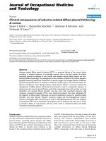
3.3 Morphologic changes and decreased cell density
demonstrated microscopically after T0901317 treatment
As seen in Figure 3A-F, the changes are quite dramatic.
The cells were photographed and viewed at 100× magni-
fication using the Nikon TE 600 series microscope. With
increasing doses of the LXR agonist, the morphologic
changes included decreased cytoplasm with a spindle-
like formation that appears apoptotic at the highest con-
centrations. Additionally, the cell density is concomi-
tantly reduced.
Determination of pro-apoptotic effects with T0901317
treatment
We examined apoptosis in CaOV3 cells by measurement
of caspase -3 and -7 activity via flow cytometric analysis.
Figure 4A shows the percentage of cells in early apopto-
sis, as assessed by Vybrant FAM Caspase 3-and 7 Assay
Kit and 7-Amino-Actinomycin D (7 AAD) staining.
Treatment with T0901317 resulted in a significant (P <
0.05) increase of cells in early apoptosis from 2.2% ± 2 in
vehicle-treated cells to 10.7% ± 5 after a 24 hour treat-
ment (10 μM). At a higher dose of 40 μM, the cells in
early apoptosis significantly (P < 0.00004) increased to
59.5% ± 8. Additionally, caspase 3 and 7 activation was
measured via a luminescent assay (Caspase-Glo). We
found a significant (P < 0.0006) increase in caspase 3 and
7 activity in cells treated for 24 h with the LXR agonist at
a dose of 50 μM. As seen in Figure 4B, in T0901317
treated cells Caspase 3/7 activity was 287% ± 36 (5 μM),
420% ± 27 (10 μM), 580% ± 56 (20 μM), 2,406 ± 242 (40
μM) and 3,158% ± 601 (50 μM) compared to vehicle-
treated cells. We confirmed caspase 3 activation by inves-
tigating the caspase 3-precursor protein level by Western
blot analysis (Figure 4C). We noted a decreased level of
caspase 3-precursor protein after 24 hours of T0901317
treatment. We then examined the effect of T0901317
treatment on apoptotic gene induction, and we observed
a significant (P < 0.05) upregulation in gene expression of
selected pro-apoptotic genes, specifically BAX and cas-
pase-3, at the dose of 30 μM (Figure 5A-C). An induction
of the anti-apoptotic gene, BCL-2, was also demonstrated
at the 30 μM concentration. At the dose of 10 μM, a sig-
nificant (P < 0.05) induction of BAX gene expression was
demonstrated. After 48 hours, the level of BAX protein
expression increased in a dose- dependent manner (Fig-
ure 5D).
Attenuation of LXR-α/β expression by siRNA does not
reverse the anti-proliferative effect of T1317
In order to determine whether the growth inhibitory
effect of T0901317 is mediated by LXR, siRNA experi-
ments in CaOV3 cells were done to decrease expression
of LXRα/β and then assayed cellular proliferation in
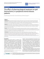
Figure 1 Expression levels of LXRα/β proteins in human ovarian
carcinoma cell lines. Whole-cell lysates of A2780, CaOV3 and SKOV3
cells were obtained and subjected to immunoblotting. Forty micro-
grams of lysate were loaded per lane. LXRα primary antibody was used
in (A) and LXRβ primary antibody was used in (B).
Rough et al. Journal of Ovarian Research 2010, 3:13
/>Page 5 of 10
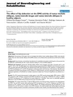
Figure 2 Characterization of antiproliferative effects of T0901317 treatment in ovarian carcinoma cells. A2780, CaOV3 and SKOV3 cells were
cultured and treated with DMSO (? blue) or T0901317 at a concentration of 5 μM ( pink), 10 μM (Њ yellow), 20 μM (X, light blue), 40 μM (X, purple)
or 50 μM (᭹ red) for 24 h, 48 h or 72 h (A-C). Proliferation status was determined by the CyQuant proliferation assay. T0901317 significantly inhibits
cellular proliferation in all cell lines in a dose-dependent and time-dependent manner. Each value is the mean ± SD of three independent experiments,
and the proliferation value is expressed as percentage of vehicle-treated cells (DMSO). (*P < 0.0001 vs. untreated cells). After culturing with vehicle
(DMSO) or with T0901317 for the indicated time-points at a concentration of 10 μM, cells were stained with propidium iodide as detailed in Material
and Methods and examined by flow cytometry to determine cell cycle phase distribution (D). After 24, 48 or 72 hours of treatment, the LXR agonist
T0901317 decreased the percentage of cells in S phase and increased the percentage of cells in the G0/G1 phase, indicating a cell cycle arrest at the
G1-S checkpoint. The percentage of cells in G0/G1 phase increases in a time-dependent manner. Results are the mean of three independent experi-
ments and are expressed as percentage of cells, presented as mean ± SD. *P < 0.001. CaOV3 cells were grown in media supplemented with 10% FBS
for 48 hours in presence of vehicle (DMSO) or the indicated concentrations of T0901317 (5 μM to 40 μM). Whole-cell extract was obtained and 60-90
μg of protein was analyzed for phospho-pRb (E), p21 (F) or p27 (G) protein levels by Western blot analysis.
Rough et al. Journal of Ovarian Research 2010, 3:13
/>Page 6 of 10
response to LXR agonist. As shown in Figure 6A and 6B,
expression of LXRα was inhibited by 70% and of LXRβ by
50%. However, inhibition of LXRα/β did not prevent the
anti-proliferative effect demonstrated after T0901317
treatment (Figure 6C).
Effect of T0901317 Treatment on an FXR-dependent gene,
short heterodimer partner (SHP) in ovarian carcinoma cells
The concentration used for our studies, 10 to 40 μM ago-
nist suggests activation of alternate receptors such as the
farnesoid-X receptor (FXR). The expression of FXR was
evident in HS68, A2780, CaOV3, and SKOV3 cells via
Western Blot analysis (Figure 7A). A 24 hour treatment
with T0901317 of CaOV3 cells resulted in significant (P <
0.05) induction in gene expression of SHP, an FXR-
dependent gene (Figure 7B).
Discussion
Ovarian cancer has an overall poor prognosis especially
in the case of chemoresistance; therefore, the develop-
ment of effective chemotherapeutic agents is of ultimate
importance [16]. Our study demonstrates a possible ther-
apeutic mechanism of T0901317 which possesses anti-
neoplastic properties in ovarian cancer cells with sup-
pression of proliferation and induction of apoptosis. This
is the first study to report these observations in human
ovarian carcinoma cells. However, the antineoplastic
properties of LXR agonists have been demonstrated in
other human carcinomas such as breast and prostate [12-
14]. LXRs are nuclear receptors that first were discovered
to have a regulatory function in control of lipid metabo-
lism. They were shown to have the ability to induce lipid
efflux from atherosclerotic plaques [17]. Subsequently,
LXR's were also demonstrated to have an additional regu-
latory role in immune cell function, specifically modula-
tion of murine macrophage response to inflammatory
stimuli [18].
Interestingly, our study demonstrates that the primary
receptor involved in induction of cell death and cell cycle
arrest is not LXR. T0901317 has been demonstrated to
have agonistic effects on receptors other than LXR, such
as the Pregnane X Receptor (PXR) and the Farnesoid X
Receptor (FXR) [19]. According to a study by Houck, et
al., the principal receptor activated at a dose of 1 μM and
below, primarily activates the Liver X Receptor, whereas
doses above 1 μM primarily activate the farnesoid X
receptor (FXR) [20]. Interestingly, a Phase I pharmacoki-
netic trial and correlative in vitro Phase II tumor kinetic
study of apomine, a FXR agonist, demonstrated inhibi-
tion of tumor growth from patients with ovarian cancer
[21]. A study by Swales, et al. demonstrated the ability of
an FXR agonist, GW4064, to induce apoptosis and inhibit
proliferation in breast cancer cells [22]. Therefore, it is
likely that FXR activation by T0901317 may lead to
induction of apoptosis and cell cycle arrest in ovarian
cancer cells. T0901317 has the ability to induce the gene
Figure 3 Effect of the LXR agonist T0901317 on cellular morphology. CaOV3 cells were cultured and treated with DMSO (1%, A) or T0901317 at
a concentration of 5 μM (B), 10 μM (C), 20 μM (D), 40 μM (E) or 50 μM (F) for a total of 48 hours. Cells were visualized microscopically (10X) and pictures
taken. The pictures clearly demonstrate a significant effect on cellular morphology. At increasing doses of the LXR agonist, the cells appeared to have
a decreased amount of cytoplasm with a concomitant decrease in cell cumber. At the doses of 40 μM and 50 μM, the cells appeared apoptotic with
necrotic debris present in the media.
Rough et al. Journal of Ovarian Research 2010, 3:13
/>Page 7 of 10
Figure 4 Induction of apoptosis with T0901317 treatment. Flow
cytometric analysis of apoptosis was utilized for determination of cas-
pase-3 and -7 activities. CaOV3 cells were treated with either vehicle
(DMSO) or T0901317 at the indicated doses (10 μM to 40 μM) for 24
hours and then stained with Vybrant FAM dye, and 7-AAD strictly fol-
lowing manufacturer's instruction. Data are mean ± SD of three differ-
ent experiments (A). Caspase 3/7 activity was also measured in CaOV3
cells after 12 hours of treatment with vehicle (DMSO) or 5 μM, 10 μM,
20 μM, 40 μM or 50 μM. A luminescent assay was used, as detailed in
Material and Methods. T0901317 significantly increases Caspase 3/7
activation. Results are the mean ± SD of three independent experi-
ments and are expressed as percentage of negative control (DMSO). (*
p < 0.006 vs. negative control, (B). The activation of caspase 3 was con-
firmed by Western Blot analysis. LXR agonist treatment enhances cas-
pase 3 activation, resulting in increased caspase 3 precursor cleavage
rate and decreased caspase 3 precursor protein levels. Decreased cas-
pase-3 precursor protein levels occur in a concentration dependent
manner (C). β-actin expression was determined by Western blot anal-
ysis and used as an endogenous control.
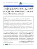
Figure 5 Effect of T0901317 treatment on apoptotic gene and
BAX protein expression. After a 24 hour treatment, cells were har-
vested for isolation of mRNA as detailed in the methods section. A sig-
nificant induction of BAX and caspase gene induction was
demonstrated, especially at the 30 μM dose. Upregulation of the anti-
apoptotic Bcl-2 gene expression was demonstrated with the 30 μM
concentration (A-C), *P < 0.05, **P < 0.001). CaOV3 cells were grown in
media supplemented with 10% FBS for 24 hours in presence of vehicle
(DMSO) or the indicated concentrations of T0901317 (5 μM to 50 μM).
Whole-cell extract was obtained and 60 μg of protein was analyzed for
BAX protein levels by Western blot analysis. β-actin expression was
used as an endogenous control (D).
Rough et al. Journal of Ovarian Research 2010, 3:13
/>Page 8 of 10
expression of short heterodimer protein (SHP), which is
involved in bile acid synthesis regulation, and is reported
to be an FXR-dependent gene [23]. Despite T0901317
being a synthetic LXR agonist, the concentration depen-
dent activation of other receptors must be taken into
account when studying this compound.
We have demonstrated the effect of T0901317 on ovar-
ian cancer cell morphology and on cellular proliferation.
These occur in a time- and dose-dependent manner,
which are similar to findings reported in a study by
Wente, et al., describing inhibition of cell proliferation in
insulinoma cells [15]. Cell cycle analysis indicated that
T0901317 induced G0/G1 cell cycle arrest with a con-
comitant decrease in both the S and G/M2 phases. A
study in human prostate cells demonstrated similar find-
ings with a decrease in the percentage of cells in the S-
phase after treatment [13]. We analyzed the expression of
p21 and p27 which are regulatory proteins involved in
G0/G1 phase arrest, via inhibition of cyclin/CDK com-
plexes that are necessary for cell cycle progression [24].
One such mechanism for cell cycle progression into the
S-phase is phosphorylation of the retinoblastoma (Rb)
protein by cyclin/CDK complexes [25]. Our study dem-
onstrates that upregulation of both p21 and p27 corre-
Figure 6 Effect of LXRα/β inhibition on cell proliferation in T0901317 treated CaOV3 cells. (A) Evaluation of LXRα and (B) LXRβ expression in
siRNA transfected cells by quantitative real time RT-PCR. (C) siRNA transfected CaOV3 cells were cultured and treated with DMSO or 20 μM T0901317
for 24 hours. The cell growth of was assessed by the Cyquant proliferation assay. Each value is the mean ± SD of three independent experiments (* p
< 0.05 vs control siRNA).
Rough et al. Journal of Ovarian Research 2010, 3:13
/>Page 9 of 10
lates with inhibition of phosphorylation of the Rb protein,
therefore causing G0/G1 cell cycle arrest and inhibition
of cellular proliferation.
We analyzed the ability of T0901317 to induce apopto-
sis in ovarian cancer cells. T0901317 has a significant
ability to induce the activity of caspase-3 and -7 leading
to apoptosis in ovarian carcinoma cells. Further evidence
is elucidated by the induction of caspase-3 and BAX gene
expression. Induction of the pro-apoptotic protein, BAX,
was upregulated in a dose-dependent manner. The BAX
protein is a member of the Bcl-2 family, and when over
expressed has the ability to accelerate apoptosis [26].
Conclusion
To our knowledge, this is the first study to report the anti-
proliferative and pro-apoptotic activity of T0901317 on
ovarian cancer cells mediated via an LXR-independent
pathway. We believe that based on our results that syn-
thetic LXR agonists warrant further studies as anti-neo-
plastic agents in the treatment of ovarian cancer.
Conflicts of interests
The authors declare that they have no competing inter-
ests.
Authors' contributions
JR carried out proliferation/apoptosis assays, knockdown experiments along
with drafting of the manuscript. SY carried out Western Blot and flow cytome-
try analysis. MAM assisted in conception of study and aided in the drafting of
the manuscript. JMD coordinated the study and provided funding of the stud-
ies. All authors read and approved the final manuscript.
Acknowledgements
We would like to take the opportunity to acknowledge Dr. Mario Rico for his
assistance in the acquisition and interpretation of data.
This study was funded by an NIH training grant (T32CA103652-04).
Author Details
1
Department of Surgery, Temple University School of Medicine, Philadelphia,
PA, USA and
2
Department of Anatomy and Cell Biology, Temple University
School of Medicine, Philadelphia, PA, USA
References
1. Edwards BK, Howe HL, Ries LA, Thun MJ, Rosenberg HM, Yancik R, Wingo
PA, Jemal A, Feigal EG: Annual report to the nation on the status of
cancer, 1973-1999, featuring implications of age and aging on U.S.
cancer burden. Cancer 2002, 94:2766-92.
2. Jemal A, Siegel T, Ward E, Hao Y, Murray T, Thun MJ: Cancer statistics. CA
Cancer J Clin 2008, 55:10-30.
3. Goff BA, Mandel LS, Melancon CH, Muntz HG: Frequency of symptoms of
ovarian cancer in women presenting to primary care clinics. JAMA
2004, 22:2705-12.
4. Burg ME Van der, van Lent M, Buyse M: The effect of debulking surgery
after induction chemotherapy on the prognosis in advanced epithelial
ovarian cancer: Gynecological Cancer Cooperative Group of the
European Organization for Research and Treatment of Cancer. N Engl J
Med 1995, 10:629-34.
5. Apfel R, Benbrook D, Lernhardt E, Ortiz MA, Salbert G, Pfahl M: A novel
orphan receptor specific for a subset of thyroid hormone-responsive
elements and its interaction with the retinoid/thyroid hormone
receptor subfamily. Mol Cell Biol 1994, 14:7025-35.
6. Lu TT, Repa JJ, Mangelsdorf DJ: Orphan nuclear receptors as eLiXiRs and
FiXeRs of sterol metabolism. J Biol Chem 2001, 276:37735-8.
7. Willy PJ, Umesono K, Ong ES, Evans RM, Heyman RA, Mangelsdorf DJ: LXR,
a nuclear receptor that defines a distinct retinoid response pathway.
Genes Dev 1995, 9:1033-45.
8. Lehmann JM, Kliewer SA, Moore LB, Smith-Oliver TA, Oliver BB, Su JL,
Sundseth SS, Winegar DA, Blanchard DE, Spencer TA, Willson TM:
Activation of the nuclear receptor LXR by oxysterols defines a new
hormone response pathway. J Biol Chem 1997, 272:3137-40.
9. Chawla A, Repa JJ, Evans RM, Mangelsdorf DJ: Nuclear receptors and
lipid physiology: opening the X-files. Science 2001, 294:1866-70.
10. Castrillo A, Tontonoz P: Nuclear receptors in macrophage biology: at the
crossroads of lipid metabolism and inflammation. Annu Rev Cell Dev Biol
2004, 20:455-80.
11. Chuu CP, Chen RY, Hiipakka RA, Kokontis JM, Warner KV, Xiang J, Liao S:
The liver X receptor agonist T0901317 acts as androgen receptor
antagonist in human prostate cancer cells. Biochem Biophys Res
Commun 2007, 57:341-6.
12. Chuu CP, Hiipakka RA, Kokontis JM, Fukuchi J, Chen RY, Liao S: Inhibition
of tumor growth and progression of LNCaP prostate cancer cells in
athymic mice by androgen and liver X receptor agonist. Cancer Res
2006, 66:6482-6.
13. Fukuchi J, Kokontis JM, Hiipakka RA, Chuu CP, Liao S: Antiproliferative
effect of liver X receptor agonists on LNCaP human prostate cancer
cells. Cancer Res 2004, 64:7686-9.
14. Vigushin DM, Dong Y, Inman L, Peyvandi N, Alao JP, Sun C, Ali S, Niesor EJ,
Bentzen CL, Coombes RC: The nuclear oxysterol receptor LXRalpha is
Received: 22 September 2009 Accepted: 26 May 2010
Published: 26 May 2010
This article is available from: 2010 Rough et al; licensee BioMed Central Ltd. This is an Open Access article distributed under the terms of the Creative Commons Attribution License ( which permits unrestricted use, distribution, and reproduction in any medium, provided the original work is properly cited.Journa l of Ovaria n Resear ch 2010, 3:13
Figure 7 Effect of T0901317 Treatment on an FXR-dependent
gene, short heterodimer partner (SHP) in ovarian carcinoma cells.
(A) Whole-cell lysates of HS68, A2780, CaOV3 and SKOV3 cells were ob-
tained and subjected to immunoblotting. Fifty micrograms of lysate
were loaded per lane and the blot was probed with anti-FXR antibody.
(B) CaOV3 cells were treated with T0901317 for 24 hours and SHP gene
mRNA expression was examined by real time RT-PCR. (*P < 0.001)
Rough et al. Journal of Ovarian Research 2010, 3:13
/>Page 10 of 10
expressed in the normal human breast and in breast cancer. Med Oncol
2004, 21:123-31.
15. Wente W, Brenner MB, Zitzer H, Gromada J, Efanov AM: Activation of liver
X receptors and retinoid X receptors induces growth arrest and
apoptosis in insulin-secreting cells. Endocrinology 2007, 148:1843-9.
16. Pfisterer J, Plante M, Vergote I, du Bois A, Hirte H, Lacave AJ: Gemcitabine
plus carboplatin compared with carboplatin in patients with platinum-
sensitive recurrent ovarian cancer: an intergroup trial of the AGO-
OVAR, the NCIC CTG, and the EORTC GCG. J Clin Onco 2006,
29:4699-707.
17. Zelcer N, Tontonoz P: Liver X receptors as integrators of metabolic and
inflammatory signaling. J Clin Invest 2006, 116:607-14.
18. Birrell M, Catley M, Hardaker E, Wong S, Willson T, McCluskie K, Leonard T,
Farrow S, Collins J, Haj-Yahai S, Belvisi M: Novel Role for the liver X nuclear
receptor in suppression of lung inflammatory responses. J Biol Chem
2007, 44:31822-90.
19. Mitro N, Vargas L, Romeo R, Koder A, Saez E: T0901317 is a potent PXR
ligand: Implications for the biology ascribed to LXR. FEBS Letters 2007,
9:1721-1726.
20. Houck KA, Borchert KM, Hepler CD, Thomas JS, Bramlett KS, Michael LF,
Burris TP: T0901317 is a dual LXR/FXR agonist. Molecular Genetics and
Metabolism 2004, 83:184-187.
21. Alberts DS, Hallum AV III, Stratton-Custis M, Garcia DJ, Gleason-Guzman M,
Salmon SE, Santabarbara P, Niesor EJ, Floret S, Bentzen CL: Phase I
Pharmacokinetic Trial and Correlative in Vitro Phase II Tumor Kinetic
Study of Apomine (SR-45023 A), a Novel Oral Biphosphonate
Anticancer Drug. Clinical Cancer Research 2001, 7:1246-1250.
22. Swales KE, Korbonits M, Carpenter R, Walsh DT, Warner TD, Bishop-Bailey
D: The Farsenoid X Receptor Is Expressed in Breast Cancer and
Regulates Apoptosis and Aromatase Expression. Cancer Research 2006,
20:10120-10126.
23. Choonjans K, Auwerx J: A Sharper Image of SHP. Nature 2002, 8:789-791.
24. Sherr CJ, Roberts JM: CDK inhibitors: positive and negative regulators of
G1-phase progression. Genes Dev 1999, 13:1501-12.
25. Connell-Crowley L, Harper JW, Goodrich DW: Cyclin D1/Cdk4 regulates
retinoblastoma protein-mediated cell cycle arrest by site-specific
phosphorylation. Mol Biol Cell 1997, 8:287-301.
26. Gayathri R, Gunadharini DN, Arunkumar A, Senthilkumar K,
Krishnamoorthy G, Banudevi S, Vignesh RC, Arunakaran J: Effects of diallyl
disulfide (DADS) on expression of apoptosis associated proteins in
androgen independent human prostate cancer cells (PC-3). Mol Cell
Biochem 2009, 320:197-203.
doi: 10.1186/1757-2215-3-13
Cite this article as: Rough et al., Anti-proliferative effect of LXR agonist
T0901317 in ovarian carcinoma cells Journal of Ovarian Research 2010, 3:13