Báo cáo hóa học: " Bilateral hemotympanum as a result of spontaneous epistaxis" doc
Bạn đang xem bản rút gọn của tài liệu. Xem và tải ngay bản đầy đủ của tài liệu tại đây (1.21 MB, 3 trang )
CAS E REP O R T Open Access
Bilateral hemotympanum as a result of
spontaneous epistaxis
Vural Fidan
1*
, Kemal Ozcan
2
, Filiz Karaca
3
Abstract
Hemotympanum is a rare condition and usually depends on a secondary reason. Therefore, idiopathic
hemotympanum is rarely seen in the literature. In this paper, we report a case of this problem.
Introduction
Hemotympanum is mo st often associated with basilar
skull fractures or nasal packing. Only six cases asso-
ciated with spontaneous epistaxis have been described
in the literature [1,2]. Because of this rare situation, we
present the case of a 51-year-old woman with bilateral
hemotympanum secondary to spontaneous epistaxis.
Initial evaluation must include an audiogram a nd radi-
ological imaging (computed tomography, magnetic reso-
nance imaging, etc.). Close follow-up of the patient is
necessary for reducing the risk of long-term sequelae
such as cholesterol granuloma [3].
Case report
A 51-year-old woman was referred to the emergency
department with a complaint of epistaxis associated
with exercise. She had been sweeping her house when
she noticed the epistaxis. Her history indicated that
after epistaxis had started, she went to the sink and
cleaned her nose with water. She had pressed on her
nose and called an ambulanc e. About 30 min after the
start of epistaxis, an ambulance and emergency doctor
arrived. The bleeding stopped while she was in the
ambulance. Her blood pressure was 125/80 mmHg. She
had an unremarkable past medical history and did not
have coagulation diathesis or trauma/barotrauma, nor
was she undergoing anticoagulant or salicylate therapy.
She complained of slight hearing loss a nd a f eeling of
fullness in both ears. The physical examination was nor-
mal except for red-blue tympanic membranes and bilat-
eral septal excoriation. There were no other petechiae
or ecchy moses on the s kin or mucous membranes. Her
hematologic, biochemical and coagulation tests were
also normal. Temporal bone fracture was ruled out by
computed tomography scan.
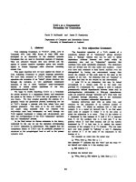
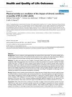
She was referred to the emergency department 2 days
after the problem had started. In our examination, we
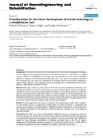
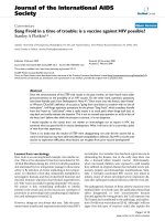
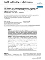
found bilateral blue ear drums (Figures 1 and 2), inac-
tive epistaxis and septal excoriation (Figure 3). An
audiogram demonstrated moderate bilateral conductive
hearin g loss, and the tympanogram findings were type b
(flat type). After consulting an otolaryngologist, we pre-
scribed amoxicillin (2 g/day). Five days after starting the
medication, the patient’s otoscopic findings and tem-
poral MRI were normal at the control visit.
Idiopathic or spontaneous hemotympanum is an
uncommon disorder characterized by a black-blue tym-
panic membrane discoloration as a result of recurrent
hemorrhage in the middle ear or mastoid in the pre-
sence of Eustachian tube obstruction. Initial evaluation
of a b lue middle ear mass includes an audiogram and
computed tomography (CT) scan with intravenous con-
trast. CT may identify congen ital vascular malformation
or bone erosion due to chronic otitis media or tumors.
A magnetic resonance imaging (MRI) scan is useful to
distinguish hemotympanum from a vascular tumor and
to avoiding angiography, which is associated with signifi-
cant morbidity. Evidenc e suggests that secretory otitis
media and spontaneous hemotympanum are different
phases of the same disease process.
Discussion
Epistaxis is common and occurs more commonly in
male than female patients. Epistaxis is noted at higher
incidence in older patients [4]. It is secondary to local
or systemic causes. Nasal trauma (surgical, digital),
* Correspondence:
1
Ear, Nose and Throat Department, District Education and Research Hospital,
25100 Erzurum, Turkey.
Full list of author information is available at the end of the article
Fidan et al . International Journal of Emergency Medicine 2011, 4:3
/>© 2011 Fidan et al. This is an Open Access article distribute d under the terms of the Creative Commons Attribution License (ht tp://
creativecommons.org/licenses/by/2.0), which permits unrestricted use, distribution, and reproduction in any medium, provided the
original work is properly cited.
foreignbodiesinthenasalpassage, topical sprays or
dust, inflammatory nasal diseases, septal deformities,
tumors and vascular a neurysms can be the local factors
[5,6]. Coagulation deficits, Osler-Weber-Rendu disease
and arteriosclerotic vascula r diseases are possible sys-
temic factors [5,6]. Also regular uptake of anticoagulants
can cause spontaneous bilateral hemotympanum [7].
The vascular supply of nasa l mucosa originates from
the external and internal carotid arteries. Kiesselbach’ s
plexus, which is on the anterior part of the septum, is
the site of most epistaxis events [6]; it is also known as
Little’s area and is rich in vascular supply [5].
Especially temporal bone fractures, nasal packing,
anticoagu lant therapy, chronic otitis media and coagula-
tion deficits are the causes of hemotympanum [8-10]. It
is most often associated with temporal traumas rather
than nasal packing [1], but occasionally nasal packing,
which can lead to peritubal lymphatic stasis, is a cause
of hemotympanum [11]. Dysfunction of the Eustachian
tube is th ought to be the reason for spontaneous hemo-
tympanum secondary to epistaxis [1]. In the case pre-
sented here, there was no history of nasal packing, so
retrograde blood reflux to the Eustachian tube could
have been the cause because there was a history of
nasal pressure that could have caused reflux to the
Eustachian tubes.
Computed tomography or magnetic resonance ima-
ging is necessary for making the differential diagnosis
concerning the etiology of epistaxis [12]. In temporal
traumas a fracture line could be visib le on the scan, and
chronic middle ear effusion can also be seen in cases of
chronic otitis media. In patients with a basilar skull frac-
ture, there can also be facial paralysis, tympanic mem-
brane perforation o r otorrhea. In patients with chronic
otitis media, retraction pockets on the tympanic mem-
brane are also visible.
All patients with hemotympanum need close follow-
up. A fl uid-filled middle ear cavity may result in con-
ductive, sensorineural or mixed hearing l oss [13]. Not
thetypeoffluidinthemiddleearbutratherthe
amount of fluid affects the rate of hearing loss [14]. To
prevent persistent effusion, physicians must treat the
patient with antimicrobial drugs [15]. The hearing defi-
cits normalize after the middle ear effusion has been
absorbed. Persistency of fluid may lead to permanent
conductive hearing loss. Myringotomy with tube place-
ment is needed for persistent effusions [16]. All patients
with hemotympanum must be followed up closely to
ensure resolution.
Conclusion
Generally temporal bone fractures, nasal packing, antic-
oagulant therapy, chronic otitis media and coagulation
deficits are the causes of hemotympanum. However,
infrequently epistaxis is the causative factor. In patients
Figure 1 Endoscopic view of right tympanic membrane.
Figure 2 Endoscopic view of left tympanic membrane.
Figure 3 Endoscopic view of septal excoriation.
Fidan et al . International Journal of Emergency Medicine 2011, 4:3
/>Page 2 of 3
with spontaneous hemotympanum secondary to epis-
taxis, emergency doctors need to work with otolaryngol-
ogists for close follow-up. Physicians must remember
that to prevent long-term sequelae of persistent hemo-
tympanum, myringotomy may be required.
Consent
Written informed consent was obtained from the patient
for publication of this case report and accompanying
images.
Author details
1
Ear, Nose and Throat Department, District Education and Research Hospital,
25100 Erzurum, Turkey.
2
Otorhinolaryngology Department, Malatya
Government Hospital, Malatya, Turkey.
3
Otorhinolaryngology Department,
Erzurum Education and Training Hospital, Erzurum, Turkey.
Authors’ contributions
VF intervened the patient in the emergency department. KO and FK were
conceived of the study, and participated in its design and coordination. All
authors read and approved the final manuscript.
Competing interests
The authors declare that they have no competing interests.
Received: 27 July 2010 Accepted: 27 January 2011
Published: 27 January 2011
References
1. Evans TC, Hecker J, Zaiser DK: Hemotympanums secondary to
spontaneous epistaxis. J Emerg Med 1988, 6:387-389.
2. Hurtado TR, Zeger WG: Hemotympanums secondary to spontaneous
epistaxis in a 7-year-old. J Emerg Med 2004, 26:61-63.
3. Plaza G, Alvarez-Linera J, Galindo N: Cholesterol granuloma of the middle
ear: cause of idiopathic hemotympanum. Acta Otorrinolaringol Esp 2000,
51:724-728.
4. Perretta LJ, Denslow BL, Brown CG: Emergency evaluation and
management of epistaxis. Emerg Med Clin North Am 1987, 5:265-277.
5. Santamaria JP, Abrunzo TS: Ear, nose and throat disorders. In Pediatric
emergency medicine: concepts and clinical practice. Edited by: Barkin RM,
Caputo GL, Jaffee DM, Knapp JF, Schafermeyer RW, Seidel JS. Mosby, St.
Louis, MO; 1997:713-716.
6. Padgham N: Epistaxis: anatomical and clinical correlates. J Laryngol Otol
1990, 104:308-311.
7. Balatsouras DG, Dimitropoulos P, Fassolis A, Kloutsos G, Economou NC,
Korres S, Kaberos A: Bilateral spontaneous hemotympanum: case report.
Head Face Med 2006, 4:31.
8. Hough JD, McGee MM: Otologic trauma. In Otolaryngology. 3 edition.
Edited by: Paparella MM, Shumrick DA. WB Saunders, Philadelphia, PA;
1991:1137-1160.
9. Lalwani AK, Jackler RK: Spontaneous hemotympanum associated with
chronic middle ear effusion. Am J Otol 1991, 12:455-458.
10. Pulec JL, DeGuine C: Hemotympanum from trauma. Ear Nose Throat J
2001, 80:486-487.
11. McCurdy JA Jr: Effects of nasal packing on Eustachian tube function. Arch
Otolaryngol 1977, 103:521-523.
12. Pulec JL, DeGuine C: Hemotympanum. Ear Nose Throat J 1996, 75:66-68.
13. Paparella MM, Jung TT, Goycoolea MV: Chronic middle ear effusion. In
Otolaryngology. 3 edition. Edited by: Paparella MM, Shumrick DA. WB
Saunders, Philadelphia, PA; 1991:1335-1336.
14. Bluestone CD, Klein JO: Intratemporal complications and sequelae of
otitis media. In Pediatric otolaryngology. 3 edition. Edited by: Bluestone CD,
Stool SE, Kenna MA. WB Saunders, Philadelphia, PA; 1996:583-635.
15. Healy GB: Antimicrobial therapy of chronic otitis media with effusions. Int
J Pediatr Otorhinolaryngol 1984, 8:13.
16. Parisier SC, McGuirt WF: Injuries of the ear and the temporal bone. In
Pediatric otolaryngology. 3 edition. Edited by: Bluestone CD, Stool SE, Kenna
MA. WB Saunders, Philadelphia, PA; 1996:700.
doi:10.1186/1865-1380-4-3
Cite this article as: Fidan et al.: Bilateral hemotympanum as a result of
spontaneous epistaxis. International Journal of Emergency Medicine 2011
4:3.
Submit your manuscript to a
journal and benefi t from:
7 Convenient online submission
7 Rigorous peer review
7 Immediate publication on acceptance
7 Open access: articles freely available online
7 High visibility within the fi eld
7 Retaining the copyright to your article
Submit your next manuscript at 7 springeropen.com
Fidan et al . International Journal of Emergency Medicine 2011, 4:3
/>Page 3 of 3