Recent Advances in Biomedical Engineering 2011 Part 12 pdf
Bạn đang xem bản rút gọn của tài liệu. Xem và tải ngay bản đầy đủ của tài liệu tại đây (7.69 MB, 40 trang )
Arterial Blood Velocity Measurement by Portable Wireless System for Healthcare Evaluation:
The related effects and signicant reference data 429
It had been reported that women had lower carotid artery distensibility compared with men
(Ylitalo et al., 2000). From the findings of present study, we agreed that women had lower
arterial elasticity using the proposed velocity indices. The difference in the velocities and its
indices were related to smaller body size in women that largely accounted for the gender
differences. However, the difference in velocity indices was also influenced by
concentrations of estrogen in hormone status of women (Krejza et al., 2001).
The gender difference in velocity waveforms in CCA found in this population was not
depended on blood pressure. It was demonstrated that the gender difference in blood
velocity waveforms of CCA are not directly linked to it pressure waveforms (Azhim et al.,
2007b).
Although the finding in the effect of increased wave reflection in arterial system on body
height was consistent, because the relation of body weight and body fat on the artery
stiffness and flow velocities were largely unknown, further investigations are needed. The
Doppler angle of insonation was important because it must be taken into account when
calculating blood flow velocity from the Doppler shift frequency. However, the velocity
indices of were independent of the insonating angle so that the assessments of
hemodynamics were more accurate and reliable.
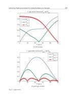
Fig. 11. Comparison of typical flow velocity waveforms in CCA for gender difference of
man (dashed line) and woman (solid line). Subject’s details were 171 cm, 65 kg, BMI: 22
kg/m
2
, age: 23 years for man and 154 cm, 48 kg, BMI: 20 kg/m
2
, age: 25 years for woman.
5. Conclusion
In the chapter, we have presented first, the portable measurement system has developed for
ambulatory and nonivansive determination of blood circulation with synchronized of blood
pressure and ECG signals, which has potential to provide the critical information in clinical
and healthcare applications. Second, there are multiple factors which have effects on blood
velocity waveforms in CCA. Regular exercise training is able to improve age-associated
decrease blood velocity in CCA with similar effect between young and older exercise-
trained. The velocity waveform patterns have no significantly change with age in entire
groups who regularly performed aerobic exercise. Gender-associated difference in the
outcome of velocities and the indices is also found in the study. Reference data for normal
velocities and the indices in CCA are determined after adjustment for the effects of age,
gender, and exercise training. Reductions in blood flow velocities are believed to have
contributed significantly to the pathophysiology of age-associated increase in not only
cardiovascular but also cerebrovascular diseases. The findings have potentially important
clinical and healthcare requirements for prevention of cardiovascular diseases.
6. References
Azhim, A.; Akioka, K.; Akutagawa, M.; Hirao, Y.; Yoshizaki, K.; Obara, S.; Nomura, M.;
Tanaka, H.; Yamaguchi, H. & Kinouchi, Y. (2007c). Effects of aging and exercise
training on the common carotid blood velocities in healthy men. Conf. Proc. IEEE
Eng. Med. Biol. Soc., vol. 1, pp. 989-99
Azhim, A.; Katai, M.; Akutagawa, M.; Hirao, Y.; Yoshizaki, K.; Obara, S.; Nomura, M.;
Tanaka, H.; Yamaguchi, H. & Kinouchi, Y. (2008) Measurement of blood flow
velocity waveforms in the carotid, brachial and femoral arteries during head-up tilt.
Journal of Biomedical & Pharmaceutical Engineering, vol. 2-1, pp. 1-6
Azhim, A.; Akioka, K.; Akutagawa, M.; Hirao, Y.; Yoshizaki, K.; Obara, S.; Nomura, M.;
Tanaka, H.; Yamaguchi, H. & Kinouchi, Y. (2007b). Effect of gender on blood flow
velocities and blood pressure: Role of body weight and height. Conf Proc IEEE Eng
Med Biol Soc., pp. 967-970
Azhim, A.; Katai, M.; Akutagawa, M.; Hirao, Y.; Yoshizaki, K.; Obara, S.; Nomura, M.;
Tanaka, H.; Yamaguchi, H. & Kinouchi, Y. (2007a). Exercise improved age-
associated changes in the carotid blood velocity waveforms. Journal of Biomedical &
Pharmaceutical Engineering, vol. 1-1, pp. 17-26
Azhim, A.; Kinouchi, Y. & Akutagawa, M. (2009). Biomedical Telemetry: Technology and
Applications, In: Telemetry: Research, Technology and Applications, Diana Barculo and
Julia Daniels, (Eds.), Nova Science Publishers, New York, ISBN: 978-1-60692-509-6
(2009)
Baskett, J. J.; XBeasley, J. J.; Murphy, G. J.; Hyams, D. E. & Gosling, R. G. (1977). Screening
for carotid junction disease by spectral analysis of Doppler signals. Cardiovasc Res.,
vol. 11(2), pp. 147-55
Chen, C. & Dicarlo, SE. (1997). Endurance exercise training-induces resting bradycardia.
Sport Med. Training Rehabil., vol. 8, pp. 37-77
Costill, D. (1986). Inside running: basics of sports physiology. Benchmark Press, Indianapolis,
pp. 15
Dahnoun, N.; Thrush, A.J.; Fothergill, J.C. & Evans, D.H. (1990). Portable directional
ultrasonic Doppler blood velocimeter for ambulatory use. Med Biol Eng Comput, vol.
28, pp. 474-482
Darne, B.; Girerd, X.; Safar, M.; Cambien, F. & Guize L. (1989) Pulsatile versus steady
component of blood pressure: a cross-sectional analysis on cardiovascular
mortality. Hypertension, vol. 13, pp. 392-400
Donofrio MT, Bremer YA, Schieken RM, Gennings C, Morton LD, Eidem BW, Cetta F,
Falkensammer CB, Huhta JC and Kleinman CS. Autoregulation of cerebral blood
Recent Advances in Biomedical Engineering430
flow in fetuses with congenital heart disease: The brain sparing effect. Pediatr
Cardiol 2003; 24: 436-443
Fujishiro, K. & Yoshimura, S. (1982). Haemodynamic change in carotid blood flow with age.
J. Jekeikai Med, vol. 29, pp. 125-138
Goldsmith, R.L.; Bloomfeld, D.M. & Rosenwinkel, E.T. (2000). Exercise and autonomic
function. Coron. Artery Dis., vol. 11, pp. 129-135
Gosling, R.G. (1977). Extraction of physiological information from spectrum-analysed
Doppler-shifted continuous wave ultrasound signals obtained non-invasively from
the arterial system. In: Institute of Electrical Engineers medical electronics monographs,
Hill D.W. & Watson B.W., (Eds), pp. 73-125, Peter Peregrinus, Stevenage
Gregova, D.; Termerova, J.; Korsa, J.; Benedikt, P.; Peisker, T.; Prochazka, B.; & Kalvach, P.
(2004) Age dependence of flow velocities in the carotid arteries. Ceska a Slovenska
Neurologie a Neurochirurgie, vol. 67 (6), pp. 409-414, 2004 (abstract in English)
He, J.; Kinouchi, Y.; Iritani, T.; Yamaguchi, H. & Miyamoto, H. (1992). Telemetering blood
flow velocity and ECG during exercise. Innov Tech Biol Med., vol. 13, pp. 567-577
He, J.; Pan, A. W.; Ozaki, T.; Kinouchi, Y. & Yamaguchi, H. (1996). Three channels telemetry
system: ECG, blood velocities of the carotid and the brachial arteries. Biomedical
Engineering Applications Basis Communications, vol. 8, pp. 364-369
Jiang, Z-L.; He, J.; Yamaguchi, H.; Tanaka, H. & Miyamoto, H. (1994). Blood flow velocity in
common carotid artery in humans during breath-holding and face immersion. Aviat
Space Environ Med., vol. 65, pp. 936-943
Jiang, Z-L.; Yamaguchi, H.; Takahashi, A.; Tanabe, S.; Utsuyama, N.; Ikehara, T.; Hosokawa,
K.; Tanaka, H.; Kinouchi, Y. & Miyamoto, H. (1995). Blood flow velocity in the
common carotid artery in humans during graded exercise on a treadmill. Eur J Appl
Physiol, vol. 70, no. 3, pp. 234-239
Johannes, S.; Michael, S.; Thomas, W.; Wolfgang, R.N.; Markus, V.; Markus, L. & Stefan F.
(2001). Quantification of blood flow in the carotid arteries comparison of Doppler
ultrasound and three different phase-contrast magnetic resonance imaging
sequences. Investigate Radiology, vol. 36-11, pp. 642-647
Kaneko, Z.; Shiraishi, J.; Inaoka, H.; Furukawa, T. & Sekiyama, M. (1978). Intra- and
extracerebral hemodynamics of migrainous headache. In: Current concepts in
migraine research, Greene, R. (Ed.), pp. 17-24, Raven, New York
Kannel, W. B. & Stokes III, J. (1985). Hypertension as a cardiovascular risk factor. In:
Handbook of Hypertension. Clinical Aspects of Hypertension, Robertson, J.I.S. (Ed.), pp.
15-34, Elsevier Science Publishing, New York
Krejza, J.; Mariak, Z.; Huba, M.; Wolczynski, S. & Lewko, J. (2001). Effect of endogenous
estrogen on blood flow through carotid arteries. Stroke, vol. 32, pp. 30-36
Lakatta, E.G. (2002). Age-associated cardiovascular changes in health: Impact on
cardiovascular disease in older persons. Heart Fail Rev, vol. 1, pp. 29-49
Latham, R. D.; Westerhof, N.; Sipkema, P.; Rubal, B. J.; Reuderink, P. & Murgo, J. P. (1985).
Regional wave travel and reflections along the human aorta: A study with six
simultaneous micromanometric pressures. Circulation, vol. 72, pp. 1257-1269
London, G.M.; Guerin, A.P.; Pannier, B.; Marchais, S.J. & Stimpel, M. (1995). Influence of sex
on arterial hemodynamics and blood pressure: Role of body height.
Hypertension,
vol. 26, pp. 514-519
Maciel, B.C.; Gallo, L.; Marin-Neto, JA; Lima-Filho, E.C. & Mancoy, J.C. Parasympathetic
contribution to bradycardia induced by endurance training in man. Cardiovasc Res
1985; 19: 642-648
Marchais, S.J.; Guerin, A.P.; Pannier, B.M.; Levy, B.I.; Safar, M.E. & London, G.M. (1993).
Wave reflections and cardiac hypertrophy in chronic uremia: Influence of body
size. Hypertension, vol. 22, pp. 876-883
Mitchell, G. F.; Parise, H.; Benjamin, E. J.; Larson, M. G.; Keyes, M. J.; Vita, J. A.; Vasan, R. S.
& Levy, D. (2004). Changes in arterial stiffness and wave reflection with advancing
age in healthy men and women: The Framingham Heart Study. Hypertension, vol.
43, pp.1239-1245
Murgo, J.; Westerhof, N.; Giolma, J. P. & Altobelli, S. (1980). Aortic impedance in normal
man: relationship to pressure waveforms. Circulation, vol. 62, pp. 105-16
Nagatomo, I.; Nomaguchi M. & Matsumoto K. (1992). Blood flow velocity waveform in the
common carotid artery and its analysis in elderly subjects. Clin Auton Res., vol. 2(3),
pp. 197-200
Nichols, W. W. & O'Rourke, M. F. (2005) McDonald's Blood Flow in Arteries: Theoretic,
Experimental and Clinical Principles. Hodder Arnold, ISBN 0-340-80941-8, London
Permal JM. Neonatal cerebral blood flow velocity measurement. Clin Perinatol 1985; vol. 12,
pp. 179-193
Planiol T and Pourcelot L. (1973). Doppler effects study of the carotid circulation, In:
Ultrasonics in medicine, Vlieger, M.; White, D.N. & McCready, V.R. (Eds), pp. 141-
147, Elsevier, New York
Pourcelot L. (1976). Diagnostic ultrasound for cerebral vascular diseases, In: Present and
future of diagnostic ultrasound, Donald, I. & Levi, S., (Eds), pp. 141-147, Kooyker,
Rotterdam
Prichard, D. R.; Martin, T. R. & Sherriff, S. B. (1979). Assessment of directional Doppler
ultrasound techniques in the diagnosis of carotid artery diseases. Journal of
Neurology, Neurosurgery, and Psychiatry, vol. 42, pp. 563-568
Rutherford, R.B; Hiatt, W.R. & Kreuter, E.W. (1977). The use of velocity wave form analysis
in the diagnosis of carotid artery occlusive. Surgery, vol. 82-5, pp. 695-702
Satomura S. (1959). Study of the flow pattern in peripheral arteries by ultrasonics. J. Acoust
Soc Jpn, vol. 15, pp. 151-158
Scheel, P.; Ruge, C. & Schoning, M. (2000). Flow velocity and flow volume measurements in
the extracranial carotid and vertebral arteries in healthy adults: Reference data of
age. Ultrasound Med Biol., vol. 26, pp. 1261-1266
Schmidt-Trucksass, A.; Grathwohl, D.; Schmid, A.; Boragk, R.; Upmeier, C.; Keul, J. &
Huonker M. (1999). Structural, functional, and hemodynamic changes of the
common carotid artery with age in male subjects. Arterioscler Thromb Vasc Biol., vol.
19, pp. 1091-1097
Tanaka, H.; Dinenno, F. A.; Monahan, K. D.; Christopher, M. C.; Christopher, A. D. & Seals,
D.R. (2000). Aging, habitual exercise, and dynamic arterial compliance. Circulation,
vol. 102, pp. 1270-1275
Ylitalo, A.; Airaksinen, K.E.; Hautanen, A. M.; Kupari, A.; Carson, M.; Virolainen, J.;
Savolainen, M.; Kauma, H.; Kesaniemi, Y.A.; White, P.C. & Huikuri, H.V. (2000).
Baroreflex sensitivity and variants of the renin angiotensin system genes. J. Am.
Coll. Cardiol., vol. 35, pp. 194-200
Arterial Blood Velocity Measurement by Portable Wireless System for Healthcare Evaluation:
The related effects and signicant reference data 431
flow in fetuses with congenital heart disease: The brain sparing effect. Pediatr
Cardiol 2003; 24: 436-443
Fujishiro, K. & Yoshimura, S. (1982). Haemodynamic change in carotid blood flow with age.
J. Jekeikai Med, vol. 29, pp. 125-138
Goldsmith, R.L.; Bloomfeld, D.M. & Rosenwinkel, E.T. (2000). Exercise and autonomic
function. Coron. Artery Dis., vol. 11, pp. 129-135
Gosling, R.G. (1977). Extraction of physiological information from spectrum-analysed
Doppler-shifted continuous wave ultrasound signals obtained non-invasively from
the arterial system. In: Institute of Electrical Engineers medical electronics monographs,
Hill D.W. & Watson B.W., (Eds), pp. 73-125, Peter Peregrinus, Stevenage
Gregova, D.; Termerova, J.; Korsa, J.; Benedikt, P.; Peisker, T.; Prochazka, B.; & Kalvach, P.
(2004) Age dependence of flow velocities in the carotid arteries. Ceska a Slovenska
Neurologie a Neurochirurgie, vol. 67 (6), pp. 409-414, 2004 (abstract in English)
He, J.; Kinouchi, Y.; Iritani, T.; Yamaguchi, H. & Miyamoto, H. (1992). Telemetering blood
flow velocity and ECG during exercise. Innov Tech Biol Med., vol. 13, pp. 567-577
He, J.; Pan, A. W.; Ozaki, T.; Kinouchi, Y. & Yamaguchi, H. (1996). Three channels telemetry
system: ECG, blood velocities of the carotid and the brachial arteries. Biomedical
Engineering Applications Basis Communications, vol. 8, pp. 364-369
Jiang, Z-L.; He, J.; Yamaguchi, H.; Tanaka, H. & Miyamoto, H. (1994). Blood flow velocity in
common carotid artery in humans during breath-holding and face immersion. Aviat
Space Environ Med., vol. 65, pp. 936-943
Jiang, Z-L.; Yamaguchi, H.; Takahashi, A.; Tanabe, S.; Utsuyama, N.; Ikehara, T.; Hosokawa,
K.; Tanaka, H.; Kinouchi, Y. & Miyamoto, H. (1995). Blood flow velocity in the
common carotid artery in humans during graded exercise on a treadmill. Eur J Appl
Physiol, vol. 70, no. 3, pp. 234-239
Johannes, S.; Michael, S.; Thomas, W.; Wolfgang, R.N.; Markus, V.; Markus, L. & Stefan F.
(2001). Quantification of blood flow in the carotid arteries comparison of Doppler
ultrasound and three different phase-contrast magnetic resonance imaging
sequences. Investigate Radiology, vol. 36-11, pp. 642-647
Kaneko, Z.; Shiraishi, J.; Inaoka, H.; Furukawa, T. & Sekiyama, M. (1978). Intra- and
extracerebral hemodynamics of migrainous headache. In: Current concepts in
migraine research, Greene, R. (Ed.), pp. 17-24, Raven, New York
Kannel, W. B. & Stokes III, J. (1985). Hypertension as a cardiovascular risk factor. In:
Handbook of Hypertension. Clinical Aspects of Hypertension, Robertson, J.I.S. (Ed.), pp.
15-34, Elsevier Science Publishing, New York
Krejza, J.; Mariak, Z.; Huba, M.; Wolczynski, S. & Lewko, J. (2001). Effect of endogenous
estrogen on blood flow through carotid arteries. Stroke, vol. 32, pp. 30-36
Lakatta, E.G. (2002). Age-associated cardiovascular changes in health: Impact on
cardiovascular disease in older persons. Heart Fail Rev, vol. 1, pp. 29-49
Latham, R. D.; Westerhof, N.; Sipkema, P.; Rubal, B. J.; Reuderink, P. & Murgo, J. P. (1985).
Regional wave travel and reflections along the human aorta: A study with six
simultaneous micromanometric pressures. Circulation, vol. 72, pp. 1257-1269
London, G.M.; Guerin, A.P.; Pannier, B.; Marchais, S.J. & Stimpel, M. (1995). Influence of sex
on arterial hemodynamics and blood pressure: Role of body height.
Hypertension,
vol. 26, pp. 514-519
Maciel, B.C.; Gallo, L.; Marin-Neto, JA; Lima-Filho, E.C. & Mancoy, J.C. Parasympathetic
contribution to bradycardia induced by endurance training in man. Cardiovasc Res
1985; 19: 642-648
Marchais, S.J.; Guerin, A.P.; Pannier, B.M.; Levy, B.I.; Safar, M.E. & London, G.M. (1993).
Wave reflections and cardiac hypertrophy in chronic uremia: Influence of body
size. Hypertension, vol. 22, pp. 876-883
Mitchell, G. F.; Parise, H.; Benjamin, E. J.; Larson, M. G.; Keyes, M. J.; Vita, J. A.; Vasan, R. S.
& Levy, D. (2004). Changes in arterial stiffness and wave reflection with advancing
age in healthy men and women: The Framingham Heart Study. Hypertension, vol.
43, pp.1239-1245
Murgo, J.; Westerhof, N.; Giolma, J. P. & Altobelli, S. (1980). Aortic impedance in normal
man: relationship to pressure waveforms. Circulation, vol. 62, pp. 105-16
Nagatomo, I.; Nomaguchi M. & Matsumoto K. (1992). Blood flow velocity waveform in the
common carotid artery and its analysis in elderly subjects. Clin Auton Res., vol. 2(3),
pp. 197-200
Nichols, W. W. & O'Rourke, M. F. (2005) McDonald's Blood Flow in Arteries: Theoretic,
Experimental and Clinical Principles. Hodder Arnold, ISBN 0-340-80941-8, London
Permal JM. Neonatal cerebral blood flow velocity measurement. Clin Perinatol 1985; vol. 12,
pp. 179-193
Planiol T and Pourcelot L. (1973). Doppler effects study of the carotid circulation, In:
Ultrasonics in medicine, Vlieger, M.; White, D.N. & McCready, V.R. (Eds), pp. 141-
147, Elsevier, New York
Pourcelot L. (1976). Diagnostic ultrasound for cerebral vascular diseases, In: Present and
future of diagnostic ultrasound, Donald, I. & Levi, S., (Eds), pp. 141-147, Kooyker,
Rotterdam
Prichard, D. R.; Martin, T. R. & Sherriff, S. B. (1979). Assessment of directional Doppler
ultrasound techniques in the diagnosis of carotid artery diseases. Journal of
Neurology, Neurosurgery, and Psychiatry, vol. 42, pp. 563-568
Rutherford, R.B; Hiatt, W.R. & Kreuter, E.W. (1977). The use of velocity wave form analysis
in the diagnosis of carotid artery occlusive. Surgery, vol. 82-5, pp. 695-702
Satomura S. (1959). Study of the flow pattern in peripheral arteries by ultrasonics. J. Acoust
Soc Jpn, vol. 15, pp. 151-158
Scheel, P.; Ruge, C. & Schoning, M. (2000). Flow velocity and flow volume measurements in
the extracranial carotid and vertebral arteries in healthy adults: Reference data of
age. Ultrasound Med Biol., vol. 26, pp. 1261-1266
Schmidt-Trucksass, A.; Grathwohl, D.; Schmid, A.; Boragk, R.; Upmeier, C.; Keul, J. &
Huonker M. (1999). Structural, functional, and hemodynamic changes of the
common carotid artery with age in male subjects. Arterioscler Thromb Vasc Biol., vol.
19, pp. 1091-1097
Tanaka, H.; Dinenno, F. A.; Monahan, K. D.; Christopher, M. C.; Christopher, A. D. & Seals,
D.R. (2000). Aging, habitual exercise, and dynamic arterial compliance. Circulation,
vol. 102, pp. 1270-1275
Ylitalo, A.; Airaksinen, K.E.; Hautanen, A. M.; Kupari, A.; Carson, M.; Virolainen, J.;
Savolainen, M.; Kauma, H.; Kesaniemi, Y.A.; White, P.C. & Huikuri, H.V. (2000).
Baroreflex sensitivity and variants of the renin angiotensin system genes. J. Am.
Coll. Cardiol., vol. 35, pp. 194-200
Recent Advances in Biomedical Engineering432
Yuhi, F. (1987). Diagnostic characteristics of intracranial lesions with ultrasonic Doppler
sonography on the common carotid artery. Med J Kagoshima Univ., vol. 39, pp. 183-
225 (abstract in English)
Zhang, D.; Hirao, Y.; Kinouchi, Y.; Yamaguchi, H. & Yoshizaki, K. (2002). Effects of
nonuniform acoustic fields in vessels and blood velocity profiles on Doppler power
spectrum and mean blood velocity. IEICE Transactions on Information and Systems,
vol. E85-D, pp. 1443-1451
Studying Ion Channel Dysfunction and Arrythmogenesis in the Human Atrium:
A Computational Approach 433
Studying Ion Channel Dysfunction and Arrythmogenesis in the Human
Atrium: A Computational Approach
Sanjay R. Kharche, Phillip R. Law, and Henggui Zhang
X
Studying Ion Channel Dysfunction and
Arrythmogenesis in the Human Atrium:
A Computational Approach
Sanjay R. Kharche, Phillip R. Law, and Henggui Zhang
The University of Manchester, Manchester, UK
1. Introduction
Human atrial fibrillation (AF) is the most common sustained clinically observed cardiac
arrhythmia causing mortality and morbidity in patients with increasing incidence in the
elderly (Aronow 2009; Wetzel, Hindricks et al. 2009). It is prevalent in the developed world
and a considerable burden on health care services in the UK and elsewhere (Stewart,
Murphy et al. 2004; Aronow 2008a; Aronow 2008b). AF is a heterogeneously occurring
disease often in complex with embolic stroke, thromboembolism, heart failure and other
conditions (Novo, Mansueto et al. 2008; Bourke and Boyle 2009; Roy, Talajic et al. 2009). The
treatment of paroxysmal AF includes pharmacological intervention primarily targeting
cellular ion channel function (Ehrlich and Nattel 2009; Viswanathan and Page 2009).
Persistent AF where episodes last for prolonged periods possibly requires electrical
cardioversion (Wijffels and Crijns 2003; Conway, Musco et al. 2009) or repeated surgical
interventions that isolate focal trigger sites that induce AF (Gaita, Riccardi et al. 2002;
Saltman and Gillinov 2009; Stabile, Bertaglia et al. 2009). A better understanding of the
underlying ion channel and structural mechanisms of AF will assist in design of improved
clinical therapy at all stages of the disease.
The structure of the human atrium is shown in Fig. 1. Mechanisms underlying the genesis of
AF are poorly understood yet. It is believed to be predominantly initiated by focal ectopic
activity in the cristae terminalis of the right atrium, and pulmonary vein ostia in the left
atrium (Haissaguerre, Jais et al. 1998). Spontaneous focal activities in the atrium could also
be generated by intracellular calcium ([Ca
2+
]
i
) dysfunction (Chou and Chen 2009). The
ectopic activity, under AF conditions, normally leads to a persistent single mother rotor of
re-entrant excitation circuits. Upon interaction with anatomical obstacles along with intra-
atrial electrical heterogeneity, the mother rotor wavefront breaks giving rise to smaller
randomly propagating electrical wavefronts resulting in rapid erratic excitation of the atria
(Moe, Rheinboldt et al. 1964) leading to uncoordinated contractions of the myocardium,
which is reflected in the abnormal P-wave and R-R intervals of clinical ECG (Rosso and
Kistler 2009). Recently a new mechanism, “AF begets AF“ (Wijffels, Kirchhof et al. 1995) due
to AF induced electrical remodelling (AFER), has been identified by which rapid excitation of
atrial tissue gives rise to persistent AF AFER produces remarkable reduction in atrial action
potential (AP) duration (APD) and effective refractive period (ERP), which are associated with
23
Recent Advances in Biomedical Engineering434
AF-induced changes in electrophysiology of ion channels. Several experimental studies have
studied the effects of AFER on individual ion channels of human atrial myocytes (Bosch,
Zeng et al. 1999; Workman, Kane et al. 2001; Bosch and Nattel 2002; Balana, Dobrev et al.
2003; Ravens and Cerbai 2008), and have identified several ion channels remodelled by
chronic AF (Bosch, Zeng et al. 1999; Workman, Kane et al. 2001) .
Another mechanism underlying the genesis of AF is ion channel dysfunction arising from
genetic mutations. There is growing interest in identifying genetic bases underlying familial
AF following the first study by Chen et al. (Chen, Xu et al. 2003). In the rare but debilitating
cases of familial AF, or lone AF, there is no apparent structural remodelling that precludes
the onset of AF. However, several clinical studies have characterised the familial nature of
several genetic defects that lead to AF (Chen, Xu et al. 2003; Xia, Jin et al. 2005; Makiyama,
Akao et al. 2008; Restier, Cheng et al. 2008; Zhang, Yin et al. 2008; Li, Huang et al. 2009;
Yang, Li et al. 2009). Hormonal imbalance during AF also causes electrical remodelling (Cai,
Gong et al. 2007; Cai, Shan et al. 2009) that facilitates AF, but is not considered in this
Chapter.
Fig. 1. 3D anatomical model of the human female atria showing internal structure and
conduction pathways (figure adapted from our previous study (Zhang, Garratt et al. 2009)).
Atrial tissue in the left (LA) and right (RA) atria is homogeneous (translucent blue). The
sino-atrial node (SAN) is the pacemaker wherefrom cardiac electrical excitations originate.
The main atrial conduction pathways, i.e. pectinate muscles (PM), cristae terminalis (CT)
and the Bachman’s bundles (BB), are the tissue types which possess electrical and structural
heterogeneity and contribute to a small proportion of total atrial mass.
Experimental and clinical electrophysiological studies are vital to improve our
understanding of AF and its underlying mechanisms. Such studies, however, require vast
resources and involve ethical considerations. In addition, the effects of cellular level
electrophysiological remodelling at multi-scale levels of cellular and spatially extended
tissues is practically impossible in a clinical or physiology laboratory environment. Recently
powerful biophysically detailed mathematical models of cardiac cells (Courtemanche,
Ramirez et al. 1998; Nygren, Fiset et al. 1998; Zhang, Holden et al. 2000; Pandit, Clark et al.
2001; ten Tusscher, Noble et al. 2004) and spatially extended tissues have been developed.
Such biophysically detailed models of cardiac cells and tissues offer cost effective
alternatives to experimental studies to investigate and dissect the effects changes in
individual ion channels on cellular AP (Zhang, Garratt et al. 2005; Zhang, Zhao et al. 2007;
Salle, Kharche et al. 2008) and tissue conduction properties (Kharche, Garratt et al. 2008;
Kharche and Zhang 2008; Keldermann, ten Tusscher et al. 2009). With the ready availability
of vast computational power, simulation offers an excellent complimentary method of
studying AF in silico (Kharche, Seemann et al. 2008; Reumann, Fitch et al. 2008; Bordas,
Carpentieri et al. 2009).
In this Chapter, we present a review of some of our recent works on studies of AFER and
gene mutations in genesis and maintenance of AF. Comprehensive computational
techniques for the quantification of the effects of AFER at cellular and tissue levels are
described. Our simulation data at a multi-scale tissue level supported the “AF begets AF”
hypothesis (Zhang, Garratt et al. 2005; Kharche, Seemann et al. 2007; Kharche, Seemann et
al. 2008; Kharche and Zhang 2008), and demonstrated the dramatic pro-fibrillatory effects of
Kir2.1 V93I gene mutation on the human atrium computational study (Kharche, Garratt et
al. 2008). Techniques of high performance computing and visualisation of the
computationally intensive 3D simulations are discussed.
2. Multi-scale simulation of the effects of AFER and lone AF
In our studies of human atrial AF, we choose the widely used biophysically detailed cell
model for human atrial AP developed by Courtemanche et al. (Courtemanche, Ramirez et al.
1998) (CRN). This 21 variable electrophysiological model consists of several sarcolemmal ion
channel currents, pumps and exchanger currents, along with a sufficiently detailed
intracellular ionic homeostasis mechanism. The model is able to reproduce human atrial AP
accurately. Electrophysiological changes due to AFER and Kir2.1 V93I gene mutation can be
immediately incorporated into this model allowing ready simulation of the resulting AP and
[Ca
2+
]
i
transients. Further, as described later in this section, the cellular models can be
incorporated into multi-cellular tissue models using reaction diffusion formulations to
simulate conduction propagation behaviour. To quantify the effects of AFER and Kir2.1
V93I gene mutation, a series of experimental protocols are computationally emulated
quantifying their effects on atrial excitation at cellular and 3D anatomically detailed models.
2.1 Single cell modelling: electrophysiological changes due to AFER and monogenic AF
AFER and Kir2.1 V93I mutation both alter the biophysical properties of sarcolemmal ion
channels underlying human atrial AP. Changes in ion channel current densities, time
kinetics and steady state properties of ion channels have been quantified by experimental
and clinical studies. The experimental data regarding AFER was obtained from two
extensive studies wherein the effects of chronic human AF on atrial ion channels properties
were studied. The study by Bosch et al. (Bosch, Zeng et al. 1999) considered patients with AF
episodes lasting for more then 1 month (AF1), while the study by Workman et al.
(Workman, Kane et al. 2001) considers patients with AF episodes lasting for more than 6
months (Workman, Kane et al. 2001) (AF2). In brief, remodelling in AF1 includes a 235%
increase of the maximal conductance of the inward rectifier potassium current I
K1
, 74%
Studying Ion Channel Dysfunction and Arrythmogenesis in the Human Atrium:
A Computational Approach 435
AF-induced changes in electrophysiology of ion channels. Several experimental studies have
studied the effects of AFER on individual ion channels of human atrial myocytes (Bosch,
Zeng et al. 1999; Workman, Kane et al. 2001; Bosch and Nattel 2002; Balana, Dobrev et al.
2003; Ravens and Cerbai 2008), and have identified several ion channels remodelled by
chronic AF (Bosch, Zeng et al. 1999; Workman, Kane et al. 2001) .
Another mechanism underlying the genesis of AF is ion channel dysfunction arising from
genetic mutations. There is growing interest in identifying genetic bases underlying familial
AF following the first study by Chen et al. (Chen, Xu et al. 2003). In the rare but debilitating
cases of familial AF, or lone AF, there is no apparent structural remodelling that precludes
the onset of AF. However, several clinical studies have characterised the familial nature of
several genetic defects that lead to AF (Chen, Xu et al. 2003; Xia, Jin et al. 2005; Makiyama,
Akao et al. 2008; Restier, Cheng et al. 2008; Zhang, Yin et al. 2008; Li, Huang et al. 2009;
Yang, Li et al. 2009). Hormonal imbalance during AF also causes electrical remodelling (Cai,
Gong et al. 2007; Cai, Shan et al. 2009) that facilitates AF, but is not considered in this
Chapter.
Fig. 1. 3D anatomical model of the human female atria showing internal structure and
conduction pathways (figure adapted from our previous study (Zhang, Garratt et al. 2009)).
Atrial tissue in the left (LA) and right (RA) atria is homogeneous (translucent blue). The
sino-atrial node (SAN) is the pacemaker wherefrom cardiac electrical excitations originate.
The main atrial conduction pathways, i.e. pectinate muscles (PM), cristae terminalis (CT)
and the Bachman’s bundles (BB), are the tissue types which possess electrical and structural
heterogeneity and contribute to a small proportion of total atrial mass.
Experimental and clinical electrophysiological studies are vital to improve our
understanding of AF and its underlying mechanisms. Such studies, however, require vast
resources and involve ethical considerations. In addition, the effects of cellular level
electrophysiological remodelling at multi-scale levels of cellular and spatially extended
tissues is practically impossible in a clinical or physiology laboratory environment. Recently
powerful biophysically detailed mathematical models of cardiac cells (Courtemanche,
Ramirez et al. 1998; Nygren, Fiset et al. 1998; Zhang, Holden et al. 2000; Pandit, Clark et al.
2001; ten Tusscher, Noble et al. 2004) and spatially extended tissues have been developed.
Such biophysically detailed models of cardiac cells and tissues offer cost effective
alternatives to experimental studies to investigate and dissect the effects changes in
individual ion channels on cellular AP (Zhang, Garratt et al. 2005; Zhang, Zhao et al. 2007;
Salle, Kharche et al. 2008) and tissue conduction properties (Kharche, Garratt et al. 2008;
Kharche and Zhang 2008; Keldermann, ten Tusscher et al. 2009). With the ready availability
of vast computational power, simulation offers an excellent complimentary method of
studying AF in silico (Kharche, Seemann et al. 2008; Reumann, Fitch et al. 2008; Bordas,
Carpentieri et al. 2009).
In this Chapter, we present a review of some of our recent works on studies of AFER and
gene mutations in genesis and maintenance of AF. Comprehensive computational
techniques for the quantification of the effects of AFER at cellular and tissue levels are
described. Our simulation data at a multi-scale tissue level supported the “AF begets AF”
hypothesis (Zhang, Garratt et al. 2005; Kharche, Seemann et al. 2007; Kharche, Seemann et
al. 2008; Kharche and Zhang 2008), and demonstrated the dramatic pro-fibrillatory effects of
Kir2.1 V93I gene mutation on the human atrium computational study (Kharche, Garratt et
al. 2008). Techniques of high performance computing and visualisation of the
computationally intensive 3D simulations are discussed.
2. Multi-scale simulation of the effects of AFER and lone AF
In our studies of human atrial AF, we choose the widely used biophysically detailed cell
model for human atrial AP developed by Courtemanche et al. (Courtemanche, Ramirez et al.
1998) (CRN). This 21 variable electrophysiological model consists of several sarcolemmal ion
channel currents, pumps and exchanger currents, along with a sufficiently detailed
intracellular ionic homeostasis mechanism. The model is able to reproduce human atrial AP
accurately. Electrophysiological changes due to AFER and Kir2.1 V93I gene mutation can be
immediately incorporated into this model allowing ready simulation of the resulting AP and
[Ca
2+
]
i
transients. Further, as described later in this section, the cellular models can be
incorporated into multi-cellular tissue models using reaction diffusion formulations to
simulate conduction propagation behaviour. To quantify the effects of AFER and Kir2.1
V93I gene mutation, a series of experimental protocols are computationally emulated
quantifying their effects on atrial excitation at cellular and 3D anatomically detailed models.
2.1 Single cell modelling: electrophysiological changes due to AFER and monogenic AF
AFER and Kir2.1 V93I mutation both alter the biophysical properties of sarcolemmal ion
channels underlying human atrial AP. Changes in ion channel current densities, time
kinetics and steady state properties of ion channels have been quantified by experimental
and clinical studies. The experimental data regarding AFER was obtained from two
extensive studies wherein the effects of chronic human AF on atrial ion channels properties
were studied. The study by Bosch et al. (Bosch, Zeng et al. 1999) considered patients with AF
episodes lasting for more then 1 month (AF1), while the study by Workman et al.
(Workman, Kane et al. 2001) considers patients with AF episodes lasting for more than 6
months (Workman, Kane et al. 2001) (AF2). In brief, remodelling in AF1 includes a 235%
increase of the maximal conductance of the inward rectifier potassium current I
K1
, 74%
Recent Advances in Biomedical Engineering436
reduction of the conductance of the L-type calcium current I
Ca,L
, 85% reduction of
conductance of the transient outward current (I
to
), a shift of -16 mV of the I
to
steady-state
activation, and a -1.6 mV shift of sodium current (I
Na
) steady state activation. Fast
inactivation kinetics of I
Ca,L
is slowed down, and was implemented as a 62% increase of the
voltage dependent inactivation time constant. Remodelling in AF2 includes a 90% increase
of I
K1
, 64% reduction of I
Ca,L
, 65% reduction of I
to
, 12% increase of the sustained outward
potassium current (I
Ksus
), and a 12% reduction of the sodium potassium pump (I
Na,K
). Both
AF1 and AF2 data have been incorporated into the CRN model in our previous study
(Zhang, Garratt et al. 2005).
Simulation of Kir2.1 V93I gene mutation was based on the recent clinical data from Xia et al.
(Xia, Jin et al. 2005) who examined several generations of a large family with hereditary AF
associated with Kir2.1 V93I gene mutation. The Kir2.1 gene primarily regulates the I
K1
channel current, which is modelled as
KKK
EVgI
11
(1)
cVb
K
KK
e
ga
agg
1
1
max1
max11
(2)
where V is the cell membrane potential; E
K
the reversal potential of the channel; g
K1max
the
maximal channel conductance; “a” is the fraction of the channel conductance that is voltage-
independent, (1-a) is the fraction of the channel conductance that is voltage-dependent, “b”
the steepness of the g
K1
-V relationship; “c” is the half point of the g
K1
-V relationship. In
simulations, we considered different conditions of the mutation from Control (Con), to
heterozygous (Het) to homozygous (Hom) cases. Parametric values of equations 1 and 2 for
different conditions of Kir2.1 V93I gene mutation are listed in Table 1, which were based on
the experimental study of Xia et al. (Xia, Jin et al., 2005).
Experimental data sets of AFER and Kir2.1 V93I gene mutation as described above were
then incorporated into the CRN human atrial AP model to simulate their effects on human
atrial excitation at cellular and tissue models. A quantitative summary of all results is given
in Table 2.
2.2 Quantifying the effects of AFER and Kir2.1 V93I gene mutation on atrial APs at
cellular level
We first quantify the functional effects of AFER and Kir2.1 V93I mutation on atrial cellular
APs. Excitable models, including human atrial cell models, are usually at resting state far
away from the oscillating state and show rate dependent adaptation upon periodic pacing,
similar to those seen experimentally (Workman, Kane et al. 2001; Cherry, Hastings et al.
2008). Therefore, the models have to be conditioned with several pulses before stable
excitations can be elicited. In case of the CRN model, it was found that 10 pulses at a pacing
cycle length (PCL) of 1 s was sufficient conditioning. Upon simulation, characteristics of AP
profiles were quantified by measuring the resting potential and APD at 90% repolarisation
(APD
90
), the overshoot and the maximal upstroke velocity, dV/dt
max
. APD
90
reflects the
overall changes in ion channel function during AP. dV/dt
max
on the other hand, not only
Quantity Con Het Hom
g
K1max
(nS/pF)
0.09 (100%) 0.13 (141% ↑) 0.16 (173% ↑)
a 0.0 0.0355 0.0575
b (mV
-1
) 0.070 0.156 0.232
c (mV) -80.0 -60.1 -54.7
Table 1. Parameters of I
K1
equations (1-2) for various Kir2.1 V93I gene mutation conditions.
Values were determined based on experimental data of Xia et al. (Xia, Jin et al. 2005) under
Con, Het and Hom conditions.
influences cellular behaviour, but also the conduction properties at tissue level (Biktashev
2002). Due to the large increase in repolarisation potassium currents and reduction in
depolarising currents, the AP profiles show large abbreviation in APD
90
under AFER and
Kir2.1 V93I gene mutation conditions. APD abbreviation under AFER conditions is due to a
integral actions of remodelled ion channels. However, in the gene mutation condition, such
an abbreviation is caused by gain-in-function of the I
K1
channel. The effects of AFER and
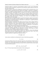
Kir2.1 V93I gene mutation on AP profiles are shown in Fig. 2.
Fig. 2. AP profiles under AFER (A) and Kir2.1 V93I gene mutation (B) conditions. AFER and
the mutation cause a dramatic abbreviation of APD.
APD restitution (APDr) measures the excitation behaviour of atrial cells subjected to
premature pulses immediately after a previous excitation (Franz, Karasik et al. 1997; Qi,
Tang et al. 1997; Kim, Kim et al. 2002; Burashnikov and Antzelevitch 2005; Cherry, Hastings
et al. 2008). Recent experimental and modelling studies have shown the correlation between
the maximal slope of APDr greater than unity and instability of re-entrant excitation waves
in 2D and 3D tissues (Xie, Qu et al. 2002; Banville, Chattipakorn et al. 2004; ten Tusscher,
Mourad et al. 2009). In our study, APDr is computed using a standard S1S2 protocol. A train
of ten conditioning stimuli (S1) at a physiological PCL were applied before the premature
pulse (S2) was applied. The time interval between the final conditioning excitation and onset
of the premature excitation emulates atrial diastolic interval (DI), or the time the atrial organ
has for recovery from the previous excitation. In the CRN model, S1 and S2 have stimulus
amplitude of 2 nA and duration of 2 ms. A plot of the DI against APD
90
gives APDr, as
shown in Fig. 3 for Control, AFER and Kir2.1 V93I gene mutation conditions. At large DI,
Studying Ion Channel Dysfunction and Arrythmogenesis in the Human Atrium:
A Computational Approach 437
reduction of the conductance of the L-type calcium current I
Ca,L
, 85% reduction of
conductance of the transient outward current (I
to
), a shift of -16 mV of the I
to
steady-state
activation, and a -1.6 mV shift of sodium current (I
Na
) steady state activation. Fast
inactivation kinetics of I
Ca,L
is slowed down, and was implemented as a 62% increase of the
voltage dependent inactivation time constant. Remodelling in AF2 includes a 90% increase
of I
K1
, 64% reduction of I
Ca,L
, 65% reduction of I
to
, 12% increase of the sustained outward
potassium current (I
Ksus
), and a 12% reduction of the sodium potassium pump (I
Na,K
). Both
AF1 and AF2 data have been incorporated into the CRN model in our previous study
(Zhang, Garratt et al. 2005).
Simulation of Kir2.1 V93I gene mutation was based on the recent clinical data from Xia et al.
(Xia, Jin et al. 2005) who examined several generations of a large family with hereditary AF
associated with Kir2.1 V93I gene mutation. The Kir2.1 gene primarily regulates the I
K1
channel current, which is modelled as
KKK
EVgI
11
(1)
cVb
K
KK
e
ga
agg
1
1
max1
max11
(2)
where V is the cell membrane potential; E
K
the reversal potential of the channel; g
K1max
the
maximal channel conductance; “a” is the fraction of the channel conductance that is voltage-
independent, (1-a) is the fraction of the channel conductance that is voltage-dependent, “b”
the steepness of the g
K1
-V relationship; “c” is the half point of the g
K1
-V relationship. In
simulations, we considered different conditions of the mutation from Control (Con), to
heterozygous (Het) to homozygous (Hom) cases. Parametric values of equations 1 and 2 for
different conditions of Kir2.1 V93I gene mutation are listed in Table 1, which were based on
the experimental study of Xia et al. (Xia, Jin et al., 2005).
Experimental data sets of AFER and Kir2.1 V93I gene mutation as described above were
then incorporated into the CRN human atrial AP model to simulate their effects on human
atrial excitation at cellular and tissue models. A quantitative summary of all results is given
in Table 2.
2.2 Quantifying the effects of AFER and Kir2.1 V93I gene mutation on atrial APs at
cellular level
We first quantify the functional effects of AFER and Kir2.1 V93I mutation on atrial cellular
APs. Excitable models, including human atrial cell models, are usually at resting state far
away from the oscillating state and show rate dependent adaptation upon periodic pacing,
similar to those seen experimentally (Workman, Kane et al. 2001; Cherry, Hastings et al.
2008). Therefore, the models have to be conditioned with several pulses before stable
excitations can be elicited. In case of the CRN model, it was found that 10 pulses at a pacing
cycle length (PCL) of 1 s was sufficient conditioning. Upon simulation, characteristics of AP
profiles were quantified by measuring the resting potential and APD at 90% repolarisation
(APD
90
), the overshoot and the maximal upstroke velocity, dV/dt
max
. APD
90
reflects the
overall changes in ion channel function during AP. dV/dt
max
on the other hand, not only
Quantity Con Het Hom
g
K1max
(nS/pF)
0.09 (100%) 0.13 (141% ↑) 0.16 (173% ↑)
a 0.0 0.0355 0.0575
b (mV
-1
) 0.070 0.156 0.232
c (mV) -80.0 -60.1 -54.7
Table 1. Parameters of I
K1
equations (1-2) for various Kir2.1 V93I gene mutation conditions.
Values were determined based on experimental data of Xia et al. (Xia, Jin et al. 2005) under
Con, Het and Hom conditions.
influences cellular behaviour, but also the conduction properties at tissue level (Biktashev
2002). Due to the large increase in repolarisation potassium currents and reduction in
depolarising currents, the AP profiles show large abbreviation in APD
90
under AFER and
Kir2.1 V93I gene mutation conditions. APD abbreviation under AFER conditions is due to a
integral actions of remodelled ion channels. However, in the gene mutation condition, such
an abbreviation is caused by gain-in-function of the I
K1
channel. The effects of AFER and
Kir2.1 V93I gene mutation on AP profiles are shown in Fig. 2.
Fig. 2. AP profiles under AFER (A) and Kir2.1 V93I gene mutation (B) conditions. AFER and
the mutation cause a dramatic abbreviation of APD.
APD restitution (APDr) measures the excitation behaviour of atrial cells subjected to
premature pulses immediately after a previous excitation (Franz, Karasik et al. 1997; Qi,
Tang et al. 1997; Kim, Kim et al. 2002; Burashnikov and Antzelevitch 2005; Cherry, Hastings
et al. 2008). Recent experimental and modelling studies have shown the correlation between
the maximal slope of APDr greater than unity and instability of re-entrant excitation waves
in 2D and 3D tissues (Xie, Qu et al. 2002; Banville, Chattipakorn et al. 2004; ten Tusscher,
Mourad et al. 2009). In our study, APDr is computed using a standard S1S2 protocol. A train
of ten conditioning stimuli (S1) at a physiological PCL were applied before the premature
pulse (S2) was applied. The time interval between the final conditioning excitation and onset
of the premature excitation emulates atrial diastolic interval (DI), or the time the atrial organ
has for recovery from the previous excitation. In the CRN model, S1 and S2 have stimulus
amplitude of 2 nA and duration of 2 ms. A plot of the DI against APD
90
gives APDr, as
shown in Fig. 3 for Control, AFER and Kir2.1 V93I gene mutation conditions. At large DI,
Recent Advances in Biomedical Engineering438
APDr curves have negligible slopes and show AP profiles under physiological rates of
pacing. At low DI, however, the slopes are noticeable. Under AFER conditions, the
computed APDr slopes under various conditions are much greater than under Control
conditions (Table 2).
Fig. 3. APDr profiles under AFER (A) and Kir2.1 V93I gene mutation (B) conditions. At large
DI, APDr curves reflect the changes in APD
90
under Control (Con) and AF (AF1, AF2, Het
and Hom) conditions. At low DI, the maximal slopes of APDr curves indicate the
instabilities in 2D and 3D simulations. Quantitative details are given in Table 2.
Fig. 4. ERP restitution curves under AFER (A) and Kir2.1 V93I gene mutation (B) conditions.
Shortening of atrial APD and effective refractory period (ERP) are well recognised features
of atrial electrical activities during AF. ERP is generally measured by using cellular or tissue
preparations (Workman, Kane et al. 2001; Laurent, Moe et al. 2008). In our studies, we
adopted the cell based experimental protocol as described by Workman et al. (Workman,
Kane et al. 2001) where the cell was stimulated 10 times at various PCLs. A premature
stimulus S2 was then applied. The maximal time interval between S1 and S2 where the final
excitation has AP amplitude of 80% as compared to the premature pulses is defined as the
ERP. Due to the rate dependent adaptability of atrial AP, we usually compute ERP at several
PCL values to obtain an ERP restitution curve. Results are shown in Fig. 4. It can be seen
that AF reduces ERP (Table 2). Such a reduction is in qualitative agreement with
experimental observations and clinical data (Workman, Kane et al. 2001; Li, Hertervig et al.
2002; Oliveira, da Silva et al. 2007).
2.3 1D and 2D tissue modelling
Human atrial tissue is spatially and electrically homogeneous tissue (Jalife 2003; Seemann,
Hoper et al. 2006). The primary sources of heterogeneity in the human atrium are the
conduction pathways as shown in Fig. 1, which contribute only a small fraction to total atrial
mass. Therefore, it is reasonable to take human atrial tissue as homogeneous in simulations
of the effects of AFER and Kir2.1 V93I gene mutation on atrial excitations (Kharche, Garratt
et al. 2008; Kharche, Seemann et al. 2008).
To simulate atrial excitation at the tissue level, the CRN atrial cell AP model is incorporated
into tissue models using a mono-domain reaction diffusion partial differential equation,
2
( )
( ) ( )
ion
V r
D V r I r
t
(3)
where D is the homogeneous diffusion constant mimicking the intracellular gap junctional
coupling,
2
is the Laplacian operator and I
ion
is the total reactive current at any given
spatial location r in the tissue associated with the ion channels of the atrial cell at r. We take
D to be 0.03125 mm
2
/ms to give physiological value of conduction velocity (CV) of 0.265
mm/ms, which falls in the range of physiological measurements. Such a formulation is
sufficient for our purposes as we do not consider any extracellular potentials, fluids or
indeed mechanical activity, for which more complex bi-domain formulations have to be
adopted (Potse, Dube et al. 2006; Whiteley 2007; Vigmond, Weber dos Santos et al. 2008;
Linge, Sundnes et al. 2009; Morgan, Plank et al. 2009).
To quantify the functional effects of AFER and Kir2.1 V93I gene mutation on atrial CV
restitution (CVr) and temporal vulnerability (VW), models of 1D homogeneous atrial strand
were used. CVr is computed by conditioning the 1D strand (S1) after which a premature
pulse is applied. The CV of the second propagation as a function of the inter-pulse duration,
or PCL, is termed as CVr. CV of propagations is computed from the central region of the
strands as shown in Fig 5A. CVr for AFER and the gene mutation conditions are shown in
Fig. 5, B and C, where the stimulation protocol is also illustrated. As can be seen, AF reduces
solitary wave CV, i.e. CV at large PCL, or low pacing rates. Such CV reduction is not due to
any changes in the inter-cellular coupling in the tissue, but solely due to the changes of atrial
cell AP profiles. Our simulation data revealed that atrial tissue has better ability to sustain
atrial conduction at fast pacing rates under AFER or gene mutation conditions than under
Control conditions.
Studying Ion Channel Dysfunction and Arrythmogenesis in the Human Atrium:
A Computational Approach 439
APDr curves have negligible slopes and show AP profiles under physiological rates of
pacing. At low DI, however, the slopes are noticeable. Under AFER conditions, the
computed APDr slopes under various conditions are much greater than under Control
conditions (Table 2).
Fig. 3. APDr profiles under AFER (A) and Kir2.1 V93I gene mutation (B) conditions. At large
DI, APDr curves reflect the changes in APD
90
under Control (Con) and AF (AF1, AF2, Het
and Hom) conditions. At low DI, the maximal slopes of APDr curves indicate the
instabilities in 2D and 3D simulations. Quantitative details are given in Table 2.
Fig. 4. ERP restitution curves under AFER (A) and Kir2.1 V93I gene mutation (B) conditions.
Shortening of atrial APD and effective refractory period (ERP) are well recognised features
of atrial electrical activities during AF. ERP is generally measured by using cellular or tissue
preparations (Workman, Kane et al. 2001; Laurent, Moe et al. 2008). In our studies, we
adopted the cell based experimental protocol as described by Workman et al. (Workman,
Kane et al. 2001) where the cell was stimulated 10 times at various PCLs. A premature
stimulus S2 was then applied. The maximal time interval between S1 and S2 where the final
excitation has AP amplitude of 80% as compared to the premature pulses is defined as the
ERP. Due to the rate dependent adaptability of atrial AP, we usually compute ERP at several
PCL values to obtain an ERP restitution curve. Results are shown in Fig. 4. It can be seen
that AF reduces ERP (Table 2). Such a reduction is in qualitative agreement with
experimental observations and clinical data (Workman, Kane et al. 2001; Li, Hertervig et al.
2002; Oliveira, da Silva et al. 2007).
2.3 1D and 2D tissue modelling
Human atrial tissue is spatially and electrically homogeneous tissue (Jalife 2003; Seemann,
Hoper et al. 2006). The primary sources of heterogeneity in the human atrium are the
conduction pathways as shown in Fig. 1, which contribute only a small fraction to total atrial
mass. Therefore, it is reasonable to take human atrial tissue as homogeneous in simulations
of the effects of AFER and Kir2.1 V93I gene mutation on atrial excitations (Kharche, Garratt
et al. 2008; Kharche, Seemann et al. 2008).
To simulate atrial excitation at the tissue level, the CRN atrial cell AP model is incorporated
into tissue models using a mono-domain reaction diffusion partial differential equation,
2
( )
( ) ( )
ion
V r
D V r I r
t
(3)
where D is the homogeneous diffusion constant mimicking the intracellular gap junctional
coupling,
2
is the Laplacian operator and I
ion
is the total reactive current at any given
spatial location r in the tissue associated with the ion channels of the atrial cell at r. We take
D to be 0.03125 mm
2
/ms to give physiological value of conduction velocity (CV) of 0.265
mm/ms, which falls in the range of physiological measurements. Such a formulation is
sufficient for our purposes as we do not consider any extracellular potentials, fluids or
indeed mechanical activity, for which more complex bi-domain formulations have to be
adopted (Potse, Dube et al. 2006; Whiteley 2007; Vigmond, Weber dos Santos et al. 2008;
Linge, Sundnes et al. 2009; Morgan, Plank et al. 2009).
To quantify the functional effects of AFER and Kir2.1 V93I gene mutation on atrial CV
restitution (CVr) and temporal vulnerability (VW), models of 1D homogeneous atrial strand
were used. CVr is computed by conditioning the 1D strand (S1) after which a premature
pulse is applied. The CV of the second propagation as a function of the inter-pulse duration,
or PCL, is termed as CVr. CV of propagations is computed from the central region of the
strands as shown in Fig 5A. CVr for AFER and the gene mutation conditions are shown in
Fig. 5, B and C, where the stimulation protocol is also illustrated. As can be seen, AF reduces
solitary wave CV, i.e. CV at large PCL, or low pacing rates. Such CV reduction is not due to
any changes in the inter-cellular coupling in the tissue, but solely due to the changes of atrial
cell AP profiles. Our simulation data revealed that atrial tissue has better ability to sustain
atrial conduction at fast pacing rates under AFER or gene mutation conditions than under
Control conditions.
Recent Advances in Biomedical Engineering440
Fig. 5. (A) Electrical waves in a 1D strand where the first wave conditions the tissue, whilst
the second wave is initiated after an interval S2. CV is computed according to when the
second wave is at x1 (t1) and x2 (t2). (B) CVr under AFER conditions. (C) CVr under Kir2.1
V93I gene mutation conditions.
Fig. 6. Atrial excitation wave evoked by a S2 stimulus, applied at a time delay after the
conditioning excitation wave, can be either bi-directional blocked (Ai) if the time delay is too
soon, or bi-directional conduction (Aii) if the time delay is too late, or uni-directional
conduction block (Aiii) if the time delay falls in the VW. Computed VW under AFER
conditions (B) and Kir2.1 V93I gene mutation conditions (C).
Fig. 7. Computed SVW from 2D tissue models by applying a premature stimulus in the
repolarisation tail of a conditioning pulse so as to evoke a figure of 8 re-entry (Ai, Aii and Bi,
Bii). The minimal length of the premature stimulus such that the evoked reentry sustains is
termed as SVW. (C) SVW under AFER conditions. (D) SVW under Kir2.1 V93I gene
mutation conditions. AFER and the gene mutation cause a dramatic reduction of SVW
allowing the tissue to sustain re-entry with reduced substrate size.
Uni-directional conduction block in atria can lead to genesis of re-entrant excitation waves.
Temporal vulnerability or vulnerability window (VW) measures the vulnerability of cardiac
tissue to genesis of uni-directional conduction block. VW is computed by allowing a single
solitary wave to propagate from one end of the 1D tissue to the other. After certain duration
and in the repolarisation phase in the middle of the tissue, a premature pulse is applied. The
time window during which the premature pulse elicits uni-directional propagation block is
termed as the VW. Fig. 6 illustrates the protocol and also shows the measured VW under
AFER and Kir2.1 V93I gene mutation conditions.
The effects of AFER and the Kir2.1 gene mutation on atrial tissue’s spatial vulnerability are
quantified by using 2D homogeneous models of human atrial tissue. Spatial vulnerability
(SVW) is computed as the minimal atrial substrate size that can sustain re-entrant waves. To
this end, a sufficiently long pulse as shown in Fig. 7 is applied in the repolarisation tail of
the conditioning pulse, giving rise to a figure of “8” re-entrant waves. The minimum length
that sustains such re-entry is termed as SVW. The results for AFER and gene mutation
conditions are given in Fig. 7.
Effects of the AFER and Kir2.1 V93I gene mutation on the dynamical behaviours of re-
entrant excitation waves are also studied. In 2D tissues, re-entrant wave simulations are
performed in a tissue with a size of 37.5 cm x 37.5 cm. In simulations, re-entrant waves are
initiated by using a cross-field stimulation protocol. After allowing a planar wave to
sufficiently propagate through the 2D sheet, a cross-field stimulus is applied so as to initiate
re-entry (Kharche, Seemann et al. 2007). Upon initiation of a re-entrant wave in the middle
of the tissue, the re-entrant waves are allowed to evolve for several seconds. Results are
shown in Fig. 8. Under Control conditions, the 2D re-entrant waves self-terminate.
However, under AFER and Kir2.1 V93I gene mutation conditions, re-entrant waves become
persistent. During the simulation, time series of APs from representative locations were also
Studying Ion Channel Dysfunction and Arrythmogenesis in the Human Atrium:
A Computational Approach 441
Fig. 5. (A) Electrical waves in a 1D strand where the first wave conditions the tissue, whilst
the second wave is initiated after an interval S2. CV is computed according to when the
second wave is at x1 (t1) and x2 (t2). (B) CVr under AFER conditions. (C) CVr under Kir2.1
V93I gene mutation conditions.
Fig. 6. Atrial excitation wave evoked by a S2 stimulus, applied at a time delay after the
conditioning excitation wave, can be either bi-directional blocked (Ai) if the time delay is too
soon, or bi-directional conduction (Aii) if the time delay is too late, or uni-directional
conduction block (Aiii) if the time delay falls in the VW. Computed VW under AFER
conditions (B) and Kir2.1 V93I gene mutation conditions (C).
Fig. 7. Computed SVW from 2D tissue models by applying a premature stimulus in the
repolarisation tail of a conditioning pulse so as to evoke a figure of 8 re-entry (Ai, Aii and Bi,
Bii). The minimal length of the premature stimulus such that the evoked reentry sustains is
termed as SVW. (C) SVW under AFER conditions. (D) SVW under Kir2.1 V93I gene
mutation conditions. AFER and the gene mutation cause a dramatic reduction of SVW
allowing the tissue to sustain re-entry with reduced substrate size.
Uni-directional conduction block in atria can lead to genesis of re-entrant excitation waves.
Temporal vulnerability or vulnerability window (VW) measures the vulnerability of cardiac
tissue to genesis of uni-directional conduction block. VW is computed by allowing a single
solitary wave to propagate from one end of the 1D tissue to the other. After certain duration
and in the repolarisation phase in the middle of the tissue, a premature pulse is applied. The
time window during which the premature pulse elicits uni-directional propagation block is
termed as the VW. Fig. 6 illustrates the protocol and also shows the measured VW under
AFER and Kir2.1 V93I gene mutation conditions.
The effects of AFER and the Kir2.1 gene mutation on atrial tissue’s spatial vulnerability are
quantified by using 2D homogeneous models of human atrial tissue. Spatial vulnerability
(SVW) is computed as the minimal atrial substrate size that can sustain re-entrant waves. To
this end, a sufficiently long pulse as shown in Fig. 7 is applied in the repolarisation tail of
the conditioning pulse, giving rise to a figure of “8” re-entrant waves. The minimum length
that sustains such re-entry is termed as SVW. The results for AFER and gene mutation
conditions are given in Fig. 7.
Effects of the AFER and Kir2.1 V93I gene mutation on the dynamical behaviours of re-
entrant excitation waves are also studied. In 2D tissues, re-entrant wave simulations are
performed in a tissue with a size of 37.5 cm x 37.5 cm. In simulations, re-entrant waves are
initiated by using a cross-field stimulation protocol. After allowing a planar wave to
sufficiently propagate through the 2D sheet, a cross-field stimulus is applied so as to initiate
re-entry (Kharche, Seemann et al. 2007). Upon initiation of a re-entrant wave in the middle
of the tissue, the re-entrant waves are allowed to evolve for several seconds. Results are
shown in Fig. 8. Under Control conditions, the 2D re-entrant waves self-terminate.
However, under AFER and Kir2.1 V93I gene mutation conditions, re-entrant waves become
persistent. During the simulation, time series of APs from representative locations were also
Recent Advances in Biomedical Engineering442
recorded to allow analysis of dominant frequency of the re-entry. It is shown that the rates
of atrial re-entrant excitation waves increased markedly from Control conditions to AF ER
and gene mutation conditions. Traced trajectory of the core tips of re-entrant excitation
illustrated the increased stability and persistence of the re-entrant waves under AFER and
gene mutation conditions. These results are shown in Fig. 9.
2.4 Simulation of re-entrant waves in a 3D realistic geometry
The 3D anatomically detailed spatial model of human female atria as shown in Fig. 1 was
developed in a previous study (Seemann, Hoper et al. 2006). It is based on the anatomical
geometry of the human atria reconstructed from the visible human project (Ackerman, 1991;
Ackerman and Banvard 2000). The anatomical model consists of electrically homogeneous
atrial tissue, the SAN and conduction pathways. The SAN is the main pacemaker
wherefrom cardiac electrical excitation originates. The conduction pathways are electrically
and structurally heterogeneous and assist in normal conduction of electrical excitation in the
human atrium. In our studies, we however study re-entrant waves and therefore do not
consider SAN electrical activity, nor the heterogeneity associated with the conduction
pathways. All cells in our 3D anatomical model simulations are considered to be electrically
homogeneous.
Fig. 8. Representative frames at regular intervals from 2D homogeneous re-entrant waves
simulations under Control, AFER and Kri2.1 V93I gene mutation conditions. Re-entry self-
terminates under Control conditions (top row), but becomes persistent under AFER and
gene mutation conditions.
Re-entrant waves were initiated and allowed to propagate through the electrically and
anatomically homogeneous model under Control, AFER and gene mutation conditions. The
re-entrant waves were initiated using a protocol similar to the 2D case at a place in the right
atrium to reduce boundary effects and interference from anatomical obstacles. The right
atrium was chosen to be ideal as it offers minimal anatomical defects interfering with the
initial evolution of the re-entrant waves. Results from the 3D simulations under Control and
AFER and gene mutation conditions are shown in Fig. 10.
Under Control conditions, re-entry self-terminated at around 4.2 s. AFER however rendered
re-entry to be persistent. Again, if we study representative AP profiles during the
simulation, we can see that AF increases the dominant frequency. The dominant frequency
of oscillations in Control case is low at less than 3 Hz. In contrast, under AFER conditions,
the re-entry is persistent with rapid excitation rate. AFER increases stability of the mother
rotor under AF2 conditions. Due to the anatomical defects, the mother rotor degenerates
into smaller persistent erratic propagating wavelets, with a dominant frequency more than
10 Hz. Similar results were obtained under the Kir2.1 V93I gene mutation conditions as
shown in Fig. 11.
Fig. 9. Dynamical behaviours of 2D re-entrant waves as shown in Fig. 8 with core tip traces
(left column), representative AP profiles (middle column) and dominant frequency of the
AP profiles (right column) under various AFER and gene mutation conditions. Re-entrant
waves are more stable and cause high rate of atrial tissue excitation under AFER and gene
mutation conditions.
Our simulations have also shown another important mechanism by which re-entry becomes
persistent without effects of AFER or gene mutation. Upon initiation of re-entry close to a
blood vessel ostium, the electrical wave readily becomes anchored, as seen in Fig. 12. Such
anchoring of an electrical propagation also gives rise to persistent and rapid excitation of
atrial tissue.
2.5 Numerical considerations, algorithms and visualisation
Time integration of the CRN cellular models was carried out at a constant time step of 0.005
ms as given in the original CRN model. In the spatial 1D and 2D models, a space step of 0.1
mm was used in an explicit central Euler spatial integration scheme. The inter-node distance
Studying Ion Channel Dysfunction and Arrythmogenesis in the Human Atrium:
A Computational Approach 443
recorded to allow analysis of dominant frequency of the re-entry. It is shown that the rates
of atrial re-entrant excitation waves increased markedly from Control conditions to AF ER
and gene mutation conditions. Traced trajectory of the core tips of re-entrant excitation
illustrated the increased stability and persistence of the re-entrant waves under AFER and
gene mutation conditions. These results are shown in Fig. 9.
2.4 Simulation of re-entrant waves in a 3D realistic geometry
The 3D anatomically detailed spatial model of human female atria as shown in Fig. 1 was
developed in a previous study (Seemann, Hoper et al. 2006). It is based on the anatomical
geometry of the human atria reconstructed from the visible human project (Ackerman, 1991;
Ackerman and Banvard 2000). The anatomical model consists of electrically homogeneous
atrial tissue, the SAN and conduction pathways. The SAN is the main pacemaker
wherefrom cardiac electrical excitation originates. The conduction pathways are electrically
and structurally heterogeneous and assist in normal conduction of electrical excitation in the
human atrium. In our studies, we however study re-entrant waves and therefore do not
consider SAN electrical activity, nor the heterogeneity associated with the conduction
pathways. All cells in our 3D anatomical model simulations are considered to be electrically
homogeneous.
Fig. 8. Representative frames at regular intervals from 2D homogeneous re-entrant waves
simulations under Control, AFER and Kri2.1 V93I gene mutation conditions. Re-entry self-
terminates under Control conditions (top row), but becomes persistent under AFER and
gene mutation conditions.
Re-entrant waves were initiated and allowed to propagate through the electrically and
anatomically homogeneous model under Control, AFER and gene mutation conditions. The
re-entrant waves were initiated using a protocol similar to the 2D case at a place in the right
atrium to reduce boundary effects and interference from anatomical obstacles. The right
atrium was chosen to be ideal as it offers minimal anatomical defects interfering with the
initial evolution of the re-entrant waves. Results from the 3D simulations under Control and
AFER and gene mutation conditions are shown in Fig. 10.
Under Control conditions, re-entry self-terminated at around 4.2 s. AFER however rendered
re-entry to be persistent. Again, if we study representative AP profiles during the
simulation, we can see that AF increases the dominant frequency. The dominant frequency
of oscillations in Control case is low at less than 3 Hz. In contrast, under AFER conditions,
the re-entry is persistent with rapid excitation rate. AFER increases stability of the mother
rotor under AF2 conditions. Due to the anatomical defects, the mother rotor degenerates
into smaller persistent erratic propagating wavelets, with a dominant frequency more than
10 Hz. Similar results were obtained under the Kir2.1 V93I gene mutation conditions as
shown in Fig. 11.
Fig. 9. Dynamical behaviours of 2D re-entrant waves as shown in Fig. 8 with core tip traces
(left column), representative AP profiles (middle column) and dominant frequency of the
AP profiles (right column) under various AFER and gene mutation conditions. Re-entrant
waves are more stable and cause high rate of atrial tissue excitation under AFER and gene
mutation conditions.
Our simulations have also shown another important mechanism by which re-entry becomes
persistent without effects of AFER or gene mutation. Upon initiation of re-entry close to a
blood vessel ostium, the electrical wave readily becomes anchored, as seen in Fig. 12. Such
anchoring of an electrical propagation also gives rise to persistent and rapid excitation of
atrial tissue.
2.5 Numerical considerations, algorithms and visualisation
Time integration of the CRN cellular models was carried out at a constant time step of 0.005
ms as given in the original CRN model. In the spatial 1D and 2D models, a space step of 0.1
mm was used in an explicit central Euler spatial integration scheme. The inter-node distance
Recent Advances in Biomedical Engineering444
of 0.1 mm represents human atrial size which is close to physiological values. In the 3D
models, the space step was taken to be 0.33 mm, which allowed use of a time step of 0.5 ms.
These choices gave stable solutions independent of integration parameters.
The 2D and 3D spatial models are large with 140625 and more than 26 x 10
6
nodes
respectively. Parallelisation is therefore an important part of cardiac simulations. Solvers that
used shared memory parallelism (OpenMP) and large distributed memory parallelism (MPI)
were developed in our laboratory. Scaling of the solvers is shown in Fig. 13. In addition to
parallelisation, novel cardiac specific algorithms that exploit peculiarities of the model were
developed (Kharche, Seemann et al. 2008). The full geometrical model demands very large
amounts of contiguous memory. 3D Atrial tissue geometry occupies about 8% geometry of the
total data set, due to atrium being thin walled with large holes of atrial chambers and vena
caves. We re-structured the computer code such that only atrial nodes, i.e. only 8% of the total
26 million nodes and related information are stored in the computer memory. This improved
efficacy of memory usage. By re-numbering the real atrial nodes we are not storing any data
points that are not atrium. The memory required is reduced to less than 10 GB in the 3D case,
and the required computer floating point operations (flops) are also reduced.
Fig. 10. 3D re-entry under Control (top panels), AF1 (middle panels) and AF2 (bottom
panels). Re-entry self-terminates under Control conditions in 4.2 s. Under AF1 conditions,
the narrow wavelength re-entrant wave breaks up due to interaction with anatomical
obstacles and gives rise to rapid erratic electrical propagations which are persistent. AF2
caused the re-entrant rotor to be stable and gave rise to a mother rotor.
The 3D simulations produce large data sets of more than 30 GB. Traditionally this output is
then post-processed to obtain measures quantifying the simulation, e.g. scroll wave filament
meander, and to visualise the dynamics of the electrical propagations. Each output file
consists of a binary data file of approximately 150 MB size. Efficient visualisation of the 3D
data shown in Figs. 10 and 12 was carried using the RAVE package (Grimstead, Kharche et
al. 2007) developed elsewhere. We have also developed visualisation techniques based on
the visualisation package Advanced Visualisation System (AVS) developed by Manchester
Visualisation Centre. This is versatile high level graphical software with a high level of
functionality. Images in Fig. 11 were produced using diamond shaped glyphs, each of which
was colour coded with a scalar value, namely the value of voltage at that location.
For smaller visualisation jobs, e.g. 2D visualisation, we have used MATLAB due to its
functionality and transparent scripting. Development of visualisation scripts using
MATLAB is relatively straightforward with a high level of functionality. MATLAB is also
available to our laboratory locally. Having successfully developed 2D visualisation pipelines
using MATLAB, AVS as a high level visual programming environment is also versatile and
the results obtained using MATLAB can be replicated by AVS.
Fig. 11. 3D re-entry under Control (top panels), various Kir2.1 V93I gene mutation
conditions (Het, middle panels; Hom, bottom panels). With Kri2.1 V93I gene mutation,
condition, the re-entry became erratic leading to rapid excitation of atrial tissue.
Fig. 12. Anchoring of re-entrant wave to pulmonary vein (PV). Location of PV is marked by
the arrow in the first panel of the second column.
Studying Ion Channel Dysfunction and Arrythmogenesis in the Human Atrium:
A Computational Approach 445
of 0.1 mm represents human atrial size which is close to physiological values. In the 3D
models, the space step was taken to be 0.33 mm, which allowed use of a time step of 0.5 ms.
These choices gave stable solutions independent of integration parameters.
The 2D and 3D spatial models are large with 140625 and more than 26 x 10
6
nodes
respectively. Parallelisation is therefore an important part of cardiac simulations. Solvers that
used shared memory parallelism (OpenMP) and large distributed memory parallelism (MPI)
were developed in our laboratory. Scaling of the solvers is shown in Fig. 13. In addition to
parallelisation, novel cardiac specific algorithms that exploit peculiarities of the model were
developed (Kharche, Seemann et al. 2008). The full geometrical model demands very large
amounts of contiguous memory. 3D Atrial tissue geometry occupies about 8% geometry of the
total data set, due to atrium being thin walled with large holes of atrial chambers and vena
caves. We re-structured the computer code such that only atrial nodes, i.e. only 8% of the total
26 million nodes and related information are stored in the computer memory. This improved
efficacy of memory usage. By re-numbering the real atrial nodes we are not storing any data
points that are not atrium. The memory required is reduced to less than 10 GB in the 3D case,
and the required computer floating point operations (flops) are also reduced.
Fig. 10. 3D re-entry under Control (top panels), AF1 (middle panels) and AF2 (bottom
panels). Re-entry self-terminates under Control conditions in 4.2 s. Under AF1 conditions,
the narrow wavelength re-entrant wave breaks up due to interaction with anatomical
obstacles and gives rise to rapid erratic electrical propagations which are persistent. AF2
caused the re-entrant rotor to be stable and gave rise to a mother rotor.
The 3D simulations produce large data sets of more than 30 GB. Traditionally this output is
then post-processed to obtain measures quantifying the simulation, e.g. scroll wave filament
meander, and to visualise the dynamics of the electrical propagations. Each output file
consists of a binary data file of approximately 150 MB size. Efficient visualisation of the 3D
data shown in Figs. 10 and 12 was carried using the RAVE package (Grimstead, Kharche et
al. 2007) developed elsewhere. We have also developed visualisation techniques based on
the visualisation package Advanced Visualisation System (AVS) developed by Manchester
Visualisation Centre. This is versatile high level graphical software with a high level of
functionality. Images in Fig. 11 were produced using diamond shaped glyphs, each of which
was colour coded with a scalar value, namely the value of voltage at that location.
For smaller visualisation jobs, e.g. 2D visualisation, we have used MATLAB due to its
functionality and transparent scripting. Development of visualisation scripts using
MATLAB is relatively straightforward with a high level of functionality. MATLAB is also
available to our laboratory locally. Having successfully developed 2D visualisation pipelines
using MATLAB, AVS as a high level visual programming environment is also versatile and
the results obtained using MATLAB can be replicated by AVS.
Fig. 11. 3D re-entry under Control (top panels), various Kir2.1 V93I gene mutation
conditions (Het, middle panels; Hom, bottom panels). With Kri2.1 V93I gene mutation,
condition, the re-entry became erratic leading to rapid excitation of atrial tissue.
Fig. 12. Anchoring of re-entrant wave to pulmonary vein (PV). Location of PV is marked by
the arrow in the first panel of the second column.
Recent Advances in Biomedical Engineering446
Model Quantity Con AF1 AF2 Het Hom
Cell
Resting
potential (mV)
-80.5 -85.2 -83.8 -84.59 -85.07
APD
90
(ms) 313.0 108.5 147.6 196.2 137.2
Overshoot
(mV)
22.9 24.6 25.0 24.3 24.1
dV/dt
max
(mV/ms)
147.2 86.5 97.2 113.4 98.1
APDr
maximal slope
0.91 4.63 1.56 2.2 0.46
ERP (ms)
(stimulus
interval ~ 1 s)
318.0 142.0 192.0 232.0 150.0
1D
CV (mm/ms) 0.27 0.25 0.26 0.26 0.25
VW (ms) 15.4 14.8 14 13.1 12.9
Wavelength
(mm)
84.51 27.13 38.22 67.3 56.5
2D
LS (s) 1.8 > 10 > 10 >10 >10
DF (Hz) <3.0 10.0 7.0 8-15 12
Tip meander
area (mm
2
)
615.0 48.0 72.0 101.2 76.1
SVW (mm) 99.1 20.5 34.7 26.2 17.0
3D
LS (s) 4.2 > 6 > 6 >6 >6
DF (Hz) 3.0 6.7 6.1 10.2 13.5
Table 2. Quantitative summary of the effects of AFER and Kir2.1 V93I gene mutation on
atrial excitations.
3. Conclusions and future work
Our simulation results have shown that both the AFER and Kir2.1 V93I mutation shortened
atrial APD and increased the maximal slopes of APDr. They reduced atrial ERP and the
intra-atrial CV, all of which facilitated high rate atrial excitation and conduction as observe
d experimentally and clinically in AF patients. Due to the large increase in repolarisation
currents, the both the AFER and Kir2.1 V93I gene mutation reduced tissue’s temporal VW.
However, they also reduced the minimal substrate size required to sustain re-entry.
Collectively of all these suggested the pro-arrhythmic effects of AFER and Kir2.1 gene
mutation. Our results also showed AFER and the gene mutation increased the stability of re-
entry, leading them to be persistent.
Fig. 13. (A) Scaling of the shared memory (OpenMP) solver. (B) Scaling of the distributed
memory (MPI) solver.
These data have provided the first evidence in support of the hypothesis of “AF begetting
AF”. The methods described above characterise several aspects of the AFER and Kir2.1 gene
mutation on generating and sustaining AF. Future studies may consider mechanism
involving malfunctioning of intracellular [Ca
2+
]
i
handling (Hove-Madsen, Prat-Vidal et al.
2006), spontaneous firing at atrial blood vessel ostia, interaction between SAN and atria. In
addition to macro re-entrant waves, micro re-entry is also an important factor responsible
for AF (Markowitz, Nemirovksy et al. 2007). Inclusion of the electrical and spatial
heterogeneities in the various tissue sub-types in the atrium will further our understanding
the genesis of AF, especially the micro-entry due to heterogeneity boundaries.
Computational methods and algorithms can be further improved. This is especially relevant
for patient specific simulations where real time results are vital. An immediate aspect of the
current simulation-visualisation pipeline that can be addressed is that of incorporating the
visualisation, at least partly, into the simulation process. This will enormously improve
efficacy of the 3D simulations.
4. References
Aronow, W. S. (2008a). "Management of atrial fibrillation Part 1." Compr Ther 34(3-4): 126-
33.
Aronow, W. S. (2008b). "Management of atrial fibrillation Part 2." Compr Ther 34(3-4): 134-
42.
Aronow, W. S. (2009). "Management of atrial fibrillation in the elderly." Minerva Med 100(1):
3-24.
Balana, B., D. Dobrev, et al. (2003). "Decreased ATP-sensitive K(+) current density during
chronic human atrial fibrillation." J Mol Cell Cardiol 35(12): 1399-405.
Banville, I., N. Chattipakorn, et al. (2004). "Restitution dynamics during pacing and
arrhythmias in isolated pig hearts." J Cardiovasc Electrophysiol 15(4): 455-63.
Studying Ion Channel Dysfunction and Arrythmogenesis in the Human Atrium:
A Computational Approach 447
Model Quantity Con AF1 AF2 Het Hom
Cell
Resting
potential (mV)
-80.5 -85.2 -83.8 -84.59 -85.07
APD
90
(ms) 313.0 108.5 147.6 196.2 137.2
Overshoot
(mV)
22.9 24.6 25.0 24.3 24.1
dV/dt
max
(mV/ms)
147.2 86.5 97.2 113.4 98.1
APDr
maximal slope
0.91 4.63 1.56 2.2 0.46
ERP (ms)
(stimulus
interval ~ 1 s)
318.0 142.0 192.0 232.0 150.0
1D
CV (mm/ms) 0.27 0.25 0.26 0.26 0.25
VW (ms) 15.4 14.8 14 13.1 12.9
Wavelength
(mm)
84.51 27.13 38.22 67.3 56.5
2D
LS (s) 1.8 > 10 > 10 >10 >10
DF (Hz) <3.0 10.0 7.0 8-15 12
Tip meander
area (mm
2
)
615.0 48.0 72.0 101.2 76.1
SVW (mm) 99.1 20.5 34.7 26.2 17.0
3D
LS (s) 4.2 > 6 > 6 >6 >6
DF (Hz) 3.0 6.7 6.1 10.2 13.5
Table 2. Quantitative summary of the effects of AFER and Kir2.1 V93I gene mutation on
atrial excitations.
3. Conclusions and future work
Our simulation results have shown that both the AFER and Kir2.1 V93I mutation shortened
atrial APD and increased the maximal slopes of APDr. They reduced atrial ERP and the
intra-atrial CV, all of which facilitated high rate atrial excitation and conduction as observe
d experimentally and clinically in AF patients. Due to the large increase in repolarisation
currents, the both the AFER and Kir2.1 V93I gene mutation reduced tissue’s temporal VW.
However, they also reduced the minimal substrate size required to sustain re-entry.
Collectively of all these suggested the pro-arrhythmic effects of AFER and Kir2.1 gene
mutation. Our results also showed AFER and the gene mutation increased the stability of re-
entry, leading them to be persistent.
Fig. 13. (A) Scaling of the shared memory (OpenMP) solver. (B) Scaling of the distributed
memory (MPI) solver.
These data have provided the first evidence in support of the hypothesis of “AF begetting
AF”. The methods described above characterise several aspects of the AFER and Kir2.1 gene
mutation on generating and sustaining AF. Future studies may consider mechanism
involving malfunctioning of intracellular [Ca
2+
]
i
handling (Hove-Madsen, Prat-Vidal et al.
2006), spontaneous firing at atrial blood vessel ostia, interaction between SAN and atria. In
addition to macro re-entrant waves, micro re-entry is also an important factor responsible
for AF (Markowitz, Nemirovksy et al. 2007). Inclusion of the electrical and spatial
heterogeneities in the various tissue sub-types in the atrium will further our understanding
the genesis of AF, especially the micro-entry due to heterogeneity boundaries.
Computational methods and algorithms can be further improved. This is especially relevant
for patient specific simulations where real time results are vital. An immediate aspect of the
current simulation-visualisation pipeline that can be addressed is that of incorporating the
visualisation, at least partly, into the simulation process. This will enormously improve
efficacy of the 3D simulations.
4. References
Aronow, W. S. (2008a). "Management of atrial fibrillation Part 1." Compr Ther 34(3-4): 126-
33.
Aronow, W. S. (2008b). "Management of atrial fibrillation Part 2." Compr Ther
34(3-4): 134-
42.
Aronow, W. S. (2009). "Management of atrial fibrillation in the elderly." Minerva Med 100(1):
3-24.
Balana, B., D. Dobrev, et al. (2003). "Decreased ATP-sensitive K(+) current density during
chronic human atrial fibrillation." J Mol Cell Cardiol
35(12): 1399-405.
Banville, I., N. Chattipakorn, et al. (2004). "Restitution dynamics during pacing and
arrhythmias in isolated pig hearts." J Cardiovasc Electrophysiol 15(4): 455-63.
Recent Advances in Biomedical Engineering448
Biktashev, V. N. (2002). "Dissipation of the excitation wave fronts." Phys Rev Lett
89(16):
168102.
Bordas, R., B. Carpentieri, et al. (2009). "Simulation of cardiac electrophysiology on next-
generation high-performance computers." Philos Transact A Math Phys Eng Sci
367(1895): 1951-69.
Bosch, R. F. and S. Nattel (2002). "Cellular electrophysiology of atrial fibrillation."
Cardiovasc Res
54(2): 259-69.
Bosch, R. F., X. Zeng, et al. (1999). "Ionic mechanisms of electrical remodeling in human
atrial fibrillation." Cardiovasc Res
44(1): 121-31.
Bourke, T. and N. G. Boyle (2009). "Atrial fibrillation and congestive heart failure." Minerva
Med 100(2): 137-43.
Burashnikov, A. and C. Antzelevitch (2005). "Role of repolarization restitution in the
development of coarse and fine atrial fibrillation in the isolated canine right atria." J
Cardiovasc Electrophysiol 16(6): 639-45.
Cai, B., L. Shan, et al. (2009). "Homocysteine modulates sodium channel currents in human
atrial myocytes." Toxicology
256(3): 201-6.
Cai, B. Z., D. M. Gong, et al. (2007). "Homocysteine inhibits potassium channels in human
atrial myocytes." Clin Exp Pharmacol Physiol 34(9): 851-5.
Chen, Y. H., S. J. Xu, et al. (2003). "KCNQ1 gain-of-function mutation in familial atrial
fibrillation." Science
299(5604): 251-4.
Cherry, E. M., H. M. Hastings, et al. (2008). "Dynamics of human atrial cell models:
restitution, memory, and intracellular calcium dynamics in single cells." Prog
Biophys Mol Biol 98(1): 24-37.
Chou, C. C. and P. S. Chen (2009). "New concepts in atrial fibrillation: neural mechanisms
and calcium dynamics." Cardiol Clin
27(1): 35-43, viii.
Conway, E. L., S. Musco, et al. (2009). "Drug therapy for atrial fibrillation." Cardiol Clin
27(1): 109-23, ix.
Courtemanche, M., R. J. Ramirez, et al. (1998). "Ionic mechanisms underlying human atrial
action potential properties: insights from a mathematical model." Am J Physiol
275(1 Pt 2): H301-21.
Ehrlich, J. R. and S. Nattel (2009). "Novel approaches for pharmacological management of
atrial fibrillation." Drugs
69(7): 757-74.
Franz, M. R., P. L. Karasik, et al. (1997). "Electrical remodeling of the human atrium: similar
effects in patients with chronic atrial fibrillation and atrial flutter." J Am Coll
Cardiol 30(7): 1785-92.
Gaita, F., R. Riccardi, et al. (2002). "Surgical approaches to atrial fibrillation." Card
Electrophysiol Rev 6(4): 401-5.
Grimstead, I. J., S. Kharche, et al. (2007). Viewing 0.3Tb Heart Simulation Data At Your
Desk. EG UK Theory and Practice of Computer Graphics
D. D. Ik Soo Lim.
Haissaguerre, M., P. Jais, et al. (1998). "Spontaneous initiation of atrial fibrillation by ectopic
beats originating in the pulmonary veins." N Engl J Med
339(10): 659-66.
Hove-Madsen, L., C. Prat-Vidal, et al. (2006). "Adenosine A2A receptors are expressed in
human atrial myocytes and modulate spontaneous sarcoplasmic reticulum calcium
release." Cardiovasc Res
72(2): 292-302.
Jalife, J. (2003). "Experimental and clinical AF mechanisms: bridging the divide." J Interv
Card Electrophysiol 9(2): 85-92.
Keldermann, R. H., K. H. ten Tusscher, et al. (2009). "A computational study of mother rotor
VF in the human ventricles." Am J Physiol Heart Circ Physiol 296(2): H370-9.
Kharche, S., C. J. Garratt, et al. (2008). "Atrial proarrhythmia due to increased inward
rectifier current (I(K1)) arising from KCNJ2 mutation a simulation study." Prog
Biophys Mol Biol 98(2-3): 186-97.
Kharche, S., G. Seemann, et al. (2007). Scroll Waves in 3D Virtual Human Atria: A
Computational Study. LNCS. F. B. S. a. G. Seemann. 4466: 129–138.
Kharche, S., G. Seemann, et al. (2008). "Simulation of clinical electrophysiology in 3D human
atria: a high-performance computing and high-performance visualization
application." Concurrency and Computation: Practice and Experience 20(11): 10.
Kharche, S. and H. Zhang (2008). "Simulating the effects of atrial fibrillation induced
electrical remodeling: a comprehensive simulation study." Conf Proc IEEE Eng Med
Biol Soc 2008: 593-6.
Kim, B. S., Y. H. Kim, et al. (2002). "Action potential duration restitution kinetics in human
atrial fibrillation." J Am Coll Cardiol 39(8): 1329-36.
Laurent, G., G. Moe, et al. (2008). "Experimental studies of atrial fibrillation: a comparison of
two pacing models." Am J Physiol Heart Circ Physiol 294(3): H1206-15.
Li, Q., H. Huang, et al. (2009). "Gain-of-function mutation of Nav1.5 in atrial fibrillation
enhances cellular excitability and lowers the threshold for action potential firing."
Biochem Biophys Res Commun 380(1): 132-7.
Li, Z., E. Hertervig, et al. (2002). "Dispersion of refractoriness in patients with paroxysmal
atrial fibrillation. Evaluation with simultaneous endocardial recordings from both
atria." J Electrocardiol 35(3): 227-34.
Linge, S., J. Sundnes, et al. (2009). "Numerical solution of the bidomain equations." Philos
Transact A Math Phys Eng Sci 367(1895): 1931-50.
Makiyama, T., M. Akao, et al. (2008). "A novel SCN5A gain-of-function mutation M1875T
associated with familial atrial fibrillation." J Am Coll Cardiol 52(16): 1326-34.
Markowitz, S. M., D. Nemirovksy, et al. (2007). "Adenosine-insensitive focal atrial
tachycardia: evidence for de novo micro-re-entry in the human atrium." J Am Coll
Cardiol 49(12): 1324-33.
Moe, G. K., W. C. Rheinboldt, et al. (1964). "A Computer Model of Atrial Fibrillation." Am
Heart J 67: 200-20.
Morgan, S. W., G. Plank, et al. (2009). "Low energy defibrillation in human cardiac tissue: a
simulation study." Biophys J 96(4): 1364-73.
Novo, G., P. Mansueto, et al. (2008). "Risk factors, atrial fibrillation and thromboembolic
events." Int Angiol 27(5): 433-8.
Nygren, A., C. Fiset, et al. (1998). "Mathematical model of an adult human atrial cell: the role
of K+ currents in repolarization." Circ Res 82(1): 63-81.
Oliveira, M. M., N. da Silva, et al. (2007). "Enhanced dispersion of atrial refractoriness as an
electrophysiological substrate for vulnerability to atrial fibrillation in patients with
paroxysmal atrial fibrillation." Rev Port Cardiol 26(7-8): 691-702.
Pandit, S. V., R. B. Clark, et al. (2001). "A mathematical model of action potential
heterogeneity in adult rat left ventricular myocytes." Biophys J 81(6): 3029-51.
Potse, M., B. Dube, et al. (2006). "A comparison of monodomain and bidomain reaction-
diffusion models for action potential propagation in the human heart." IEEE Trans
Biomed Eng 53(12 Pt 1): 2425-35.
Studying Ion Channel Dysfunction and Arrythmogenesis in the Human Atrium:
A Computational Approach 449
Biktashev, V. N. (2002). "Dissipation of the excitation wave fronts." Phys Rev Lett 89(16):
168102.
Bordas, R., B. Carpentieri, et al. (2009). "Simulation of cardiac electrophysiology on next-
generation high-performance computers." Philos Transact A Math Phys Eng Sci
367(1895): 1951-69.
Bosch, R. F. and S. Nattel (2002). "Cellular electrophysiology of atrial fibrillation."
Cardiovasc Res 54(2): 259-69.
Bosch, R. F., X. Zeng, et al. (1999). "Ionic mechanisms of electrical remodeling in human
atrial fibrillation." Cardiovasc Res 44(1): 121-31.
Bourke, T. and N. G. Boyle (2009). "Atrial fibrillation and congestive heart failure." Minerva
Med 100(2): 137-43.
Burashnikov, A. and C. Antzelevitch (2005). "Role of repolarization restitution in the
development of coarse and fine atrial fibrillation in the isolated canine right atria." J
Cardiovasc Electrophysiol 16(6): 639-45.
Cai, B., L. Shan, et al. (2009). "Homocysteine modulates sodium channel currents in human
atrial myocytes." Toxicology 256(3): 201-6.
Cai, B. Z., D. M. Gong, et al. (2007). "Homocysteine inhibits potassium channels in human
atrial myocytes." Clin Exp Pharmacol Physiol 34(9): 851-5.
Chen, Y. H., S. J. Xu, et al. (2003). "KCNQ1 gain-of-function mutation in familial atrial
fibrillation." Science 299(5604): 251-4.
Cherry, E. M., H. M. Hastings, et al. (2008). "Dynamics of human atrial cell models:
restitution, memory, and intracellular calcium dynamics in single cells." Prog
Biophys Mol Biol 98(1): 24-37.
Chou, C. C. and P. S. Chen (2009). "New concepts in atrial fibrillation: neural mechanisms
and calcium dynamics." Cardiol Clin 27(1): 35-43, viii.
Conway, E. L., S. Musco, et al. (2009). "Drug therapy for atrial fibrillation." Cardiol Clin
27(1): 109-23, ix.
Courtemanche, M., R. J. Ramirez, et al. (1998). "Ionic mechanisms underlying human atrial
action potential properties: insights from a mathematical model." Am J Physiol
275(1 Pt 2): H301-21.
Ehrlich, J. R. and S. Nattel (2009). "Novel approaches for pharmacological management of
atrial fibrillation." Drugs 69(7): 757-74.
Franz, M. R., P. L. Karasik, et al. (1997). "Electrical remodeling of the human atrium: similar
effects in patients with chronic atrial fibrillation and atrial flutter." J Am Coll
Cardiol 30(7): 1785-92.
Gaita, F., R. Riccardi, et al. (2002). "Surgical approaches to atrial fibrillation." Card
Electrophysiol Rev 6(4): 401-5.
Grimstead, I. J., S. Kharche, et al. (2007). Viewing 0.3Tb Heart Simulation Data At Your
Desk. EG UK Theory and Practice of Computer Graphics D. D. Ik Soo Lim.
Haissaguerre, M., P. Jais, et al. (1998). "Spontaneous initiation of atrial fibrillation by ectopic
beats originating in the pulmonary veins." N Engl J Med 339(10): 659-66.
Hove-Madsen, L., C. Prat-Vidal, et al. (2006). "Adenosine A2A receptors are expressed in
human atrial myocytes and modulate spontaneous sarcoplasmic reticulum calcium
release." Cardiovasc Res 72(2): 292-302.
Jalife, J. (2003). "Experimental and clinical AF mechanisms: bridging the divide." J Interv
Card Electrophysiol 9(2): 85-92.
Keldermann, R. H., K. H. ten Tusscher, et al. (2009). "A computational study of mother rotor
VF in the human ventricles." Am J Physiol Heart Circ Physiol
296(2): H370-9.
Kharche, S., C. J. Garratt, et al. (2008). "Atrial proarrhythmia due to increased inward
rectifier current (I(K1)) arising from KCNJ2 mutation a simulation study." Prog
Biophys Mol Biol 98(2-3): 186-97.
Kharche, S., G. Seemann, et al. (2007). Scroll Waves in 3D Virtual Human Atria: A
Computational Study. LNCS
. F. B. S. a. G. Seemann. 4466: 129–138.
Kharche, S., G. Seemann, et al. (2008). "Simulation of clinical electrophysiology in 3D human
atria: a high-performance computing and high-performance visualization
application." Concurrency and Computation: Practice and Experience
20(11): 10.
Kharche, S. and H. Zhang (2008). "Simulating the effects of atrial fibrillation induced
electrical remodeling: a comprehensive simulation study." Conf Proc IEEE Eng Med
Biol Soc 2008: 593-6.
Kim, B. S., Y. H. Kim, et al. (2002). "Action potential duration restitution kinetics in human
atrial fibrillation." J Am Coll Cardiol
39(8): 1329-36.
Laurent, G., G. Moe, et al. (2008). "Experimental studies of atrial fibrillation: a comparison of
two pacing models." Am J Physiol Heart Circ Physiol
294(3): H1206-15.
Li, Q., H. Huang, et al. (2009). "Gain-of-function mutation of Nav1.5 in atrial fibrillation
enhances cellular excitability and lowers the threshold for action potential firing."
Biochem Biophys Res Commun
380(1): 132-7.
Li, Z., E. Hertervig, et al. (2002). "Dispersion of refractoriness in patients with paroxysmal
atrial fibrillation. Evaluation with simultaneous endocardial recordings from both
atria." J Electrocardiol
35(3): 227-34.
Linge, S., J. Sundnes, et al. (2009). "Numerical solution of the bidomain equations." Philos
Transact A Math Phys Eng Sci 367(1895): 1931-50.
Makiyama, T., M. Akao, et al. (2008). "A novel SCN5A gain-of-function mutation M1875T
associated with familial atrial fibrillation." J Am Coll Cardiol
52(16): 1326-34.
Markowitz, S. M., D. Nemirovksy, et al. (2007). "Adenosine-insensitive focal atrial
tachycardia: evidence for de novo micro-re-entry in the human atrium." J Am Coll
Cardiol 49(12): 1324-33.
Moe, G. K., W. C. Rheinboldt, et al. (1964). "A Computer Model of Atrial Fibrillation." Am
Heart J 67: 200-20.
Morgan, S. W., G. Plank, et al. (2009). "Low energy defibrillation in human cardiac tissue: a
simulation study." Biophys J
96(4): 1364-73.
Novo, G., P. Mansueto, et al. (2008). "Risk factors, atrial fibrillation and thromboembolic
events." Int Angiol
27(5): 433-8.
Nygren, A., C. Fiset, et al. (1998). "Mathematical model of an adult human atrial cell: the role
of K+ currents in repolarization." Circ Res
82(1): 63-81.
Oliveira, M. M., N. da Silva, et al. (2007). "Enhanced dispersion of atrial refractoriness as an
electrophysiological substrate for vulnerability to atrial fibrillation in patients with
paroxysmal atrial fibrillation." Rev Port Cardiol
26(7-8): 691-702.
Pandit, S. V., R. B. Clark, et al. (2001). "A mathematical model of action potential
heterogeneity in adult rat left ventricular myocytes." Biophys J
81(6): 3029-51.
Potse, M., B. Dube, et al. (2006). "A comparison of monodomain and bidomain reaction-
diffusion models for action potential propagation in the human heart." IEEE Trans
Biomed Eng 53(12 Pt 1): 2425-35.
Recent Advances in Biomedical Engineering450
Qi, A., C. Tang, et al. (1997). "Characteristics of restitution kinetics in repolarization of rabbit
atrium." Can J Physiol Pharmacol
75(4): 255-62.
Ravens, U. and E. Cerbai (2008). "Role of potassium currents in cardiac arrhythmias."
Europace
10(10): 1133-7.
Restier, L., L. Cheng, et al. (2008). "Mechanisms by which atrial fibrillation-associated
mutations in the S1 domain of KCNQ1 slow deactivation of IKs channels." J Physiol
586(Pt 17): 4179-91.
Reumann, M., B. G. Fitch, et al. (2008). "Large scale cardiac modeling on the Blue Gene
supercomputer." Conf Proc IEEE Eng Med Biol Soc
2008: 577-80.
Rosso, R. and P. Kistler (2009). "Focal atrial tachycardia." Heart
.
Roy, D., M. Talajic, et al. (2009). "Atrial fibrillation and congestive heart failure." Curr Opin
Cardiol 24(1): 29-34.
Salle, L., S. Kharche, et al. (2008). "Mechanisms underlying adaptation of action potential
duration by pacing rate in rat myocytes." Prog Biophys Mol Biol
96(1-3): 305-20.
Saltman, A. E. and A. M. Gillinov (2009). "Surgical approaches for atrial fibrillation." Cardiol
Clin 27(1): 179-88, x.
Seemann, G., C. Hoper, et al. (2006). "Heterogeneous three-dimensional anatomical and
electrophysiological model of human atria." Philos Transact A Math Phys Eng Sci
364(1843): 1465-81.
Stabile, G., E. Bertaglia, et al. (2009). "Role of pulmonary veins isolation in persistent atrial
fibrillation ablation: the pulmonary vein isolation in persistent atrial fibrillation
(PIPA) study." Pacing Clin Electrophysiol
32 Suppl 1: S116-9.
Stewart, S., N. F. Murphy, et al. (2004). "Cost of an emerging epidemic: an economic analysis
of atrial fibrillation in the UK." Heart
90(3): 286-92.
ten Tusscher, K. H., A. Mourad, et al. (2009). "Organization of ventricular fibrillation in the
human heart: experiments and models." Exp Physiol
94(5): 553-62.
ten Tusscher, K. H., D. Noble, et al. (2004). "A model for human ventricular tissue." Am J
Physiol Heart Circ Physiol 286(4): H1573-89.
Vigmond, E. J., R. Weber dos Santos, et al. (2008). "Solvers for the cardiac bidomain
equations." Prog Biophys Mol Biol 96(1-3): 3-18.
Viswanathan, M. N. and R. L. Page (2009). "Pharmacological therapy for atrial fibrillation:
current options and new agents." Expert Opin Investig Drugs
18(4): 417-31.
Wetzel, U., G. Hindricks, et al. (2009). "Atrial fibrillation in the elderly." Minerva Med
100(2):
145-50.
Whiteley, J. P. (2007). "Physiology driven adaptivity for the numerical solution of the
bidomain equations." Ann Biomed Eng
35(9): 1510-20.
Wijffels, M. C. and H. J. Crijns (2003). "Recent advances in drug therapy for atrial
fibrillation." J Cardiovasc Electrophysiol
14(9 Suppl): S40-7.
Wijffels, M. C., C. J. Kirchhof, et al. (1995). "Atrial fibrillation begets atrial fibrillation. A
study in awake chronically instrumented goats." Circulation 92(7): 1954-68.
Workman, A. J., K. A. Kane, et al. (2001). "The contribution of ionic currents to changes in
refractoriness of human atrial myocytes associated with chronic atrial fibrillation."
Cardiovasc Res
52(2): 226-35.
Xia, M., Q. Jin, et al. (2005). "A Kir2.1 gain-of-function mutation underlies familial atrial
fibrillation." Biochem Biophys Res Commun 332(4): 1012-9.
Xie, F., Z. Qu, et al. (2002). "Electrical refractory period restitution and spiral wave reentry in
simulated cardiac tissue." Am J Physiol Heart Circ Physiol 283(1): H448-60.
Yang, Y., J. Li, et al. (2009). "Novel KCNA5 loss-of-function mutations responsible for atrial
fibrillation." J Hum Genet.
Zhang, H., C. J. Garratt, et al. (2009). "Remodelling of cellular excitation (reaction) and
intercellular coupling (diffusion) by chronic atrial fibrillation represented by a
reaction-diffusion system." Physica D: Nonlinear Phenomena 238(11-12): 8.
Zhang, H., C. J. Garratt, et al. (2005). "Role of up-regulation of IK1 in action potential
shortening associated with atrial fibrillation in humans." Cardiovasc Res 66(3): 493-
502.
Zhang, H., A. V. Holden, et al. (2000). "Mathematical models of action potentials in the
periphery and center of the rabbit sinoatrial node." Am J Physiol Heart Circ Physiol
279(1): H397-421.
Zhang, H., Y. Zhao, et al. (2007). "Computational evaluation of the roles of Na+ current, iNa,
and cell death in cardiac pacemaking and driving." Am J Physiol Heart Circ Physiol
292(1): H165-74.
Zhang, S., K. Yin, et al. (2008). "Identification of a novel KCNQ1 mutation associated with
both Jervell and Lange-Nielsen and Romano-Ward forms of long QT syndrome in a
Chinese family." BMC Med Genet 9: 24.
Studying Ion Channel Dysfunction and Arrythmogenesis in the Human Atrium:
A Computational Approach 451
Qi, A., C. Tang, et al. (1997). "Characteristics of restitution kinetics in repolarization of rabbit
atrium." Can J Physiol Pharmacol 75(4): 255-62.
Ravens, U. and E. Cerbai (2008). "Role of potassium currents in cardiac arrhythmias."
Europace 10(10): 1133-7.
Restier, L., L. Cheng, et al. (2008). "Mechanisms by which atrial fibrillation-associated
mutations in the S1 domain of KCNQ1 slow deactivation of IKs channels." J Physiol
586(Pt 17): 4179-91.
Reumann, M., B. G. Fitch, et al. (2008). "Large scale cardiac modeling on the Blue Gene
supercomputer." Conf Proc IEEE Eng Med Biol Soc 2008: 577-80.
Rosso, R. and P. Kistler (2009). "Focal atrial tachycardia." Heart.
Roy, D., M. Talajic, et al. (2009). "Atrial fibrillation and congestive heart failure." Curr Opin
Cardiol 24(1): 29-34.
Salle, L., S. Kharche, et al. (2008). "Mechanisms underlying adaptation of action potential
duration by pacing rate in rat myocytes." Prog Biophys Mol Biol 96(1-3): 305-20.
Saltman, A. E. and A. M. Gillinov (2009). "Surgical approaches for atrial fibrillation." Cardiol
Clin 27(1): 179-88, x.
Seemann, G., C. Hoper, et al. (2006). "Heterogeneous three-dimensional anatomical and
electrophysiological model of human atria." Philos Transact A Math Phys Eng Sci
364(1843): 1465-81.
Stabile, G., E. Bertaglia, et al. (2009). "Role of pulmonary veins isolation in persistent atrial
fibrillation ablation: the pulmonary vein isolation in persistent atrial fibrillation
(PIPA) study." Pacing Clin Electrophysiol 32 Suppl 1: S116-9.
Stewart, S., N. F. Murphy, et al. (2004). "Cost of an emerging epidemic: an economic analysis
of atrial fibrillation in the UK." Heart 90(3): 286-92.
ten Tusscher, K. H., A. Mourad, et al. (2009). "Organization of ventricular fibrillation in the
human heart: experiments and models." Exp Physiol 94(5): 553-62.
ten Tusscher, K. H., D. Noble, et al. (2004). "A model for human ventricular tissue." Am J
Physiol Heart Circ Physiol 286(4): H1573-89.
Vigmond, E. J., R. Weber dos Santos, et al. (2008). "Solvers for the cardiac bidomain
equations." Prog Biophys Mol Biol 96(1-3): 3-18.
Viswanathan, M. N. and R. L. Page (2009). "Pharmacological therapy for atrial fibrillation:
current options and new agents." Expert Opin Investig Drugs 18(4): 417-31.
Wetzel, U., G. Hindricks, et al. (2009). "Atrial fibrillation in the elderly." Minerva Med 100(2):
145-50.
Whiteley, J. P. (2007). "Physiology driven adaptivity for the numerical solution of the
bidomain equations." Ann Biomed Eng 35(9): 1510-20.
Wijffels, M. C. and H. J. Crijns (2003). "Recent advances in drug therapy for atrial
fibrillation." J Cardiovasc Electrophysiol 14(9 Suppl): S40-7.
Wijffels, M. C., C. J. Kirchhof, et al. (1995). "Atrial fibrillation begets atrial fibrillation. A
study in awake chronically instrumented goats." Circulation 92(7): 1954-68.
Workman, A. J., K. A. Kane, et al. (2001). "The contribution of ionic currents to changes in
refractoriness of human atrial myocytes associated with chronic atrial fibrillation."
Cardiovasc Res 52(2): 226-35.
Xia, M., Q. Jin, et al. (2005). "A Kir2.1 gain-of-function mutation underlies familial atrial
fibrillation." Biochem Biophys Res Commun 332(4): 1012-9.
Xie, F., Z. Qu, et al. (2002). "Electrical refractory period restitution and spiral wave reentry in
simulated cardiac tissue." Am J Physiol Heart Circ Physiol
283(1): H448-60.
Yang, Y., J. Li, et al. (2009). "Novel KCNA5 loss-of-function mutations responsible for atrial
fibrillation." J Hum Genet
.
Zhang, H., C. J. Garratt, et al. (2009). "Remodelling of cellular excitation (reaction) and
intercellular coupling (diffusion) by chronic atrial fibrillation represented by a
reaction-diffusion system." Physica D: Nonlinear Phenomena
238(11-12): 8.
Zhang, H., C. J. Garratt, et al. (2005). "Role of up-regulation of IK1 in action potential
shortening associated with atrial fibrillation in humans." Cardiovasc Res
66(3): 493-
502.
Zhang, H., A. V. Holden, et al. (2000). "Mathematical models of action potentials in the
periphery and center of the rabbit sinoatrial node." Am J Physiol Heart Circ Physiol
279(1): H397-421.
Zhang, H., Y. Zhao, et al. (2007). "Computational evaluation of the roles of Na+ current, iNa,
and cell death in cardiac pacemaking and driving." Am J Physiol Heart Circ Physiol
292(1): H165-74.
Zhang, S., K. Yin, et al. (2008). "Identification of a novel KCNQ1 mutation associated with
both Jervell and Lange-Nielsen and Romano-Ward forms of long QT syndrome in a
Chinese family." BMC Med Genet
9: 24.
Recent Advances in Biomedical Engineering452
Discovery of Biorhythmic Stories behind Daily Vital Signs and Its Application 453
Discovery of Biorhythmic Stories behind Daily Vital Signs and Its
Application
Wenxi Chen
X
Discovery of Biorhythmic Stories behind Daily
Vital Signs and Its Application
Wenxi Chen
Biomedical Information Technology Laboratory, the University of Aizu
Japan
1. Introduction
The historical development of the study of biorhythms and the physiological background, as
well as functionality of biorhythmic phenomena in human beings, is introduced. The latest
achievements in modern chronomedicine, as well as their applications in daily health care
and medical practice, are reviewed. Our challenges in monitoring vital signs during sleep in
a daily life environment, and discovery of various inherent biorhythmic stories using data
mining mathematics are described. Several representative results are presented. Finally,
potential applications and future perspectives of biorhythm studies are extensively
discussed.
1.1 Historical review
Biorhythmic phenomena are innate, cyclical biological processes or functions existing in all
forms of life on earth, including human beings, which respond dynamically to various
endogenous and exogenous conditions that surround us (Wikipedia, 2009b). The worldwide
history of biorhythmic studies and their application in medical practice can be traced back
more than 2000 years, to around a few centuries B.C. Since written records exist in China
from more than 4000 years ago, numerous unearthed cultural relics and archaeological
materials show that the philosophy of yin and yang and the concept of rhythmic alternation
had dominated almost every aspect of Chinese society and people’s behaviour (Sacred Lotus
Arts, 2009).
Following the philosophy of yin and yang, the earliest existing medical book, “The Medical
Classic of Emperor Huang”, was formulated from a dialogue between Emperor Huang and
a medical professional, Uncle Qi, based on the theory of yin and yang, and compiled from a
series of medical achievements by many medical practitioners between 770–221 B.C. The
first publication of the book was confirmed to have occurred no later than 26 B.C. and no
earlier than 99 B.C. (Wang, 2005).
The book was a medical treatise consisting of a collection of 162 papers in two parts:
“Miraculous Meridian and Acupuncture” and “Medical Issues and Fundamental Principles”.
Each part has nine volumes, and each volume has nine papers, because the number nine is
the highest number in Chinese culture, and here, implies that the book covers all aspects of
medical matters (Zhang et al., 1995).
24