Genetic Engineering Basics New Applications and Responsibilities Part 3 pptx
Bạn đang xem bản rút gọn của tài liệu. Xem và tải ngay bản đầy đủ của tài liệu tại đây (3.38 MB, 20 trang )
Expression of Non-Native Genes in a Surrogate Host Organism
29
Andersson, S. G. E., & Kurland, C. G. (1990). Codon preferences in free-living
microorganisms. Microbiological Reviews, 54, 2, pp. 198-210.
Angov, E. (2011). Codon usage: Nature's roadmap to expression and folding of proteins.
Biotechnology Journal, 6, 6, pp. 650-659.
Baird, S. D., Turcotte, M., Korneluk, R. G., & Holcik, M. (2006). Searching for IRES. RNA - A
Publication of the RNA Society, 12, 10, pp. 1755-1785.
Bernardi, G. (1995). The human genome: Organization and evolutionary history. Annual
Review of Genetics, 29, 445-476.
Boeger, H., Bushnell, D. A., Davis, R., Griesenbeck, J., Lorch, Y., Strattan, J. S., et al.
(2005). Structural basis of eukaryotic gene transcription. FEBS Letters, 579, 4,
pp. 899-903.
Boylan, M., Pelletier, J., & Meighen, E. A. (1989). Fused bacterial luciferase subunits catalyze
light emission in eukaryotes and prokaryotes. Journal of Biological Chemistry, 264, 4,
pp. 1915-1918.
Bulmer, M. (1987). Coevolution of codon usage and transfer RNA abundance. Nature, 325,
6106, pp. 728-730.
Burgess-Brown, N. A., Sharma, S., Sobott, F., Loenarz, C., Oppermann, U., & Gileadi, O.
(2008). Codon optimization can improve expression of human genes in
Escherichia coli: A multi-gene study. Protein Expression and Purification, 59, 1, pp.
94-102.
Chamary, J. V., Parmley, J. L., & Hurst, L. D. (2006). Hearing silence: non-neutral evolution
at synonymous sites in mammals. Nature Reviews Genetics, 7, 2, pp. 98-108.
Close, D. M., Hahn, R., Patterson, S. S., Ripp, S., & Sayler, G. S. (2011). Comparison of
human optimized bacterial luciferase, firefly luciferase, and green fluorescent
protein for continuous imaging of cell culture and animal models. Journal of
Biomedical Optics, 16, 4, pp. e12441.
Close, D. M., Patterson, S. S., Ripp, S., Baek, S. J., Sanseverino, J., & Sayler, G. S. (2010).
Autonomous bioluminescent expression of the bacterial luciferase gene cassette
(lux) in a mammalian cell line. PLoS ONE, 5, 8, pp. e047003.
Close, D. M., Ripp, S., & Sayler, G. S. (2009). Reporter proteins in whole-cell optical
bioreporter detection systems, biosensor integrations, and biosensing applications.
Sensors, 9, 11, pp. 9147-9174.
de Felipe, P. (2002). Polycistronic viral vectors. Current Gene Therapy, 2, 3, pp. 355-378.
Desmit, M. H., & Vanduin, J. (1990). Secondary structure of the ribosome binding site
determines translation efficiency - A quantitative analysis. Proceedings of the
National Academy of Sciences of the United States of America, 87, 19, pp. 7668-7672.
Dong, H. J., Nilsson, L., & Kurland, C. G. (1996). Co-variation of tRNA abundance and
codon usage in Escherichia coli at different growth rates. Journal of Molecular Biology,
260, 5, pp. 649-663.
Dvir, A., Conaway, J. W., & Conaway, R. C. (2001). Mechanism of transcription initiation
and promoter escape by RNA polymerase II. Current Opinion in Genetics &
Development, 11, 2, pp. 209-214.
Dvir, A., Conaway, R. C., & Conaway, J. W. (1996). Promoter escape by RNA polymerase II -
A role for an ATP cofactor in suppression of arrest by polymerase at promoter-
proximal sites. Jou
rnal of Biological Chemistry, 271, 38, pp. 23352-23356.
Genetic Engineering – Basics, New Applications and Responsibilities
30
Ebright, R. H. (2000). RNA polymerase: Structural similarities between bacterial RNA
polymerase and eukaryotic RNA polymerase II. Journal of Molecular Biology, 304, 5,
pp. 687-698.
Escher, A., Okane, D. J., Lee, J., & Szalay, A. A. (1989). Bacterial luciferase alpha-beta fusion
protein is fully active as a monomer and highly sensitive in vivo to elevated
temperature. Proceedings of the National Academy of Sciences of the United States of
America, 86, 17, pp. 6528-6532.
Eyre-Walker, A., & Hurst, L. D. (2001). The evolution of isochores. Nature Reviews Genetics, 2,
7, pp. 549-555.
Falaschi, A. (2000). Eukaryotic DNA replication: a model for a fixed double replisome.
Trends in Genetics, 16, 2, pp. 88-92.
Graf, M., Bojak, A., Deml, L., Bieler, K., Wolf, H., & Wagner, R. (2000). Concerted action of
multiple cis-acting sequences is required for Rev dependence of late human
immunodeficiency virus type 1 gene expression. Journal of Virology, 74, 22, pp.
10822-10826.
Grantham, R., Gautier, C., Gouy, M., Jacobzone, M., & Mercier, R. (1981). Codon catalog
usage is a genome strategy modulated for gene expressivity. Nucleic Acids Research,
9, 1, pp. R43-R74.
Gu, W. J., Zhou, T., & Wilke, C. O. (2010). A universal trend of reduced mRNA stability near
the translation-initiation site in prokaryotes and eukaryotes. PLoS Computational
Biology, 6, 2, pp. e1000664.
Gupta, R. K., Patterson, S. S., Ripp, S., & Sayler, G. S. (2003). Expression of the Photorhabdus
luminescens lux genes (luxA, B, C, D, and E) in Saccharomyces cerevisiae. FEMS Yeast
Research, 4, 3, pp. 305-313.
Gustafsson, C., Govindarajan, S., & Minshull, J. (2004). Codon bias and heterologous protein
expression. Trends in Biotechnology, 22, 7, pp. 346-353.
Harraghy, N., Gaussin, A., & Mermod, N. (2008). Sustained transgene expression using
MAR elements. Current Gene Therapy, 8, 5, pp. 353-366.
Hastings, J., & Nealson, K. (1977). Bacterial bioluminescence. Annual Reviews in Microbiology,
31, 1, pp. 549-595.
Hershberg, R., & Petrov, D. A. (2008). Selection on codon bias. Annual Review of Genetics, 42,
1, pp. 287-299.
Jackson, R. J. (1988). RNA translation - Picornaviruses break the rules. Nature, 334, 6180, pp.
292-293.
Jang, S. K., Krausslich, H. G., Nicklin, M. J. H., Duke, G. M., Palmenberg, A. C., & Wimmer,
E. (1988). A segment of the 5' nontranslated region of encephalomyocarditis virus
RNA directs internal entry of ribosomes during in vitro translation. Journal of
Virology, 62, 8, pp. 2636-2643.
Kane, J. F. (1995). Effects of rare codon clusters on high-level expression of heterologous
proteins in Escherichia coli
. Current Opinion in
Biotechnology, 6, 5, pp. 494-500.
Keck, J. L., & Berger, J. M. (2000). DNA replication at high resolution. Chemistry & Biology, 7,
3, pp. R63-71.
Kim, S., & Lee, S. B. (2006). Rare codon clusters at 5'-end influence heterologous expression
of archaeal gene in Escherichia coli. Protein Expression and Purification, 50, 1, pp. 49-
57.
Expression of Non-Native Genes in a Surrogate Host Organism
31
Kirchner, G., Roberts, J. L., Gustafson, G. D., & Ingolia, T. D. (1989). Active bacterial
luciferase from a fused gene: Expression of a Vibrio harveyi luxAB translational
fusion in bacteria, yeast and plant cells. Gene, 81, 2, pp. 349-354.
Kolitz, S. E., & Lorsch, J. R. (2010). Eukaryotic initiator tRNA: Finely tuned and ready for
action. FEBS Letters, 584, 2, pp. 396-404.
Koncz, C., Olsson, O., Langridge, W. H. R., Schell, J., & Szalay, A. A. (1987). Expression and
assembly of functional bacterial luciferase in plants. Proceedings of the National
Academy of Sciences USA, 84, 1, pp. 131-135.
Kozak, M. (1986). Point mutations define a sequence flanking the AUG initiator codon that
modulates translation by eukaryotic ribosomes. Cell, 44, 2, pp. 283-292.
Kozak, M. (1987). An analysis of 5'-noncoding sequences from 699 vertebrate messenger
RNAs. Nucleic Acids Research, 15, 20, pp. 8125-8148.
Kubo, M., & Imanaka, T. (1989). mRNA secondary structure in an open reading frame
reduces translation efficiency in Bacillus subtilis. Journal of Bacteriology, 171, 7, pp.
4080-4082.
Kudla, G., Lipinski, L., Caffin, F., Helwak, A., & Zylicz, M. (2006). High guanine and
cytosine content increases mRNA levels in mammalian cells. PLoS Biology, 4, 6, pp.
933-942.
Kudla, G., Murray, A. W., Tollervey, D., & Plotkin, J. B. (2009). Coding-sequence
determinants of gene expression in Escherichia coli. Science, 324, 5924, pp. 255-
258.
Kurland, C. G. (1991). Codon bias and gene expression. FEBS Letters, 285, 2, pp. 165-169.
Kwaks, T. H. J., & Otte, A. P. (2006). Employing epigenetics to augment the expression of
therapeutic proteins in mammalian cells. Trends in Biotechnology, 24, 3, pp. 137-142.
Lafontaine, D. L. J., & Tollervey, D. (2001). The function and synthesis of ribosomes. Nature
Reviews Molecular Cell Biology, 2, 7, pp. 514-520.
Lavergne, J. P., Reboud, A. M., Sontag, B., Guillot, D., & Reboud, J. P. (1992). Binding of
GDP to a ribosomal protein after elongation factor-2 dependent GTP hydrolysis.
Biochimica et Biophysica Acta - Gene Structure and Expression, 1132, 3, pp. 284-289.
Levine, M., & Tjian, R. (2003). Transcription regulation and animal diversity. Nature, 424,
6945, pp. 147-151.
Li, Q. L., Peterson, K. R., Fang, X. D., & Stamatoyannopoulos, G. (2002). Locus control
regions. Blood, 100, 9, pp. 3077-3086.
Li, S., MacLaughlin, F. C., Fewell, J. G., Gondo, M., Wang, J., Nicol, F., et al. (2001). Muscle-
specific enhancement of gene expression by incorporation of SV/40 enhancer in the
expression plasmid. Gene Therapy, 8, 6, pp. 494-497.
Lucchini, S., Rowley, G., Goldberg, M. D., Hurd, D., Harrison, M., & Hinton, J. C. D. (2006).
H-NS mediates the silencing of laterally acquired genes in bacteria. PLoS Pathogens,
2, 8, pp. 746-752.
Lupez-Lastra, M., Rivas, A., & BarrÌa, M. (2005). Protein synthesis in eukaryotes: the
growing biological relevance of cap-independent translation initiation. Biol
ogical
Research, 38, 121-146.
McDowall, K. J., Linchao, S., & Cohen, S. N. (1994). A+U content rather than a particular
nucleotide order determines the specificity of RNase E cleavage. Journal of Biological
Chemistry, 269, 14, pp. 10790-10796.
Genetic Engineering – Basics, New Applications and Responsibilities
32
Meighen, E. A. (1991). Molecular biology of bacterial bioluminescence. Microbiological
Reviews, 55, 1, pp. 123-142.
Moreira, D., Kervestin, S., Jean-Jean, O., & Philippe, H. (2002). Evolution of eukaryotic
translation elongation and termination factors: Variations of evolutionary rate and
genetic code deviations. Molecular Biology and Evolution, 19, 2, pp. 189-200.
Morita, S., Kojima, T., & Kitamura, T. (2000). Plat-E: An efficient and stable system for
transient packaging of retroviruses. Gene Therapy, 7, 12, pp. 1063-1066.
Murakami, K. S., & Darst, S. A. (2003). Bacterial RNA polymerases: The whole story. Current
Opinion in Structural Biology, 13, 1, pp. 31-39.
Navarre, W. W., Porwollik, S., Wang, Y. P., McClelland, M., Rosen, H., Libby, S. J., et al.
(2006). Selective silencing of foreign DNA with low GC content by the H-NS
protein in Salmonella. Science, 313, 5784, pp. 236-238.
Nilsson, J., & Nissen, P. (2005). Elongation factors on the ribosome. Current Opinion in
Structural Biology, 15, 3, pp. 349-354.
Norrman, K., Fischer, Y., Bonnamy, B., Sand, F. W., Ravassard, P., & Semb, H. (2010).
Quantitative comparison of constitutive promoters in human ES cells. PLoS ONE, 5,
8, pp. e12413.
Oldfield, S., & Proud, C. G. (1993). Phosphorylation of elongation factor-2 from the
lepidopteran insect, spodoptera frugiperda. FEBS Letters, 327, 1, pp. 71-74.
Oshima, T., Ishikawa, S., Kurokawa, K., Aiba, H., & Ogasawara, N. (2006). Escherichia coli
histone-like protein H-NS preferentially binds to horizontally acquired DNA in
association with RNA polymerase. DNA Research, 13, 4, pp. 141-153.
Pal-Bhadra, M., Bhadra, U., & Birchler, J. A. (2002). RNAi related mechanisms affect both
transcriptional and posttranscriptional transgene silencing in Drosophila. Molecular
Cell, 9, 2, pp. 315-327.
Patterson, S. S., Dionisi, H. M., Gupta, R. K., & Sayler, G. S. (2005). Codon optimization of
bacterial luciferase (lux) for expression in mammalian cells. Journal of Industrial
Microbiology & Biotechnology, 32, 3, pp. 115-123.
Pazzagli, M., Devine, J. H., Peterson, D. O., & Baldwin, T. O. (1992). Use of bacterial and
firefly luciferases as reporter genes in DEAE-dextran mediated transfection of
mammalian cells. Analytical Biochemistry, 204, 2, pp. 315-323.
Pestova, T. V., Kolupaeva, V. G., Lomakin, I. B., Pilipenko, E. V., Shatsky, I. N., Agol, V. I., et
al. (2001). Molecular mechanisms of translation initiation in eukaryotes. Proceedings
of the National Academy of Sciences of the United States of America, 98, 13, pp. 7029-
7036.
Pikaart, M. I., Recillas-Targa, F., & Felsenfeld, G. (1998). Loss of transcriptional activity of a
transgene is accompanied by DNA methylation and histone deacetylation and is
prevented by insulators. Genes & Development, 12, 18, pp. 2852-2862.
Plotkin, J. B., & Kudla, G. (2011). Synonymous but not the same: the causes and
consequences of codon bias. Nature Reviews Genetics, 12, 1, pp. 32-42.
Pribnow, D. (1975). Nucleotide sequence of an RNA polymerase binding site at an early T7
promoter. Proceedings of the National Academy of Sciences of the United States of
America, 72, 3,
pp. 784-788.
Expression of Non-Native Genes in a Surrogate Host Organism
33
Qin, J., Zhang, L., Clift, K., Hulur, I., Xiang, A., Ren, B., et al. (2010). Systematic comparison
of constitutive promoters and the doxycycline-inducible promoter. PLoS One, 5, 5,
pp. e10611.
Ramakrishnan, V. (2002). Ribosome structure and the mechanism of translation. Cell, 108, 4,
pp. 557-572.
Recillas-Targa, F., Valadez-Graham, V., & Farre, C. M. (2004). Prospects and implications of
using chromatin insulators in gene therapy and transgenesis. Bioessays, 26, 7, pp.
796-807.
Richardson, J. P. (2003). Loading Rho to terminate transcription. Cell, 114, 2, pp. 157-159.
Riis, B., Rattan, S. I. S., Clark, B. F. C., & Merrick, W. C. (1990). Eukaryotic protein elongation
factors. Trends in Biochemical Sciences, 15, 11, pp. 420-424.
Riu, E. R., Chen, Z. Y., Xu, H., He, C. Y., & Kay, M. A. (2007). Histone modifications are
associated with the persistence or silencing of vector-mediated transgene
expression in vivo. Molecular Therapy, 15, 7, pp. 1348-1355.
Rosano, G. L., & Ceccarelli, E. A. (2009). Rare codon content affects the solubility of
recombinant proteins in a codon bias-adjusted Escherichia coli strain. Microbial Cell
Factories, 8, 1, pp. 41.
Rosenberg, M., & Court, D. (1979). Regulatory sequences involved in the promotion
and termination of RNA transcription. Annual Review of Genetics, 13, 1, pp. 319-
353.
Schreiber, S. L. (2005). Small molecules: The missing link in the central dogma. Nature
Chemical Biology, 1, 2, pp. 64-66.
So, A. G., & Downey, K. M. (1992). Eukaryotic DNA replication. Critical Reviews in
Biochemistry and Molecular Biology, 27, 1-2, pp. 129-155.
Szymczak, A. L., & Vignali, D. A. A. (2005). Development of 2A peptide-based strategies in
the design of multicistronic vectors. Expert Opinion on Biological Therapy, 5, 5, pp.
627-638.
Wahle, E. (1995). 3'-End cleavage and polyadenylation of mRNA precursors. Biochimica et
Biophysica Acta - Gene Structure and Expression, 1261, 2, pp. 183-194.
Watson, J., Baker, T., Bell, S., Gann, A., Levine, M., & Losick, R. (2008). Molecular Biology of
the Gene (6 ed.). Cold Spring Harbor: Cold Spring Harbor Laboratory Press.
Williams, S., Mustoe, T., Mulcahy, T., Griffiths, M., Simpson, D., Antoniou, M., et al. (2005).
CpG-island fragments from the HNRPA2B1/CBX3 genomic locus reduce silencing
and enhance transgene expression from the hCMV promoter/enhancer in
mammalian cells. BMC Biotechnology, 5, 1, pp. 17.
Wilson, G. G., & Murray, N. E. (1991). Restriction and modification systems. Annual Review
of Genetics, 25, 1, pp. 585-627.
Wu, X. Q., Jornvall, H., Berndt, K. D., & Oppermann, U. (2004). Codon optimization reveals
critical factors for high level expression of two rare codon genes in Escherichia coli:
RNA stability and secondary structure but not tRNA abundance. Biochemical and
Biophysical Research Communications, 313
, 1, pp. 89-96.
Yew, N. S., Wysokenski, D. M., Wang, K. X., Ziegler, R. J., Marshall, J., McNeilly, D., et al.
(1997). Optimization of plasmid vectors for high-level expression in lung epithelial
cells. Human Gene Therapy, 8, 5, pp. 575-584.
Genetic Engineering – Basics, New Applications and Responsibilities
34
Zhang, G., Hubalewska, M., & Ignatova, Z. (2009). Transient ribosomal attenuation
coordinates protein synthesis and co-translational folding. Nature Structural &
Molecular Biology, 16, 3, pp. 274-280.
Zolotukhin, S., Potter, M., Hauswirth, W., Guy, J., & Muzyczka, N. (1996). A "humanized"
green fluorescent protein cDNA adapted for high-level expression in mammalian
cells. Journal of Virology, 70, 7, pp. 4646-4654.
Zur Megede, J., Chen, M. C., Doe, B., Schaefer, M., Greer, C. E., Selby, M., et al. (2000).
Increased expression and immunogenicity of sequence-modified human
immunodeficiency virus type 1 gag gene. Journal of Virology, 74, 6, pp. 2628.
Zvereva, M., Shcherbakova, D., & Dontsova, O. (2010). Telomerase: Structure, functions, and
activity regulation. Biochemistry, 73, 13, pp. 1563-1583.
2
Gateway Vectors for Plant Genetic Engineering:
Overview of Plant Vectors, Application for
Bimolecular Fluorescence Complementation
(BiFC) and Multigene Construction
Yuji Tanaka
1
, Tetsuya Kimura
2
, Kazumi Hikino
3
, Shino Goto
3,4
,
Mikio Nishimura
3,4
, Shoji Mano
3,4
and Tsuyoshi Nakagawa
1
1
Department of Molecular and Functional Genomics,
Center for Integrated Research in Science, Shimane University,
2
Department of Sustainable Resource Science,
Graduate School of Bioresources, Mie University,
3
Department of Cell Biology, National Institute for Basic Biology,
4
Department of Basic Biology, School of Life Science,
The Graduate University for Advanced Studies,
Japan
1. Introduction
Transgenic technologies for the genetic engineering of plants are very important for basic
plant research and biotechnology. For example, promoter analysis with a reporter such as
green fluorescent protein (GFP) is typically used to determine the expression pattern of
genes of interest in basic plant research. Moreover, downregulation or controlled expression
studies of target genes are used to determine the function of these genes. In plant
biotechnology, overexpression of heterologous genes by transgenic methods is widely used
to improve industrially important crop plants. Recently, genome projects focusing on
various higher plants have provided abundant sequence information, and genome-wide
studies of gene function and gene regulation are being carried out. In these areas of
research, transgenic analyses using genetically modified plants will become more essential.
For example, high-throughput promoter analysis to examine the temporal and spatial
regulation of gene expression, the subcellular localization of the gene products based on
reporter genes, and ectopic expression of cDNA clones and RNAi will reveal the functions
of a variety of genes. For gene manipulation in plants, the binary system of Agrobacterium-
mediated transformation is most widely used. This system consists of two plasmids derived
from Ti plasmids, namely disarmed Ti plasmids and binary vectors (Bevan, 1984). The
former contains most genes for T-DNA transfer from Agrobacterium tumefaciens to plants,
whereas the latter is composed of a functional T-DNA and minimal elements for replication
both in Escherichia coli and in A. tumefaciens. Most of the widely used binary vectors
established in the 1990s were constructed by a traditional restriction endonuclease based
method. Therefore, it was time consuming and laborious to construct modified genes on
Genetic Engineering – Basics, New Applications and Responsibilities
36
binary vectors using the limited number of available restriction sites because of their large
size and the existence of many restriction sites outside their cloning sites. To overcome this
disadvantage and perform high-throughput analysis of plant genes, a new cloning system to
realize rapid and efficient construction of modified genes on binary vectors was desired. The
Gateway cloning system provided by Invitrogen (Carlsbad, CA, USA) is one of these
solutions. We have constructed a variety of Gateway compatible Ti binary vectors for plant
transgenic research.
2. Basic Ti-binary vector for Agrobacterium-mediated transformation and
Gateway cloning
Transformation mediated by the soil bacterium A. tumefaciens is widely used for gene
manipulation of plants. This bacterium has huge Ti-plasmids (larger than 200 kb) and the
ability to transfer the T-DNA region of the Ti-plasmid to infect plant chromosomes. The
natural Ti-mediated transformation system can be applied to transfer novel genes into a
plant genome. To be useful for gene manipulation, binary vectors possessing the T-DNA
region were developed. The vectors must possess a plant selection marker gene, a bacterial
antibiotic resistance gene, a site for cloning foreign genes, T-DNA border sequences for gene
transfer to the plant genome, an origin of replication (ori) for a broad host range of the
plasmid and an ori for E. coli. Although binary vectors are much smaller than native Ti–
plasmids, they are still large and cause difficulties in gene cloning by traditional methods.
Gateway Technology (available from Invitrogen) is based on the site-specific recombination
system between phage lambda and E. coli DNA. This system was modified to improve its
specificity and efficiency to utilize it as a universal cloning system. The advantages of
Gateway cloning are as follows: it is free from the need for restriction endonucleases and
DNA ligase, has a simple and uniform protocol, and offers highly efficient and reliable
cloning and easy manipulation of fusion constructs. Therefore, the development of a variety
of Gateway cloning compatible vectors for many purposes will expand the usefulness of this
system in plant research.
2.1 Ti-binary vector for Agrobacterium-mediated plant transformation
A. tumefaciens harboring a Ti-plasmid can transfer a specific segment of the plasmid, the
T-DNA region, which is bounded by a right border (RB) and a left border (LB) sequence, to
the genome of an infected plant (Figure 1). Expression of the T-DNA genes causes the
overproduction of phytohormones in the infected cells, which causes crown gall tumors.
Although T-DNA genes are required for crown gall tumor formation, other genes called the
vir genes outside of the T-DNA region are essential for transfer of T-DNA into the host plant
genome. These vir genes work even when they reside on another plasmid in A. tumefaciens.
Based on these findings, a Ti-binary vector system was developed to overcome the difficulty
of manipulating the original Ti plasmids in vitro by recombinant DNA methods due to their
huge size (Bevan, 1984). A wide range of shuttle vectors for E. coli and A. tumefaciens was
constructed that contain T-DNA border sequences flanking multiple restriction sites for
foreign DNA cloning and marker genes for selection in plant cells. Using this vector system,
DNA manipulation and vector construction can be done in E. coli; the vector is then
transferred to A. tumefaciens harboring an artificial Ti-plasmid in which the T-DNA has been
deleted. The vector is maintained stably in A. tumefaciens, and the cloned foreign DNA and
Gateway Vectors for Plant Genetic Engineering: Overview of Plant Vectors,
Application for Bimolecular Fluorescence Complementation (BiFC) and Multigene Construction
37
marker gene between RB and LB can be transferred to the host plant genome by the
transformation system encoded by vir genes on the T-DNA deletion Ti-plasmid. In early
studies, several dicot plants were transformed by an Agrobacterium method. However,
various dicot and monocot plants can now be transformed by co-cultivation of leaf slices or
cultured calli with chemicals inducing expression of vir genes. Transformed cells are
selected by marker gene phenotype such as antibiotic resistance and regenerated to
transgenic plants. The most important model plant, Arabidopsis thaliana, can be easily
transformed by A. tumefaciens using a floral dip procedure.
Fig. 1. Ti-binary vector system for Agrobacterium-mediated plant transformation. A binary
vector, in which a target gene and plant selection marker gene are cloned between the two
border sequences (RB and LB), is transformed into A. tumefaciens harboring a disarmed
Ti-plasmid without the T-DNA region. Plant cells are infected by the transformed
A. tumefaciens and then the target gene and marker gene are transferred into a plant
chromosome by the vir genes on Ti-plasmid
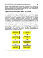
2.2 Outline of Gateway cloning
Gateway cloning technology is based on the lambda phage infection system, in which site-
specific reversible recombination reactions occur during phage integration into and excision
from E. coli genome (Figure 2). In this process, the attP site (242 bp) of lambda phage and the
attB site (25 bp) of E. coli recombine (in a BP reaction) and the lambda phage genome is
integrated into the E. coli genome. After the recombination reaction, the lambda phage
genome is flanked by the attL (100 bp) and attR (168 bp) sites. In the reverse reaction, the
Genetic Engineering – Basics, New Applications and Responsibilities
38
phage DNA is excised from the E. coli genome by recombination between the attL and attR
sites (in an LR reaction). The BP reaction needs two proteins, the phage integrase (Int) and
the E. coli integration host factor (IHF). The mixture of these two proteins is called BP
clonase in the Gateway system. In the LR reaction, Int, IHF and one more phage protein,
excisionase (Xis), are required, and this mixture is called LR clonase. The Gateway cloning
method uses these att sites and clonases for construction of recombinant DNA in vitro.
Fig. 2. BP and LR reactions in lambda phage infection of E. coli. The site-specific reversible
BP and LR recombination reactions occur during lambda phage integration into and
excision from the E. coli genome
Basic strategies for application of Gateway technology to plasmid construction are shown in
Figure 3. For the basic Gateway system, four pairs of modified att sites were generated for
directional cloning. They are attB1 and attB2, attP1 and attP2, attL1 and attL2, and attR1 and
attR2; a recombination reaction can occur only in the combinations of attB1 and attP1, attB2
and attP2, attL1 and attR1, or attL2 and attR2, since recombination strictly depends on att
sequences (Hartley et al., 2000; Walhout et al., 2000). In addition to these att sites, the
negative selection marker ccdB, the protein product of which inhibits DNA gyrase, and a
chloramphenicol-resistance (Cm
r
) marker are used for selection and maintenance of
Gateway vectors. Usually, att1 is located at the 5‘ end of the open reading frame (ORF) and
att2 is located at the 3‘ end. This orientation is maintained in all cloning steps. First, the gene
of interest should be cloned in an entry vector by TOPO cloning (pENTR/D-TOPO), a BP
reaction (pDONR221), or restriction endonuclease and ligase (pENTR1A). Each vector is
available from Invitrogen. To make an entry clone by a BP reaction, the attB1 and attB2
sequences are added to the 5‘ and 3‘ ends, respectively, of the ORF by adapter PCR. The
product (attB1-ORF-attB2) is subjected to a BP reaction with a donor vector, pDONR221,
which possesses an attP1-ccdB-Cm
r
-attP2 cassette. Because of the negative selection marker
ccdB between attP1 and attP2, only transformants harboring the recombined vectors carrying
attL1-ORF-attL2 (the entry clone) can grow on the selection plate. Once the entry clone is in
hand, the ORF is transferred to a destination vector that possesses an attR1-Cm
r
-ccdB-attR2
cassette. Since destination vectors also contain ccdB between attR1 and attR2, and have a
selection marker gene that is different from the entry clone, only the recombined destination
vectors carrying attB1-ORF-attB2 will be selected. Gateway cloning is designed so that the
smallest att sequence, attB (25 bp), appears in the final product to minimize the length of
cloning junctions after the clonase reaction. In N- or C-terminal fusion constructs, the ORF is
linked to a tag with eight or more amino acids encoded by the attB1 or attB2 sites. Because
Gateway Vectors for Plant Genetic Engineering: Overview of Plant Vectors,
Application for Bimolecular Fluorescence Complementation (BiFC) and Multigene Construction
39
the reading frame of attB1 and attB2 is unified in the Gateway system, any entry clone
incorporated into a destination vector is correctly fused to the tag sequence.
Fig. 3. Schematic illustration of Gateway cloning. An entry clone is constructed by TOPO
directional cloning, a BP reaction or restriction digestion and ligation. For construction using
the BP reaction, the ORF region is amplified by adapter PCR and the resulting attB1-ORF-attB2
fragment is cloned into pDONR221 by a BP reaction to generate an entry clone containing
attL1-ORF-attL2. Subsequently, the ORF is cloned into destination vectors by an LR reaction
to generate expression clones including tagged fusion constructs. For D-TOPO cloning,
CACC is added to the ORF by adapter PCR, and the resulting CACC-ORF fragment is
cloned into pENTR/D-TOPO. B1, attB1; B2, attB2; P1, attP1; P2, attP2; L1, attL1; L2, attL2; R1,
attR1; R2, attR2; Pro, promoter; Ter, terminator; Cm
r
, chloramphenicol resistance marker;
ccdB, negative selection marker in E. coli.; Km
r
, kanamycin-resistance marker
3. Binary vectors compatible with Gateway cloning
A large number of binary vectors compatible with Gateway cloning, known as destination
vectors, have been developed and are summarized in a review (Karimi et al., 2007b).
Gateway compatible binary vectors for promoter analysis have the general structure attR1-
Genetic Engineering – Basics, New Applications and Responsibilities
40
Cm
r
-ccdB-attR2-tag-terminator, and after an LR reaction with an attL1-promoter-attL2 entry
clone, they yield an attB1-promoter-attB2-tag-terminator binary construct. Gateway
compatible binary vectors for expression of tagged fusion proteins have the general
structure promoter-tag-attR1-Cm
r
-ccdB-attR2-terminator (for N-terminal fusions) or
promoter-attR1-Cm
r
-ccdB-attR2-tag-terminator (for C-terminal fusions). After an LR reaction
with an attL1-ORF-attL2 entry clone, they respectively yield promoter-tag-attB1-ORF-attB2-
terminator or promoter-attB1-ORF-attB2-tag-terminator. The tag added to the N-terminus of
the ORF is linked by the peptide encoded by the attB1 sequence (XSLYKKAGX), and the tag
added to the C-terminus is linked by the peptide encoded by the attB2 sequence
(XPAFLYKVX). Gateway compatible binary vectors for RNAi analysis (Helliwell &
Waterhouse, 2003; Hilson et al., 2004; Karimi et al., 2002; Miki & Shimamoto, 2004) generally
have the inverted structure of cassettes: promoter-attR1-ccdB-attR2-linker-attR2-ccdB-attR1-
terminator. By an LR reaction with an attL1-trigger-attL2 entry clone, the trigger sequence is
incorporated into both sites in opposite orientations, yielding a promoter-attB1-trigger-
attB2-linker-attB2-(complementary trigger)-attB1-terminator construct. When the construct is
introduced into plants, hairpin RNA is expressed and processed into small interfering RNA
that functions in gene silencing.
Among many Gateway compatible binary vector series, the pW (Karimi et al., 2002), pMDC
(Brand et al., 2006; Curtis & Grossniklaus, 2003) and pEarleyGate (Earley et al., 2006) series
contain vectors available for many kinds of experiments in plants. The pW series consists of
vectors for overexpression or antisense repression by the cauliflower mosaic virus 35S
promoter (P
35S
), for promoter analysis using luciferase (LUC), β-glucuronidase (GUS), or
GFP-GUS as reporters, and for construction of gene fusions with GFP, cyan fluorescent
protein (CFP), yellow fluorescent protein (YFP) or red fluorescent protein (RFP). The pMDC
series consists of vectors for cloning, for overexpression by P
35S
, for inducible expression by
heat shock or estrogen treatment, for promoter analysis using GFP-6xHis or GUS as
reporter, and for gene fusions with GFP, GFP-6xHis, or GUS. The pEarleyGate is a BASTA
®
-
resistance binary vector series consisting of vectors for overexpression by P
35S
, for promoter
analysis using HA, FLAG, Myc, or AcV5, and for gene fusions with YFP, HA, FLAG, Myc,
AcV5, tandem affinity purification (TAP) tags, YFP-HA, or GFP-HA.
The vectors described above are useful tools; however, sometimes it is necessary to use a
different series if an existing one does not have a vector of the required type. In order to
carry out most experiments within the same series (having a unified backbone and a unified
junction sequence), we constructed a comprehensive Gateway compatible binary vector
system carrying many reporters and tags based on the same backbone, as mentioned in next
section.
4. Development of Gateway binary vector (pGWB) series
To make Gateway compatible binary vectors efficiently, we first tried to establish a
systematic method for construction of a vector series. For this purpose, we designed a
construction method for introducing a tag sequence by blunt end ligation to save time and
labor caused by restriction sites in the tag sequence. Based on this notion, platform vectors
pUGW0 and pUGW2 (Nakagawa et al., 2007a) were made using pUC119 as the backbone.
As described below, many Gateway binary vector (pGWB) series were constructed from
intermediate plasmid pUGWs, which were made with pUGW0 or pUGW2. The
Gateway Vectors for Plant Genetic Engineering: Overview of Plant Vectors,
Application for Bimolecular Fluorescence Complementation (BiFC) and Multigene Construction
41
characteristics and accession nos. of each pGWB are summarized in Information of Gaeway
Binary Vectors (pGWBs) (
4.1 Platform vectors pUGW0 and pUGW2 for construction of pGWB series
The platform vectors pUGW0 and pUGW2 include P
35S
and the nopaline synthase
terminator (Tnos), as shown in Figure 4. A pUGW0 was the starting vector for N-terminal
fusions, with the structure HindIII-P
35S
-XbaI-ATG-Aor51HI-attR1-Cm
r
-ccdB-attR2-SacI-Tnos.
A tag (reporter or epitope tag) sequence amplified by blunt-end PCR was introduced into
the Aor51HI site (blunt end) to yield HindIII-P
35S
-XbaI-ATG-tag-attR1-Cm
r
-ccdB-attR2-SacI-
Tnos. In the case of a small epitope tag, an oligonucleotide could be introduced directly into
the Aor51HI site. Translation is initiated at the ATG just upstream of the Aor51HI site.
pUGW2 was the starting vector for C-terminal fusions, with the structure HindIII-XbaI-
HindIII-P
35S
-XbaI-attR1-Cm
r
-ccdB-attR2-Aor51HI-SacI-Tnos. Tag sequences were introduced
by the same method used for pUGW0. The P
35S
region could be easily removed by digestion
with XbaI followed by self-ligation for construction of promoter-less pUGWs. Because there
is no need to digest the tag fragment with restriction enzymes to introduce it into the
Aor51HI site of pUGW0 and pUGW2, any tag fragment can be cloned by the same method.
With these simple procedures, a pUGW series containing a variety of tags was efficiently
generated. They were sources of Gateway cassettes including tag sequences, and were used
for construction of a Gateway binary vector (pGWB). Moreover, the pUGWs are Gateway
compatible plant vectors useful for transient expression analysis after particle bombardment
or protoplast transformation. Because of their small size and high copy number in E. coli,
preparation and handling of pUGW plasmids are very easy.
Fig. 4. Procedure for construction of pUGWs. pUGW0 and pUGW2 are the starting vectors
for construction of new pUGW derivatives. The tag sequence amplified by blunt-end PCR is
introduced into the Aor51HI site of pUGW0 or pUGW2, which yields pUGWs for N-fusion
or C-fusion. The region between P
35S
and Tnos is indicated. The nucleotide sequence
corresponding to the region from attR1 to attR2 is underlined. Cm
r
, chloramphenicol
resistance marker; ccdB, negative selection marker in E. coli.; P
35S
, 35S promoter
4.2 The pGWB series (pGWBxx and pGWB2xx) based on the pBI plasmid
Initially, pGWB was constructed on the backbone of modified pBI carrying a nopaline
synthase promoter (Pnos) driven neomycin phosphotransferase II (NPTII) and P
35S
-driven
Genetic Engineering – Basics, New Applications and Responsibilities
42
hygromycin phosphotransferase (HPT), which confer kanamycin-resistance (Km
r
) and
hygromycin-resistance (Hyg
r
), respectively, to plants (Mita et al., 1995). The initial pGWB
series (pGWBxx) consists of 36 vectors designed for simple cloning of genes (pGWB1), for
overexpression of ORF clones (pGWB2), and for fusion with a variety of tags (pGWB3
through pGWB45) as shown in the Complete List of pGWB (http://shimane-
u.org/nakagawa/gbv.htm). GUS, TAP and LUC are available for C-fusion, and 10 other
tags, sGFP, 6xHis, FLAG, 3xHA, 4xMyc, 10xMyc, GST, T7, enhanced yellow fluorescent
protein (EYFP), and enhanced cyan fluorescent protein (ECFP), are available for both N- and
C-fusion. The promoter-less C-fusion vectors can be used for promoter analysis. By an LR
reaction with a promoter entry clone, a binary construct of promoter:tag is created. The
remaining N- and C-fusion vectors contain P
35S
for constitutive expression. By an LR
reaction with an ORF entry clone, binary constructs expressing tag-ORF or ORF-tag are
easily obtained (Figure 5). With the pGWBs, promoter activity, detection of tagged proteins,
and subcellular localization of proteins can be analyzed effectively (Nakagawa et al., 2007a).
Fig. 5. Cloning into pGWB by LR reaction. The Gateway region in pGWB (top of the figure)
represents a variety of acceptor sites (R1-R2) described in the box. The pGWB series includes
plasmids with no promoter and no tag, or with no promoter and a C-tag. These are used for
expression controlled by a gene’s own promoter. The pGWB plasmids also include the
following types: a 35S promoter and no tag, a 35S promoter and a C-tag, and a 35S promoter
and an N-tag. These are used for constitutive expression using the 35S promoter. After an
LR reaction with the entry clone, the expression clones indicated in the right panel are
obtained. The tag is fused via the attB sequence. B1, attB1; B2, attB2; L1, attL1; L2, attL2;
R1, attR1; R2, attR2; Tnos, nopaline synthase terminator; M, selection marker for plant; Cm
r
,
chloramphenicol-resistance marker; ccdB, negative selection marker in E. coli.; P
35S
, 35S
promoter
We also constructed pGWBs carrying the Pnos:HPT:Tnos marker instead of P
35S
:HPT:Tnos
(pGWB1-45) to avoid a possible effect of the P
35S
sequence on the expression pattern and
Gateway Vectors for Plant Genetic Engineering: Overview of Plant Vectors,
Application for Bimolecular Fluorescence Complementation (BiFC) and Multigene Construction
43
strength of the cloned gene (Zheng et al., 2007). These vectors are named pGWB203, 204, 228
and 235, and their characters are shown at the bottom of the Complete List of pGWB
( In early experiments, when the phosphate
transporter PHT1 promoter was used for promoter analysis in A. thaliana, GUS activity in
plant extracts was 5-fold higher with pGWB3 than with pGWB203 (Nakagawa et al., 2007a).
4.3 Improved Gateway binary vector (ImpGWB) series (pGWB4xx, pGWB5xx,
pGWB6xx and pGWB7xx) based on the pPZP plasmid
We next constructed improved Gateway binary vectors (ImpGWBs) using pPZP as a
backbone (Hajdukiewicz et al., 1994). In the ImpGWB system, handling of plasmid is largely
improved, transformation efficiency in E. coli is drastically increased and much larger
amount of plasmid DNA was recovered. The structures and characters of pGWBs (pBI
backbone) and ImpGWBs (pPZP backbone) are summarized in Figure 6.
Fig. 6. Characters of pGWBs and ImpGWBs. The Gateway region in vectors represents a
variety of acceptor sites as described in the Figure 5. Pnos, nopaline synthase promoter; Tnos,
nopaline synthase terminator; P
35S
, 35S promoter; NPTII, neomycin phosphotransferase II;
HPT, hygromycin phophotransferase; bar, bialaphos resistance gene; GPT, UDP-N-
acetylglucosamine: dolichol phosphate N-acetylglucosamine-1-P transferase (Koizumi &
Iwata, 2008; Koizumi et al., 1999) gene. Km
r
, kanamycin-resistance; Hyg
r
, hygromycin-
resistance; Spc
r
, spectinomycin-resistance; BASTA
®r
, BASTA
®
-resistance; Tunicamycin
r
,
tunicamycin-resistance
At present, four kinds of ImpGWB, the Km
r
subseries (pGWB4xx) (Nakagawa et al., 2007b),
Hyg
r
subseries (pGWB5xx) (Nakagawa et al., 2007b), BASTA
®
-resistance subseries
(pGWB6xx) (Nakamura et al., 2010) and tunicamycin-resistance subseries (pGWB7xx)
(Tanaka et al., 2011), are available, and they are useful for introducing multiple transgenes
into plants by repetitive transformation. Each subseries is composed of 46 vectors as
Genetic Engineering – Basics, New Applications and Responsibilities
44
summarized in the Complete List of ImpGWB (
A set of 16 tags, sGFP, GUS, LUC, EYFP, ECFP, G3 green fluorescent protein (G3GFP),
monomeric red fluorescent protein (mRFP), TagRFP, 6xHis, FLAG, 3xHA, 4xMyc, 10xMyc,
GST, T7, and TAP, is available in ImpGWB. Because ImpGWB is highly efficient in
transformation of E. coli, this series was used for development of a new cloning system
using multiple LR reactions as described below.
4.4 R4 Gateway binary vector (R4pGWB) series (R4pGWB4xx, R4pGWB5xx,
R4pGWB6xx and R4pGWB7xx) for promoter swapping
To assemble multiple DNA fragments in the desired order, an additional four att sites (att3,
att4, att5 and att6) have been developed and applied to MultiSite Gateway cloning (Karimi
et al., 2007a; Sasaki et al., 2004). Utilization of these att sites (att1-6) expanded the availability
of cloning technology for more complex gene construction. The cloning system equipped
with these att sites is useful for swapping of promoters, ORFs and tags, and is also
applicable for cloning of multiple transgenes in one vector (Chen et al., 2006). In a typical
MultiSite Gateway system, three entry clones containing specialized att sites, attL4-
promoter-attR1, attL1-ORF-attL2, and attR2-tag-attL3 are simultaneously connected and
incorporated into a destination vector carrying attR4-Cm
r
-ccdB-attR3 acceptor sites to make
an attB4-promoter-attB1-ORF-attB2-tag-attB3 construct (Figure 7).
Fig. 7. MultiSite Gateway system. In the MultiSite Gateway system, att1, att2, att3 and att4
sequences are used for cloning of multiple DNA fragments into one vector. A promoter
entry clone (L4-Pro-R1), ORF entry clone (L1-ORF-L2), tag entry clone (R2-tag-L3) and
destination vector R4-R3 are subjected to an LR reaction. The promoter, ORF and tag
sequences are linked and incorporated into the destination vector to form a promoter:ORF-
tag clone. B1, attB1; B2, attB2; B3, attB3; B4, attB4; L1, attL1; L2, attL2; L3, attL3; L4, attL4; R1,
attR1; R2, attR2; R3, attR3; R4, attR4; P1, attP1; P2, attP2; P3, attP3; P4, attP4; P1R, attP1R;
P2R; attP2R; Cm
r
, chloramphenicol-resistance marker; ccdB, negative selection marker in
E. coli.; Pro, promoter; Km
r
, kanamycin-resistance marker
Gateway Vectors for Plant Genetic Engineering: Overview of Plant Vectors,
Application for Bimolecular Fluorescence Complementation (BiFC) and Multigene Construction
45
Although MultiSite Gateway cloning is an excellent method for building a complicated
multigene construct, it is relatively difficult to obtain the desired clone because four
recombinations at each att site are required for successful cloning. To facilitate multi-
fragment cloning, especially for promoter swapping, we developed the R4 Gateway binary
vector (R4pGWB) by reducing the number of recombinations needed from four to three
(att4, att1 and att2) (Figure 8, left) (Nakagawa et al., 2008). The R4pGWB series was made by
replacing the attR1 site of ImpGWBs (promoter-less and C-fusion type with four resistance
markers) with the attR4 site; all tags used in ImpGWB are also available in the R4pGWB
system as shown in the Complete List of R4pGWB (http://shimane-
u.org/nakagawa/gbv.htm). By an LR reaction with a promoter entry clone (attL4-promoter-
attR1), an ORF entry clone (attL1-ORF-attL2) and R4pGWB equipped with the appropriate
tag, construction of chimeric genes among promoters, ORFs, and tags (attB4-promoter-attB1-
ORF-attB2-tag) is achieved very easily. The R4pGWB system is a powerful tool to express an
ORF by any desired promoter, e.g., a promoter for strong expression, for tissue or cell
specific expression, for developmental stage specific expression, or for induction by biotic or
abiotic stimuli.
Fig. 8. R4pGWB and R4L1pGWB systems. A promoter entry clone (L4-Pro-R1) is constructed
by a BP reaction using pDONR P4-P1R and a B4-Pro-B1 fragment prepared by adapter PCR.
Left; in the R4pGWB system, a promoter entry clone (L4-Pro-R1), ORF entry clone (L1-ORF-
L2) and R4pGWB are subjected to an LR reaction. The promoter and ORF are linked and
incorporated into R4pGWB to form a promoter:ORF-tag clone. Right; in the R4L1pGWB
system, only a promoter entry clone (L4-Pro-R1) is used for an LR reaction with an
R4L1pGWB. The promoter sequence is incorporated into R4L1pGWB and fused with the tag
on the vector. With the R4L1pGWB system using a single LR reaction, a promoter:tag
construct is obtained at high efficiency. Nucleotides in red indicate B4 and B1 sequences.
Pro, promoter; B1, attB1; B2, attB2; B4, attB4; L1, attL1; L2, attL2; L4, attL4; R1, attR1; R2,
attR2; R4, attR4; P4, attP4; P1R, attP1R; M, selection marker for plant; Cm
r
, chloramphenicol-
resistance marker; ccdB, negative selection marker in E. coli.; Pro, promoter; Km
r
, kanamycin-
resistance marker
Genetic Engineering – Basics, New Applications and Responsibilities
46
4.5 R4L1 Gateway binary vector (R4L1pGWB) series (R4L1pGWB4xx and
R4L1pGWB5xx) for promoter analysis
Due to establishment of the R4pGWB system, many kinds of attL4-promoter-attR1 entry
clones were constructed and have been used as a resource for expression of ORFs in plants.
We plan to also utilize these resources of attL4-promoter-attR1 entry clones for efficient
promoter:tag experiments, and developed an R4L1 Gateway binary vector (R4L1pGWB)
(Nakamura et al., 2009) containing attR4-Cm
r
-ccdB-attL1-tag-Tnos. By the simple bipartite
LR reaction with attL4-promoter-attR1 and R4L1pGWB, an attB4-promoter-attB1-tag-Tnos
construct used for promoter assays can be easily obtained in this system (Figure 8, right).
The tags in R4L1pGWBs are G3GFP-GUS, GUS, LUC, EYFP, ECFP, G3GFP and TagRFP as
shown in the Complete List of R4L1pGWB (
5. Application of the pGWB system
Because Gateway cloning is efficient, precise, flexible and simple to use, its application will
continue to grow in plant research. In this section, we briefly describe two recent advances
in our pGWB system, a split reporter for interaction analysis and recycling cloning for
multigene constructs.
5.1 Gateway vectors for bimolecular fluorescence complementation (BiFC) assay
BiFC is based on the reconstitution of a fluorescent signal when two interacting proteins or
peptides, which are fused to either an N- or C-fragment of a split fluorescent protein,
interact. Due to its relative technical simplicity and the ability to use fluorescence
microscopes for observation, a growing number of publications describe the use of BiFC to
analyze protein-protein interactions. In addition to monitoring protein-protein interactions,
this method has expanded to wider application, such as multicolor BiFC to investigate
protein complexes (Hu & Kerppola, 2003; Kodama & Wada, 2009; Lee et al., 2008; Waadt et
al., 2008), detection in vivo (Bracha-Drori et al., 2004; Walter et al., 2004) and combined with
bioluminescence resonance energy transfer (BRET; Chen et al., 2008; Gandia et al., 2008; Xu
et al., 2007). To date, several BiFC vectors dedicated to plant research have been constructed.
Among our efforts in development of Gateway technology, we have generated various
destination vectors for BiFC assays. In this section, we introduce our Gateway technology-
based BiFC vectors, and describe their application.
5.1.1 Detection of protein-protein interactions in plant cells by BiFC assay
The investigation of protein-protein interactions provides valuable information in cell
biology. In addition to BiFC, several other techniques detect protein-protein interactions,
such as co-immunoprecipitation assays (Co-IP), in vitro binding assays, the yeast two-hybrid
system (Y2H; James et al., 1996), the mating-based split-ubiquitin system (mbSUS; Ludewig
et al., 2003; Obrdlik et al., 2004), BRET(Chen et al., 2008; Xu et al., 2007), fluorescence
resonance energy transfer (FRET; Day et al., 2001), fluorescence lifetime imaging microscopy
(FLIM; Bastiaens & Squire, 1999) and fluorescence correlation spectroscopy (FCS; Hink et al.,
2002). The imaging-based approaches such as BiFC and FRET have been utilized in plant
research because they enable detection in plant cells, in contrast to Y2H and mbSUS, which
Gateway Vectors for Plant Genetic Engineering: Overview of Plant Vectors,
Application for Bimolecular Fluorescence Complementation (BiFC) and Multigene Construction
47
are functional only in yeast cells, and because they do not require specific antibodies or
purification of proteins, unlike Co-IP and in vitro binding assays.
The BiFC assay is one of the most convenient techniques among the image-based
approaches. Although FRET and FLIM are useful and powerful techniques for detection of
protein-protein interactions, FRET requires complicated analysis such as of acceptor
bleaching and an exclusive device is necessary for FLIM. Although several considerations
are required even for BiFC assays, special devices are not required for detection, and
complicated analysis is not necessary after obtaining image data. In addition, the BiFC assay
provides information on subcellular location of the interacting proteins.
We used our Gateway vector construction system (Hino et al., 2011; Nakagawa et al., 2008;
Nakagawa et al., 2007b) to make destination vectors for BiFC assays. Using these vectors, it
is easy to make constructs for detection of protein-protein interactions. These Gateway
vectors have worked well in plant cells (Goto et al., 2011; Hino et al., 2011; Singh et al., 2009).
5.1.2 Principles of the BiFC assay
In BiFC assays, a fluorescent reporter, such as CFP, GFP, YFP and RFP, is split into two non-
fluorescent fragments, N- and C- fragments (Figure 9A,B). Two proteins or peptides, which
are to be tested for interaction, are fused at the N- or C-terminus of each fragment. After
expression of both fusion genes simultaneously, if an interaction occurs between the two
proteins, the non-fluorescent fragments are reconstituted and behave as an unsplit
fluorescent protein. Therefore, the detection of fluorescence means the target proteins
interact (Figure 9A).
Once the interaction occurs, the reconstituted molecule does not dissociate into non-
fluorescent fragments, leading to enhancement of fluorescence due to accumulation of
reconstituted fluorescent proteins.
There are eight potential combinations to be tested for protein-protein interactions in a BiFC
assay, taking into account which protein of the two partners tested is fused to the N- or C-
terminal end of which N- or C- fragment (Figure 9C). However, improper fusion of a split
fragment sometimes abolishes protein function and masks information on subcellular
targeting. For example, the peroxisome targeting signal 2 (PTS2) must be fused to the N-
terminus of the split fluorescent protein (Singh et al., 2009; Figure 10B). In contrast, PTS1
must be fused to the C-terminus of a split fluorescent protein, because its location at the C-
terminus is necessary for its function. In these cases, the number of combinations tested is
fewer. However, if there is no information on protein function, all combinations should be
tested. Viewed in this light, our destination vectors are useful for construction of several
fusion genes at the same time.
5.1.3 Destination vectors for the multicolor and in vivo BiFC assays
Various BiFC vectors have been developed and used in plant research (Bracha-Drori et al.,
2004; Diaz et al., 2005; Ding et al., 2006; Goto et al., 2011; Hino et al., 2011; Loyter et al., 2005;
Maple et al., 2005; Marrocco et al., 2006; Ohad et al., 2007; Singh et al., 2009; Waadt et al.,
2008; Walter et al., 2004; Zamyatnin et al., 2006). All the vectors, including ours, use P
35S
to
Genetic Engineering – Basics, New Applications and Responsibilities
48
Fig. 9. Principles of the BiFC assay. (A) Nonfluorescent fragments (YN and YC) of a
fluorescent protein are brought together through interaction of the tested proteins or
peptides (a, b and c) to which they are fused. The interaction of the two proteins causes
reconstitution of a fluorescent signal. (B) Diagram of amino acid substitutions among CFP,
GFP, YFP and mRFP1, and the positions where they were fragmented. Although there are
alternative positions to split a fluorescent protein into two fragments (Hu & Kerppola, 2003;
Waadt et al., 2008), the CFP, GFP and YFP in our system were split between residues 174
and 175, and mRFP1, which contains an amino acid substitution of the 66th glutamine to
threonine, was split between residues 154 and 155. Amino acids in CFP and YFP that were
converted from GFP are depicted in white. In the case of RFP, amino acids that are different
from GFP are not represented, since there are many substitutions. (C) Potential combination
of two fragments. There are eight possible configurations in the BiFC assay. Each target
protein (gray and black) can be fused at its N- or C- terminus to the N- or C-terminal
fragment of the fluorescent protein (light green)
express a fusion gene. There are two ways to insert a target gene into the 5’ or 3’ end of a
split fragment of fluorescent protein gene: (1) cloning into a multicloning site using
digestion and ligation, and (2) Gateway technology (Hino et al., 2011; Walter et al., 2004).
Our BiFC vectors were developed to be compatible with Gateway technology. We generated
four kinds of destination vectors for BiFC assays (Figure 10A), enabling the transfer of a
gene of interest from the entry clone to the 5’ or 3’ end of each split fragment. Therefore,
researchers are able to easily fuse a gene of interest to the 5’ or 3’ end of the split fragment,
leading to various convenient constructs.
The BiFC vectors were initially generated using YFP (Hu et al., 2002). However, other
fluorescent proteins, BFP (Hu & Kerppola, 2003), CFP (Kodama & Wada, 2009; Lee et al.,
2008), GFP (Hu et al., 2002; Kodama & Wada, 2009), Venus, (Lee et al., 2008), Cerulean (Lee
et al., 2008), DsRed-monomer (Kodama & Wada, 2009), mRFP1 (Jach et al., 2006), mCherry
(Fan et al., 2008), and a far-red fluorescent protein, mLumin (Chu et al., 2009), have
reportedly been useful for BiFC assay. We adopted CFP, GFP, YFP and mRFP1 to generate
vectors (Figure 9B), and verified their usefulness for detection of protein-protein interactions