Proteomic Applications in Biology Part 12 pptx
Bạn đang xem bản rút gọn của tài liệu. Xem và tải ngay bản đầy đủ của tài liệu tại đây (1.03 MB, 17 trang )
Proteomic Applications in Biology
176
highlight the clear separation between the known and predicted Golgi- and ER-localized
protein clusters. Significantly a number of cell wall biosynthetic enzymes were identified
including a number of glycosyltransferases. This confirmed LOPIT as a valid method for
discriminating between Golgi- and ER-localized proteins from Arabidopsis crude
membrane fractions (Dunkley et al., 2004). Further development of the LOPIT technique
replaced ICAT with isotope tagging of Arabidopsis membrane peptide fractions for both
relative and absolute protein quantitation (iTRAQ) (Dunkley et al., 2006) (Fig. 4). The iTRAQ
method is a progression of ICAT by labeling the free primary amines of peptides with four
different iTRAQ reporter tags (114, 115, 116 and 117 m/z). They are detectable by MS/MS,
which allows for simultaneous quantification analysis of up to four peptide samples (Wiese
et al., 2007). Arabidopsis membrane peptide fractions were differentially tagged with the
four iTRAQ reporters, fractionated and analyzed by MudPIT and Q-TOF MS. The addition
of SCX to RP LC-MS/MS provided superior peptide separation and identification, resulting
in 689 Arabidopsis protein identifications. Multivariate analysis of iTRAQ-labeled MS/MS
data revealed 89 proteins in the Golgi density gradient cluster. This more extensive analysis
further validated the approach as further cell wall biosynthetic enzymes such as
glycosyltransferases and sugar interconverting enzymes were identified as well as
transporters, V-ATPase components and a variety of proteins with likely Golgi functions
(Dunkley et al., 2006). This was a significant improvement on the initial LOPIT set of ten
Arabidopsis Golgi-localized proteins by ICAT and LC-MS/MS (Dunkley et al., 2004).
To test its robustness in other biological system, LOPIT was used to investigate the
subcellular distribution of proteins from Drosophila embryos. A total of 329 Drosophila
proteins were identified and localized to three subcellular locations; the plasma membrane
(94), mitochondria (67) and the ER/Golgi (168) (Tan et al., 2009). The lack of distinction
between ER- and Golgi-residing Drosophila proteins by LOPIT underscored the significant
challenges faced when dissecting complex and heterogeneous biological samples, as
opposed to a simplified system of crude membranes from a relatively homogenous
Arabidopsis cell culture.
A similar strategy to LOPIT but employing label-free quantitation techniques is protein
correlation profiling (PCP). PCP uses quantitation of unmodified peptide ions by MS to
bypass the chemical modification step in ICAT and iTRAQ, which results in less
complicated MS/MS spectra and higher confidence in peptide identifications (Andersen et
al., 2003; Foster et al., 2006). However, it is heavily reliant on invariable conditions in 2D LC-
MS/MS for reproducible quantitation between samples. Proof of concept for PCP was first
demonstrated with purified human centrosomes (Andersen et al., 2003) and in the cellular
context with sucrose density gradient separations of mouse liver homogenate (Foster et al.,
2006). A total of 1,404 mouse liver proteins were identified by 2D LC-MS/MS (LTQ-FT) and
their MS ion distribution profiles were mapped by PCP to ten different subcellular locations.
These results were corroborated with MS ion distribution profiles and enzymatic assays of
known organelle marker proteins and immunofluorescence staining of mouse liver cells for
visual confirmation of select proteins with overlapping or non-overlapping PCPs. While this
study reported rates of 61 to 93% overlap from comparing its mitochondrial-localized
protein set with previous human and mouse mitochondrial proteomes, the rates of overlap
were considerably lower for proteins localized to the plasma membrane (49%) and Golgi
(36%). Nonetheless, they made significant inroads in characterizing the mouse Golgi
proteome and identified a series of Rab proteins, mannosyltransferases, COP components,
transporters and a diverse range of transferases (Foster et al., 2006).
The Current State of the Golgi Proteomes
177
Density gradient
centrifugation
Western blot
114
116115
117
Protein
digestion and
iTRAQ labeling
Labeled peptide
samples pooled
Intensity
1.0
0.0
114
116
115
117
iTRAQ
ion tags
m/z
b4
y5
y6
y7
Peptide identification
y4
Organelle protein
distributions and
identification by
LC-MS/MS
mitochondria
plastid
Golgi
ER
Golgi marker
plastid marker
mito marker
ER marker
Fig. 4. Outline of the LOPIT technique using crude cellular extracts. LOPIT employs
centrifugation of a self-forming iodixanol density gradient to partially resolve organelle
fractions. Western blotting of the fractions for known Golgi and ER marker proteins show
that in most cases, there is overlap between them. A series of four protein fractions are
digested with trypsin and treated with iTRAQ reagents containing the labels 114, 115, 116 or
117m/z and pooled for LC-MS/MS analysis. Ion intensity measurements of the iTRAQ
reporter ion fragments 114 to 117 m/z providing the basis of protein quantitation with
simultaneous analysis of the major b, y and other fragment ions for protein identification.
The introductions of LOPIT and related organelle purification-free methods were intended
to address the issue of separating Golgi from other endomembrane system components, but
this still remains rather difficult to achieve with complex biological systems. Refining these
methods by optimizing density gradient conditions to enhance the resolution of Golgi, along
with continuing development of multivariate techniques are seen as pivotal to expand the
set of genuine Golgi-residing proteins in semi-purified samples (Foster et al., 2006; Trotter et
al., 2010).
Proteomic Applications in Biology
178
5. Free flow electrophoresis (FFE) purification of Golgi
Free Flow Electrophoresis, though 50 years old has adapted well to contemporary research
fields, recently filling a particular niche in subcellular proteomics, in combination with mass
spectrometry. This section explores the role of FFE in isolation of the Golgi apparatus from
plant and mammalian tissues. Essentially, an electric field is applied perpendicular to a
sample as it moves up a separation chamber in a liquid medium. Subcellular components
are therefore separated according to surface charge and organelle streams collected as 96
fractions (Fig. 5). Hydrodynamic stability of the liquid is crucial; convection currents arising
from localized joule heating can disrupt organelle streams. Apparatus design has
consistently advanced along with the fields to which FFE has been applied. MicroFFE
apparatus designs (Turgeon & Bowser, 2009) have overcome some of the imperfections
inherent in the technique. Entirely liquid phase and continuous, FFE is appropriate for large
scale, preparative fractionation of cells, organelles, proteins and peptides. The apparatus can
be operated in two modes: zonal electrophoresis (ZE), or isoelectric focusing (IEF) mode.
ZE-FFE is becoming recognized for its impressive separation and purification capacity of
plant, mammalian and yeast organelles (reviewed by Islinger et al., 2010).
The first use of FFE for Golgi was applied to mammalian Golgi membranes and lead to
separation of sub-Golgi compartments, demonstrated by a series of enzyme assays
(Hartelschenk et al., 1991). However, this was prior to the proteomic era and was never
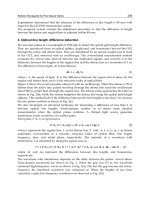
Fig. 5. A schematic diagram of a large scale FFE setup with dimensions shown on the right.
The diagram outlines a late commercially model available through BD Diagnostics, with
counter flow at sample outlets and stabilization buffers at the extreme anodic and cathodic
carrier buffer inlets (Islinger et al., 2010). MicroFFE apparatus are similar with 56.5 mm ×35
mm × 30 mm dimensions (Turgeon & Bowser, 2009).
The Current State of the Golgi Proteomes
179
revisited with modern mass spectrometry tools. Plant homogenates were first subjected to
FFE some decades ago (Kappler et al., 1986; Sandelius et al., 1986; Bardy et al., 1998) but
these first forays demonstrated little potential for Golgi isolation. With plant Golgi
antibodies then, as now, commercially unavailable, enzyme assays were the primary means
of determining fraction composition. Profiling by enzyme assays was not sufficiently precise
or efficient for tracking lower-abundance Golgi proteins amidst a relatively complex
background of contaminants, although the distribution of enzyme activities reported by
(Sandelius et al., 1986) are broadly consistent with later proteomic analyses.
The first isolation of plant Golgi membranes has depended on both FFE and proteomic
advances (Parsons and Heazlewood, unpublished data). Semi-high throughput mass
spectrometry was used to track the electrophoretic migration of Golgi membranes. The
proteins identified in individual fractions were matched against markers protein lists for
each subcellular location, including the cytosol, compiled from SUBA, the SUBcellular
Arabidopsis database (Heazlewood et al., 2007). This allowed simultaneous monitoring of
over 50 proteins in most fractions without recourse to antibodies or enzyme assays. Overlaid
on the total protein output for all 96 fractions, marker lists revealed a detailed picture of
organelle migration (Fig. 6). Once the shoulder peak corresponding to the purest Golgi
fractions had been identified, parameters could be fine tuned, exploiting the
electronegativity of Golgi vesicles and enhancing the cathodic migration of this area relative
to the main protein peak. Total protein output from this targeted Golgi purification study
showed a broader main protein peak and a prominent shoulder on the cathodic edge when
compared to earlier studies on plant homogenates (Kappler et al., 1986; Bardy et al., 1998).
Careful balancing of the carrier buffer flow rate to voltage ratio maximized the separation
range of organelles whilst organelle streams remained focussed. Cathodic migration
increased with voltage but was limited by increasing the flow rate as exposure time to the
electric field was shorter. Lateral diffusion of organelle streams dictated the lower flow rate
Total protein
Golgi
ER
glycosyltransferases
Mitochondria
Peroxisome
Fig. 6. Golgi membrane migration profile after FFE separation. A portion of the total protein
output, measured at 280 nm (fractions 1 to 48) is shown. Around 50 proteins were identified
in each fraction scanned using semi-high throughput LC-MS/MS. Overlaid are matches
from marker protein lists compiled from the SUBA subcellular database and the ~50
identified proteins from each fraction. Many glycosyltransferases are located in the Golgi
and were used as a further guide for Golgi membrane migration.
Proteomic Applications in Biology
180
limit. Golgi fractions with minimal contamination were identified through continued
monitoring and selected for detailed proteomic characterization (Parsons and Heazlewood,
unpublished data).
The application of FFE, mass spectrometry and proteomic data as tools for Golgi isolation
and characterization marked a precedent for plant Golgi proteomics. Previously, relatively
few plant Golgi proteins had been identified by proteomic techniques (Dunkley et al., 2006).
The application of FFE to isolate high purity Golgi fractions resulted in a Golgi proteome of
425 proteins identified in at least two of three biological replicates. This included over 50
glycosyltransferases, 25 transporters, the entire V-ATPase complex, a variety of trafficking
components, methyltransferases and acetyltransferases (Parsons and Heazlewood,
unpublished data). While proteins identified in a single preparation were excluded from the
final proteome, they nevertheless present a useful resource for functional analysis of the
plant Golgi apparatus. With so little Golgi proteomic data resources, common contaminants
originating from the Golgi in other proteomes were difficult to identify. This therefore
represents both significant progress in our potential to understand Golgi processes and
consolidation of the current state of subcellular protein localization in plants. As an example
the ectoapyrase protein APY1 is currently classified as a plasma membrane protein involved
in extracellular signaling through the hydrolysis of phosphate from ATP (Wu et al., 2007).
The APY1 protein was identified in all three replicates and YFP tagging confirmed its Golgi
localization. Heterologous expression of this protein in the yeast nucleoside diphosphatase
(NDPase) mutant gda1, rescued the glycosylation phenotype in this mutant, thus
functionally characterizing the APY1 protein as a Golgi-resident NDPase (Parsons and
Heazlewood, unpublished data). Since most glycosylation occurs in the Golgi, the APY1
protein represents a resident and functional Golgi protein, rather than a transitory plasma
membrane localized protein. Furthermore, plasma membrane and Golgi compartments are
easily separated using FFE (Bardy et al., 1998) with Golgi and ER compartments partially
separated (Fig. 6). Thus, selectively pre-enriching organelles and tailoring FFE parameters
for maximal separation has considerable potential in distinguishing between resident and
transitory proteins in the secretory system. Some proteins observed after FFE purification of
the plasma membrane were present in all three Golgi preparations and can be readily
classified as ‘transient proteins’ rather than contaminants (Parsons and Heazlewood,
unpublished data).
Given the success achieving high purity fractions (Taylor et al., 1997a) and sub-
compartmental resolution of Golgi structures (Hartelschenk et al., 1991), it is surprising that
a corresponding proteomic study has not been undertaken in rats. FFE was foremost
amongst techniques compared for purification of mouse mitochondria (Hartwig et al., 2009)
whilst impressive results were achieved after separating populations of PM vesicles
(Cutillas et al., 2005), suggests that FFE still has much to contribute to both Golgi and other
subcellular proteomes. In Arabidopsis, the Golgi proteome was characterized from only two
to three fractions out of approximately 15 fractions over which Golgi proteins were detected.
Further studies suggested this reflects medial to trans-Golgi separation (Parsons and
Heazlewood, unpublished data). Could FFE separate the remainder of the Golgi from
contaminating membranes or even Golgi sub-compartments? Chemical modification of
Golgi compartments holds some promise; addition of ATP was found to enhance migration
of membrane compartments towards the cathode (Barkla et al., 2007). Unfortunately no
mass spectrometry was undertaken in this study. A low ionic strength two-component
buffer system permits separation at lower currents, reducing convection from joule heating,
The Current State of the Golgi Proteomes
181
as could the use of microFFE setups, enhancing sub-compartment separation. FFE has
already enhanced our knowledge of Golgi proteomics but its role is clearly far from over
and there is much potential for further advances using FFE.
6. Comparative analysis of the Golgi proteomes
The characterization of the Golgi apparatus and associated secretory components by mass
spectrometry has been undertaken on a range of species. While most of these organisms
represent model systems with extensive genetic resources and well annotated genomes,
analyses have been undertaken in less tractable systems, namely pine trees (Mast et al.,
2010). Nonetheless, with the exception of work undertaken in rat, only a handful of
analyses have focused on the proteomic characterization of the Golgi and its associated
membranes from model systems. This is in contrast to the extensive series of proteomic
studies undertaken on organelles from many of these systems. For example, in the model
plant Arabidopsis over ten separate proteomic analyses have been undertaken on plasma
membrane fractions, six studies on mitochondria and eight analyses of the plastid
(Heazlewood et al., 2007). These facts further highlight the technical challenges when
attempting to isolate high purity Golgi fractions and associated structures, even from well
studied model systems. Overall, searches of the literature were able to identify over
twenty separate studies that have employed proteomics techniques to address the
characterization of the Golgi apparatus and associated secretory components. These
studies have been undertaken using a diverse collection of isolation and enrichment
techniques over the past decade and have employed a range of proteomics approaches
including 2-DE (Taylor et al., 1997b; Morciano et al., 2005), 1-DE (Peng et al., 2008), iTRAQ
(Dunkley et al., 2006), spectral counting (Foster et al., 2006) and MudPIT (Wu et al., 2004).
These studies also covered the range of protein identification methods namely Peptide
Mass Fingerprinting (Morciano et al., 2005), Edman degradation (Bell et al., 2001) and
MS/MS (Gilchrist et al., 2006).
The protein identifications outlined in these works were extracted from the published
manuscripts and online supplementary material to produce a collection of proteins
identified in each study. Protein sequences were obtained from GenBank or UniProt for each
accession and consolidated at the species level using BLAST analysis tool against minimally
redundant protein sets where available. These comprised the International Protein Index
(Kersey et al., 2004) for human, mouse, rat and bovine, The Arabidopsis Information
Resource (Swarbreck et al., 2008) for Arabidopsis, the Saccharomyces Genome Database
(Cherry et al., 1997) for yeast, FlyBase (Tweedie et al., 2009) for Drosophila and the Rice
Genome Annotation Project (Ouyang et al., 2007) for rice. This enabled the classification of
the total number of proteins identified from the Golgi apparatus and associated membranes
based on each isolation method and by each species (Table 1). Finally, the total number of
non-redundant proteins currently assigned to the Golgi apparatus and associated
membrane components for each species could also be ascertained (Table 1). Where possible,
we relied on annotation information and classifications outlined in each manuscript to
determine whether a protein should be included in the final lists. This included early
endosome, secretory and unknowns (when efforts to classify contaminants had been
undertaken). The largest number of proteins assigned to the Golgi of any one species is that
of rat. This reflects the number of individual studies and the fact that this represented the
major system used to study the Golgi proteome for a number of years.
Proteomic Applications in Biology
182
Species Density
centrifugation
Immuno-
affinity
Free Flow
Electrophoresis
Correlation
Analysis
Total
Pine 10 10
Human 24 18
Rice 49 43
Drosophila 168 168
Bovine 252 238
Yeast 241 52 276
Mouse 2711
a
56 490 428
Arabidopsis 145 425 92 534
Rat 1117 57 996
Table 1. The total number of proteins, by species and technique, currently identified by
proteomic approaches from the Golgi apparatus and associated membrane systems.
a
The
analysis of mouse microsomes by density centrifugation (Kislinger et al., 2006) has not been
included in the final total for this species as it represents a crude microsomal fraction.
The set of non-redundant protein sequences compiled from the proteomic analyses of the
Golgi were assembled for cross species orthology analysis. In order to remove identical
genes and splice variants, these sequences were first clustered at 95% sequence identity and
only one representative from each cluster carried over for subsequent analysis. Following
this, the sequences were clustered at 30% identity. All clustering was performed with the
program uCLUST (Edgar, 2010). A protein was mapped to an ortholog of another species if
at least one representative of that species was present in the same cluster. Proteins were
considered paralogs when two or more sequences from the same species were present in a
cluster in which sequences from no other species were present (Fig. 7)
After homology matching, a number of gene families were found across the Golgi
proteomes of most species. These included Rab GTPases, heat-shock proteins, alpha-
mannosidases, thioredoxins, and cyclophilins. Apart from the Rab GTPases, which mediate
vesicle trafficking, the other families are involved in protein folding and protein
glycosylation. There were a number of large clusters containing only Arabidopsis genes and
these clusters were contained glycosyltransferases associated with synthesis of the plant cell
wall (Scheller & Ulvskov, 2010). In addition, there was a cluster of pine sequences
containing laccases, which may be associated with the synthesis of lignin in woody tissue
(Ranocha et al., 2002). In general, when only a few proteins had been reported in a species,
those proteins were more likely to have orthologs in the other species in the set. This
suggests that the most easily detected proteins in proteomics studies are abundant proteins
involved in core Golgi-related functions that have not diverged as greatly over evolutionary
history as the less abundant and harder to find proteins.
The Current State of the Golgi Proteomes
183
Fig. 7. Orthology interaction map of the non-redundant Golgi proteome sets. The size of the
species circle indicates the number of proteins identified in the Golgi proteome of that
species. The pink shading indicates the number of paralogs for a given species. The lines
indicate orthology connections between the species with the thickness indicating the
number of proteins. The Scale refers to the number of proteins represented by the thickness
of the line.
7. Conclusion
The characterization of the Golgi proteome from various systems represents an important
technical and biological achievement. Its central role within the cell in functions ranging
from cell wall biosynthesis to protein glycosylation to secretion is of significant importance.
Knowledge about these functions contributes to both our fundamental understanding of
complex eukaryotic systems to their exploitation in areas of biofuels (cell wall manipulation)
and agriculture (milk production). While there is clearly more basic knowledge required to
understand the functionally complex roles of the Golgi apparatus, advances made by work
outlined in this chapter demonstrate that the first decade of proteomics has been fruitful and
improvements to isolation and analysis methods are promising for the field going forward.
8. Acknowledgment
The work conducted by the Joint BioEnergy Institute was supported by the Office of Science,
Office of Biological and Environmental Research, of the U.S. Department of Energy under
Contract No. DE-AC02-05CH11231. GD and EP were supported by start-up funds from the
University of California, Davis.
Proteomic Applications in Biology
184
9. References
Andersen, J.S.; Wilkinson, C.J.; Mayor, T.; Mortensen, P.; Nigg, E.A. & Mann, M. (2003).
Proteomic characterization of the human centrosome by protein correlation
profiling. Nature, Vol.426, No.6966, pp. 570-574
Asakura, T.; Hirose, S.; Katamine, H.; Sato, M.; Hujiwara, M.; Shimamoto, K.; Hori, F. &
Mitsui, T. (2006). Rice Golgi proteome: Analysis of GFP-syp31 labeled cis Golgi
membrane. Plant and Cell Physiology, Vol.47, pp. S26-S26
Bae, M.S.; Cho, E.J.; Choi, E.Y. & Park, O.K. (2003). Analysis of the Arabidopsis nuclear
proteome and its response to cold stress. Plant Journal, Vol.36, No.5, pp. 652-663
Bardy, N.; Carrasco, A.; Galaud, J.P.; Pont-Lezica, R. & Canut, H. (1998). Free-flow
electrophoresis for fractionation of Arabidopsis thaliana membranes. Electrophoresis,
Vol.19, No.7, pp. 1145-1153
Barkla, B.J.; Vera-Estrella, R. & Pantoja, O. (2007). Enhanced separation of membranes
during free flow zonal electrophoresis in plants. Analytical Chemistry, Vol.79, No.14,
pp. 5181-5187
Bell, A.W.; Ward, M.A.; Blackstock, W.P.; Freeman, H.N.; Choudhary, J.S.; Lewis, A.P.;
Chotai, D.; Fazel, A.; Gushue, J.N.; Paiement, J.; Palcy, S.; Chevet, E.; Lafreniere-
Roula, M.; Solari, R.; Thomas, D.Y.; Rowley, A. & Bergeron, J.J. (2001). Proteomics
characterization of abundant Golgi membrane proteins. Journal of Biological
Chemistry, Vol.276, No.7, pp. 5152-5165
Blondeau, F.; Ritter, B.; Allaire, P.D.; Wasiak, S.; Girard, M.; Hussain, N.K.; Angers, A.;
Legendre-Guillemin, V.; Roy, L.; Boismenu, D.; Kearney, R.E.; Bell, A.W.; Bergeron, J.J.
& McPherson, P.S. (2004). Tandem MS analysis of brain clathrin-coated vesicles reveals
their critical involvement in synaptic vesicle recycling. Proceedings of the National
Academy of Sciences of the United States of America, Vol.101, No.11, pp. 3833-3838
Breuza, L.; Halbeisen, R.; Jeno, P.; Otte, S.; Barlowe, C.; Hong, W. & Hauri, H.P. (2004).
Proteomics of endoplasmic reticulum-Golgi intermediate compartment (ERGIC)
membranes from brefeldin A-treated HepG2 cells identifies ERGIC-32, a new
cycling protein that interacts with human Erv46. Journal of Biological Chemistry,
Vol.279, No.45, pp. 47242-47253
Burre, J.; Zimmermann, H. & Volknandt, W. (2007). Immunoisolation and subfractionation
of synaptic vesicle proteins. Analytical Biochemistry, Vol.362, No.2, pp. 172-181
Cherry, J.M.; Ball, C.; Weng, S.; Juvik, G.; Schmidt, R.; Adler, C.; Dunn, B.; Dwight, S.; Riles,
L.; Mortimer, R.K. & Botstein, D. (1997). Genetic and physical maps of
Saccharomyces cerevisiae. Nature, Vol.387, No.6632, pp. 67-73
Cutillas, P.R.; Biber, J.; Marks, J.; Jacob, R.; Stieger, B.; Cramer, R.; Waterfield, M.;
Burlingame, A.L. & Unwin, R.J. (2005). Proteomic analysis of plasma membrane
vesicles isolated from the rat renal cortex. Proteomics, Vol.5, No.1, pp. 101-112
Dalton, A.J. & Felix, M.D. (1953). Studies on the Golgi Substance of the Epithelial Cells of the
Epididymis and Duodenum of the Mouse. American Journal of Anatomy, Vol.92,
No.2, pp. 277-305
de Curtis, I.; Howell, K.E. & Simons, K. (1988). Isolation of a fraction enriched in the trans-
Golgi network from baby hamster-kidney cells. Experimental Cell Research, Vol.175,
No.2, pp. 248-265
Dominguez, M.; Fazel, A.; Dahan, S.; Lovell, J.; Hermo, L.; Claude, A.; Melançon, P. &
Bergeron, J.J.M. (1999). Fusogenic domains of golgi membranes are sequestered
into specialized regions of the stack that can be released by mechanical
fragmentation. The Journal of Cell Biology, Vol.145, No.4, pp. 673-688
The Current State of the Golgi Proteomes
185
Drakakaki, G.; van de Ven, W.; Pan, S.; Miao, Y.; Wang, J.; Keinath, N.K.; Weatherly, B.;
Jiang, L.; Schumacher, K.; Hicks, G. & Raikhel, N. (2011). Isolation and proteomic
analysis of the SYP61 compartment reveal its role in exocytic trafficking in
Arabidopsis. Cell Research, doi: 10.1038/cr.2011.1129
Dröscher, A. (1998). Camillo Golgi and the discovery of the Golgi apparatus. Histochemistry
and Cell Biology, Vol.109, No.5, pp. 425-430
Dunkley, T.P.; Watson, R.; Griffin, J.L.; Dupree, P. & Lilley, K.S. (2004). Localization of
organelle proteins by isotope tagging (LOPIT). Molecular & Cellular Proteomics,
Vol.3, No.11, pp. 1128-1134
Dunkley, T.P.J.; Hester, S.; Shadforth, I.P.; Runions, J.; Weimar, T.; Hanton, S.L.; Griffin, J.L.;
Bessant, C.; Brandizzi, F.; Hawes, C.; Watson, R.B.; Dupree, P. & Lilley, K.S. (2006).
Mapping the Arabidopsis organelle proteome. Proceedings of the National Academy of
Sciences of the United States of America, Vol.103, No.17, pp. 6518-6523
Edgar, R.C. (2010). Search and clustering orders of magnitude faster than BLAST.
Bioinformatics, Vol.26, No.19, pp. 2460-2461
Forsmark, A.; Rossi, G.; Wadskog, I.; Brennwald, P.; Warringer, J. & Adler, L. (2011).
Quantitative proteomics of yeast post-Golgi vesicles reveals a discriminating role
for Sro7p in protein secretion. Traffic, Vol.12, No.6, pp. 740-753
Foster, L.J.; de Hoog, C.L.; Zhang, Y.; Xie, X.; Mootha, V.K. & Mann, M. (2006). A mammalian
organelle map by protein correlation profiling. Cell, Vol.125, No.1, pp. 187-199
Friso, G.; Giacomelli, L.; Ytterberg, A.J.; Peltier, J.B.; Rudella, A.; Sun, Q. & Wijk, K.J. (2004).
In-depth analysis of the thylakoid membrane proteome of Arabidopsis thaliana
chloroplasts: new proteins, new functions, and a plastid proteome database. Plant
Cell, Vol.16, No.2, pp. 478-499
Gilchrist, A.; Au, C.E.; Hiding, J.; Bell, A.W.; Fernandez-Rodriguez, J.; Lesimple, S.; Nagaya,
H.; Roy, L.; Gosline, S.J.; Hallett, M.; Paiement, J.; Kearney, R.E.; Nilsson, T. &
Bergeron, J.J. (2006). Quantitative proteomics analysis of the secretory pathway.
Cell, Vol.127, No.6, pp. 1265-1281
Golgi, C. (1898). Intorno alla struttura della cellula nervosa. Archives Italiennes de Biologie,
Vol.30, pp. 60-71
Gygi, S.P.; Rist, B.; Gerber, S.A.; Turecek, F.; Gelb, M.H. & Aebersold, R. (1999). Quantitative
analysis of complex protein mixtures using isotope-coded affinity tags. Nature
Biotechnology, Vol.17, No.10, pp. 994-999
Hanton, S.L.; Bortolotti, L.E.; Renna, L.; Stefano, G. & Brandizzi, F. (2005). Crossing the
divide transport between the endoplasmic reticulum and Golgi apparatus in
plants. Traffic, Vol.6, No.4, pp. 267-277
Hartelschenk, S.; Minnifield, N.; Reutter, W.; Hanski, C.; Bauer, C. & Morre, D.J. (1991).
Distribution of glycosyltransferases among Golgi-apparatus subfractions from liver
and hepatomas of the rat. Biochimica et Biophysica Acta, Vol.1115, No.2, pp. 108-122
Hartwig, S.; Feckler, C.; Lehr, S.; Wallbrecht, K.; Wolgast, H.; Muller-Wieland, D. & Kotzka,
J. (2009). A critical comparison between two classical and a kit-based method for
mitochondria isolation. Proteomics, Vol.9, No.11, pp. 3209-3214
Heazlewood, J.L.; Verboom, R.E.; Tonti-Filippini, J.; Small, I. & Millar, A.H. (2007). SUBA:
The Arabidopsis subcellular database. Nucleic Acids Research, Vol.35, pp. D213-D218
Heazlewood, J.L.; Tonti-Filippini, J.S.; Gout, A.M.; Day, D.A.; Whelan, J. & Millar, A.H.
(2004). Experimental analysis of the Arabidopsis mitochondrial proteome
highlights signaling and regulatory components, provides assessment of targeting
prediction programs, and indicates plant-specific mitochondrial proteins. Plant Cell,
Vol.16, No.1, pp. 241-256
Proteomic Applications in Biology
186
Hobman, T.C.; Zhao, B.; Chan, H. & Farquhar, M.G. (1998). Immunoisolation and
characterization of a subdomain of the endoplasmic reticulum that concentrates
proteins involved in COPII vesicle biogenesis. Molecular Biology of the Cell, Vol.9,
No.6, pp. 1265-1278
Inadome, H.; Noda, Y.; Adachi, H. & Yoda, K. (2005). Immunoisolaton of the yeast Golgi
subcompartments and characterization of a novel membrane protein, Svp26,
discovered in the Sed5-containing compartments. Molecular & Cellular Biology,
Vol.25, No.17, pp. 7696-7710
Islinger, M.; Eckerskorn, C. & Volkl, A. (2010). Free-flow electrophoresis in the proteomic
era: A technique in flux. Electrophoresis, Vol.31, No.11, pp. 1754-1763
Kaiser, C.A. & Schekman, R. (1990). Distinct sets of SEC genes govern transport vesicle
formation and fusion early in the secretory pathway. Cell, Vol.61, No.4, pp. 723-733
Kappler, R.; Kristen, U. & Morre, D.J. (1986). Membrane flow in plants: Fractionation of
growing pollen tubes of tobacco by preparative free-flow electrophoresis and
kinetics of labeling of endoplasmic reticulum and Golgi apparatus with
[3H]leucine. Protoplasma, Vol.132, No.1-2, pp. 38-50
Kersey, P.J.; Duarte, J.; Williams, A.; Karavidopoulou, Y.; Birney, E. & Apweiler, R. (2004).
The International Protein Index: an integrated database for proteomics
experiments. Proteomics, Vol.4, No.7, pp. 1985-1988
Kikuchi, M.; Hatano, N.; Yokota, S.; Shimozawa, N.; Imanaka, T. & Taniguchi, H. (2004).
Proteomic analysis of rat liver peroxisome: presence of peroxisome-specific
isozyme of Lon protease. Journal of Biological Chemistry, Vol.279, No.1, pp. 421-428
Kislinger, T.; Cox, B.; Kannan, A.; Chung, C.; Hu, P.; Ignatchenko, A.; Scott, M.S.; Gramolini,
A.O.; Morris, Q.; Hallett, M.T.; Rossant, J.; Hughes, T.R.; Frey, B. & Emili, A. (2006).
Global survey of organ and organelle protein expression in mouse: combined
proteomic and transcriptomic profiling. Cell, Vol.125, No.1, pp. 173-186
Kleffmann, T.; Russenberger, D.; von Zychlinski, A.; Christopher, W.; Sjolander, K.;
Gruissem, W. & Baginsky, S. (2004). The Arabidopsis thaliana chloroplast proteome
reveals pathway abundance and novel protein functions. Current Biology, Vol.14,
No.5, pp. 354-362
Klemm, R.W.; Ejsing, C.S.; Surma, M.A.; Kaiser, H.J.; Gerl, M.J.; Sampaio, J.L.; de Robillard,
Q.; Ferguson, C.; Proszynski, T.J.; Shevchenko, A. & Simons, K. (2009). Segregation
of sphingolipids and sterols during formation of secretory vesicles at the trans-
Golgi network. Journal of Cell Biology, Vol.185, No.4, pp. 601-612
Lilley, K.S. & Dunkley, T.P. (2008). Determination of genuine residents of plant
endomembrane organelles using isotope tagging and multivariate statistics.
Methods in Molecular Biology, Vol.432, pp. 373-387
Mast, S.; Peng, L.; Jordan, T.W.; Flint, H.; Phillips, L.; Donaldson, L.; Strabala, T.J. & Wagner,
A. (2010). Proteomic analysis of membrane preparations from developing Pinus
radiata compression wood. Tree Physiology, Vol.30, No.11, pp. 1456-1468
Mikami, S.; Hori, H. & Mitsui, T. (2001). Separation of distinct compartments of rice Golgi
complex by sucrose density gradient centrifugation. Plant Science, Vol.161, No.4,
pp. 665-675
Morciano, M.; Burre, J.; Corvey, C.; Karas, M.; Zimmermann, H. & Volknandt, W. (2005).
Immunoisolation of two synaptic vesicle pools from synaptosomes: a proteomics
analysis. J Neurochem, Vol.95, No.6, pp. 1732-1745
Morre, J. & Mollenhauer, H.H. (2009). The Golgi apparatus: The first 100 years. Springer,
ISBN 978-0-387-74346-2, New York, USA
The Current State of the Golgi Proteomes
187
Mosley, A.L.; Florens, L.; Wen, Z. & Washburn, M.P. (2009). A label free quantitative
proteomic analysis of the Saccharomyces cerevisiae nucleus. Journal of Proteomics,
Vol.72, No.1, pp. 110-120
Ouyang, S.; Zhu, W.; Hamilton, J.; Lin, H.; Campbell, M.; Childs, K.; Thibaud-Nissen, F.;
Malek, R.L.; Lee, Y.; Zheng, L.; Orvis, J.; Haas, B.; Wortman, J. & Buell, C.R. (2007).
The TIGR Rice Genome Annotation Resource: improvements and new features.
Nucleic Acids Research, Vol.35, pp. D883-887
Paiement, J.; Young, R.; Roy, L. & Bergeron, J.J. (2005). Isolation of rough and smooth
membrane domains of the endoplasmic reticulum from rat liver, In: Cell Biology: A
Laboratory Handbook, J. Celis, N. Carter, K. Simons, V. Small, T. Hunter, & D.M.
Shotton, (Eds.) 41-44, Elsevier Academic Press, ISBN 978-0-12-164730-8, Burlington,
MA, USA
Peng, L.; Rawson, P.; McLauchlan, D.; Lehnert, K.; Snell, R. & Jordan, T.W. (2008). Proteomic
analysis of microsomes from lactating bovine mammary gland. Journal of Proteome
Research, Vol.7, No.4, pp. 1427-1432
Ranocha, P.; Chabannes, M.; Chamayou, S.; Danoun, S.; Jauneau, A.; Boudet, A.M. & Goffner,
D. (2002). Laccase down-regulation causes alterations in phenolic metabolism and cell
wall structure in poplar. Plant Physiology, Vol.129, No.1, pp. 145-155
Richardson, P.J. & Luzio, J.P. (1986). Immunoaffinity purification of subcellular particles and
organelles. Applied Biochemistry and Biotechnology, Vol.13, No.2, pp. 133-145
Sadowski, P.G.; Dunkley, T.P.; Shadforth, I.P.; Dupree, P.; Bessant, C.; Griffin, J.L. & Lilley,
K.S. (2006). Quantitative proteomic approach to study subcellular localization of
membrane proteins. Nature Protocols, Vol.1, No.4, pp. 1778-1789
Sandelius, A.S.; Penel, C.; Auderset, G.; Brightman, A.; Millard, M. & Morre, D.J. (1986).
Isolation and highly purified fractions of plasma-membrane and tonoplast from the
same homogenate of soybean hypocotyls by Free-Flow Electrophoresis. Plant
Physiology, Vol.81, No.1, pp. 177-185
Scheller, H.V. & Ulvskov, P. (2010). Hemicelluloses. Annual Review of Plant Biology, Vol.61,
pp. 263-289
Sickmann, A.; Reinders, J.; Wagner, Y.; Joppich, C.; Zahedi, R.; Meyer, H.E.; Schonfisch, B.;
Perschil, I.; Chacinska, A.; Guiard, B.; Rehling, P.; Pfanner, N. & Meisinger, C. (2003).
The proteome of Saccharomyces cerevisiae mitochondria. Proceedings of the National
Academy of Sciences of the United States of America, Vol.100, No.23, pp. 13207-13212
Steuble, M.; Gerrits, B.; Ludwig, A.; Mateos, J.M.; Diep, T.M.; Tagaya, M.; Stephan, A.;
Schatzle, P.; Kunz, B.; Streit, P. & Sonderegger, P. (2010). Molecular characterization
of a trafficking organelle: dissecting the axonal paths of calsyntenin-1 transport
vesicles. Proteomics, Vol.10, No.21, pp. 3775-3788
Swarbreck, D.; Wilks, C.; Lamesch, P.; Berardini, T.Z.; Garcia-Hernandez, M.; Foerster, H.; Li,
D.; Meyer, T.; Muller, R.; Ploetz, L.; Radenbaugh, A.; Singh, S.; Swing, V.; Tissier, C.;
Zhang, P. & Huala, E. (2008). The Arabidopsis Information Resource (TAIR): gene
structure and function annotation. Nucleic Acids Research, Vol.36, pp. D1009-1014
Takatalo, M.S.; Kouvonen, P.; Corthals, G.; Nyman, T.A. & Rönnholm, R.H. (2006).
Identification of new Golgi complex specific proteins by direct organelle proteomic
analysis. Proteomics, Vol.6, pp. 3502-3508
Tan, D.J.; Dvinge, H.; Christoforou, A.; Bertone, P.; Martinez Arias, A. & Lilley, K.S. (2009).
Mapping organelle proteins and protein complexes in Drosophila melanogaster.
Journal of Proteome Research, Vol.8, No.6, pp. 2667-2678
Proteomic Applications in Biology
188
Taylor, R.S.; Jones, S.M.; Dahl, R.H.; Nordeen, M.H. & Howell, K.E. (1997a). Characterization
of the Golgi complex cleared of proteins in transit and examination of calcium
uptake activities. Molecular Biology of the Cell, Vol.8, No.10, pp. 1911-1931
Taylor, R.S.; Fialka, I.; Jones, S.M.; Huber, L.A. & Howell, K.E. (1997b). Two-dimensional
mapping of the endogenous proteins of the rat hepatocyte Golgi complex cleared of
proteins in transit. Electrophoresis, Vol.18, No.14, pp. 2601-2612
Taylor, R.S.; Wu, C.C.; Hays, L.G.; Eng, J.K.; Yates, J.R., 3rd & Howell, K.E. (2000).
Proteomics of rat liver Golgi complex: minor proteins are identified through
sequential fractionation. Electrophoresis, Vol.21, No.16, pp. 3441-3459
Taylor, S.W.; Fahy, E.; Zhang, B.; Glenn, G.M.; Warnock, D.E.; Wiley, S.; Murphy, A.N.;
Gaucher, S.P.; Capaldi, R.A.; Gibson, B.W. & Ghosh, S.S. (2003). Characterization of the
human heart mitochondrial proteome. Nature Biotechnology, Vol.21, No.3, pp. 281-286
Trotter, M.W.; Sadowski, P.G.; Dunkley, T.P.; Groen, A.J. & Lilley, K.S. (2010). Improved
sub-cellular resolution via simultaneous analysis of organelle proteomics data
across varied experimental conditions. Proteomics, Vol.10, No.23, pp. 4213-4219
Turck, N.; Richert, S.; Gendry, P.; Stutzmann, J.; Kedinger, M.; Leize, E.; Simon-Assmann, P.;
Van Dorsselaer, A. & Launay, J.F. (2004). Proteomic analysis of nuclear proteins
from proliferative and differentiated human colonic intestinal epithelial cells.
Proteomics, Vol.4, No.1, pp. 93-105
Turgeon, R.T. & Bowser, M.T. (2009). Micro free-flow electrophoresis: theory and
applications. Analytical and Bioanalytical Chemistry, Vol.394, No.1, pp. 187-198
Tweedie, S.; Ashburner, M.; Falls, K.; Leyland, P.; McQuilton, P.; Marygold, S.; Millburn, G.;
Osumi-Sutherland, D.; Schroeder, A.; Seal, R. & Zhang, H. (2009). FlyBase:
enhancing Drosophila Gene Ontology annotations. Nucleic Acids Research, Vol.37,
pp. D555-559
Walworth, N.C. & Novick, P.J. (1987). Purification and characterization of constitutive
secretory vesicles from yeast. Journal of Cell Biology, Vol.105, No.1, pp. 163-174
Wiese, S.; Reidegeld, K.A.; Meyer, H.E. & Warscheid, B. (2007). Protein labeling by iTRAQ: a
new tool for quantitative mass spectrometry in proteome research. Proteomics,
Vol.7, No.3, pp. 340-350
Wu, C.C.; Yates, J.R., 3rd; Neville, M.C. & Howell, K.E. (2000). Proteomic analysis of two
functional states of the Golgi complex in mammary epithelial cells. Traffic, Vol.1,
No.10, pp. 769-782
Wu, C.C.; MacCoss, M.J.; Howell, K.E. & Yates, J.R., 3rd. (2003). A method for the
comprehensive proteomic analysis of membrane proteins. Nature Biotechnology,
Vol.21, No.5, pp. 532-538
Wu, C.C.; MacCoss, M.J.; Mardones, G.; Finnigan, C.; Mogelsvang, S.; Yates, J.R., 3rd &
Howell, K.E. (2004). Organellar proteomics reveals Golgi arginine dimethylation.
Molecular Biology of the Cell, Vol.15, No.6, pp. 2907-2919
Wu, J.; Steinebrunner, I.; Sun, Y.; Butterfield, T.; Torres, J.; Arnold, D.; Gonzalez, A.; Jacob,
F.; Reichler, S. & Roux, S.J. (2007). Apyrases (nucleoside triphosphate-
diphosphohydrolases) play a key role in growth control in Arabidopsis. Plant
Physiology, Vol.144, No.2, pp. 961-975
Part 4
Comparative Approaches in Biology
9
Differentiation of Four Tuna Species
by Two-Dimensional Electrophoresis
and Mass Spectrometric Analysis
Tiziana Pepe
1
, Marina Ceruso
1
, Andrea Carpentieri
2
, Iole Ventrone
1
,
Angela Amoresano
2
, Aniello Anastasio
1
and Maria Luisa Cortesi
1
1
Dipartimento di Scienze Zootecniche e Ispezione degli Alimenti – Università di Napoli
2
Dipartimento di Chimica Organica e Biochimica – Università di Napoli
Italy
1. Introduction
Species belonging to the genus Thunnus are pelagic predator fishes, commonly known as
tuna. The species within this genus are of commercial value, and six of them are considered
the most valued in world trade (D.M., MIPAAF, 31 Gennaio 2008). Thunnus species originate
from a variety of geographic areas, and for this reason the different species can be
characterized by the presence of different biological contaminants and sensory
characteristics. The species Thunnus thynnus has a higher quality and commercial value due
to its excellent organoleptic features.
Tuna species are usually consumed as fillets or processed products. The loss of the external
anatomical and morphological features makes the authentication of a fish species difficult or
impossible and enables fraudulent substitutions (Marko et al., 2004). Species substitution is
very common in fish products, due to the profits resulting from the use of less expensive
species. For species of tuna, substitutions have both commercial and health implications
(Agusa et al., 2005; Besada et al., 2006; Storelli et al., 2010), thus, analytical techniques to
differentiate fish species are essential. The development of suitable analytical methods for
fish species identification in prepared and transformed fish products is of great interest to
enforcement agencies involved with labelling regulations and the authentication of fish in
various products to prevent the substitution of fish species (Mackie et al., 2000; Meyer et al.,
1995).
Several biochemical techniques enable the study and identification of fillet or minced fish
species. Among these methods, isoelectric focusing (IEF) (Etienne et al., 2000; Rehbein et al.,
2000; Renon et al., 2001;), capillary zone electrophoresis (Acuña et al., 2008), and
amplification of selected DNA sequences by the polymerase chain reaction (PCR) have been
used for the identification of certain groups of fish species (Espiñeira et al., 2008; Hubalkova
et al., 2008; Pepe et al., 2005, 2007; Trotta et al., 2005).
Presently, PCR is the most frequently used technique, as DNA is heat-stable and resistant to
heat treatments that may be applied to the tuna during processing. However, obtaining an
accurate species identification is very difficult if the species show a high degree of homology
as Thunnus does (Chow & Kishino, 1995; Lopez & Pardo, 2005; Michelini et al., 2007; Pardo
Proteomic Applications in Biology
192
& Begoña, 2004; Terio et al., 2010; Viñas & Tudela, 2009). The sequences usually used as
species molecular markers are the DNA mitochondrial fragments especially cytochome b (cyt
b) genes and the ribosomal 16S and 12S subunits (Kochzius et al., 2010; Russo et al., 1996;
Zehner et al., 1998). Previous studies demonstrated that these molecular markers are not
discriminating for Thunnus species, because they have few polymorphisms expressed by
point mutations (Bottero et al., 2007).
EU Commission Regulation no. 2065/2001 of 22 October 2001 has established detailed rules
for consumer information to be included on labels regarding fish species. Accordingly, it is
also necessary to develop new methods to prevent illegal species substitutions in seafood
products (EC No 2065/2001). Proteins are playing an increasing role in the international
scientific community and proteomics, the large-scale analysis of proteins expressed by a cell
or a tissue contributes greatly to the study of gene function (Pandey & Mann, 2000).
Recently, proteomics has been applied in the fishing industry with several aims, e.g., to
examine the water-soluble muscle proteins from farm and wild fish to show aquaculture
effects on seafood quality (Monti et al., 2005) or to elucidate the influence of internal organ
colonization by Moraxella sp. in internal organs of Sparus aurata (Addis et al., 2010).
Proteomics has also been considered as a tool for species identification in seafood products
with interesting results (Carrera et al., 2006, 2007; Chen et al., 2004; López et al., 2002;
Piñeiro et al., 1999, 2001).
The aim of this chapter is to examine the potential of proteomics to identify four tuna
species through characterisation of specific sarcoplasmic proteins. We investigated T.
albacares, T. alalunga, and T. obesus two dimensional gel electrophesis (2-DE) patterns and
also verified the presence of specie-specific proteins for these tuna species. Muscle extracts
from four tuna species of the genus Thunnus (T. thynnus, T. alalunga, T. albacares, T. obesus)
were evaluated by both mono and 2-DE and mass spectrometric techniques. In preliminary
results (Pepe et al., 2010), proteomics was applied for the identification of a species-specific
protein in T. thynnus by 2-DE profiles. The analysis of two dimensional gels by
ImageMaster
TM
2D Platinum software revealed the presence of a protein with a molecular
weight of approximately 70 kDa in the T. thynnus' 2-DE pattern, which was absent in the
other species. This protein, identified as Trioso fosfato isomerasi (gi46909469) through mass
spectrometric techniques might be considered a specific marker. The aim of this chapter was
to investigate T. albacares, T. alalunga, and T. obesus 2- DE patterns and verify the presence of
species-specific proteins for these tuna species.
2. Materials and methods
2.1 Fish samples
In this study, a total of four different tuna species were tested, with three specimens from
each species. The whole tuna specimens were identified, according to their anatomical and
morphological features, as belonging to T. thynnus, T. alalunga, T. albacores, and T. obesus
species at the Department of Animal Science and Food Inspection, University of Naples,
"Federico II". T. thynnus and T. alalunga specimens were fished in the Mediterranean Sea and
supplied by “Pozzuoli fish market”, T. albacares specimens were fished in the Indian Ocean
and supplied by Salerno P.I.F. (Posto di Ispezione Frontaliera), and T. obesus specimens were
fished in the South East Atlantic Ocean and were obtained from Philadelphia, Pennsylvania,
United States. Fish were frozen on board at – 20 ° C and shipped in insulated boxes to the
laboratory. Tuna muscle samples were taken and stored at -80 ºC for further analysis.