Báo cáo hóa học: " Decreased Fibroblast and Increased Osteoblast Functions on Ionic Plasma Deposited Nanostructured Ti Coatings" doc
Bạn đang xem bản rút gọn của tài liệu. Xem và tải ngay bản đầy đủ của tài liệu tại đây (306.88 KB, 6 trang )
NANO EXPRESS
Decreased Fibroblast and Increased Osteoblast Functions on Ionic
Plasma Deposited Nanostructured Ti Coatings
Ariel Cohen Æ Peishan Liu-Synder Æ Dan Storey Æ
Thomas J. Webster
Received: 2 May 2007 / Accepted: 6 June 2007 /Published online: 4 July 2007
Ó to the authors 2007
Abstract Bioactive coatings are in high demand to control
cellular functions for numerous medical devices. The
objective of this in vitro study was to characterize for the first
time fibroblast (fibrous scar tissue forming cells) adhesion
and proliferation on an important polymeric biomaterial
(silicone) coated with titanium using a novel ionic plasma
deposition (IPD) process. Fibroblasts are one of the first
anchorage-dependent cells to arrive at an implant surface
during the wound healing process. Persistent excessive
functions of fibroblasts have been linked to detrimental fi-
brous tissue formation which may cause implant failure. The
IPD process creates a surface-engineered nanostructure
(with features usually below 100 nm) by first using a vacuum
to remove all contaminants, then guiding charged metallic
ions or plasma to the surface of a medical device at ambient
temperature. Results demonstrated that compared to cur-
rently used titanium and uncoated silicone, silicone coated
with titanium using IPD significantly decreased fibroblast
adhesion and proliferation. Results also showed competi-
tively increased osteoblast (bone-forming cells) over fibro-
blast adhesion on silicone coated with titanium; in contrast,
osteoblast adhesion was not competitively increased over
fibroblast adhesion on uncoated silicone or titanium controls.
In this manner, this study strongly suggests that IPD should
be further studied for biomaterial applications in which fi-
brous tissue encapsulation is undesirable (such as for
orthopedic implants, cardiovascular components, etc.).
Keywords Nanometer Á Coatings Á Fibroblasts Á
Osteoblasts Á Orthopedic
Introduction
Bioactive coatings are in high demand to increase the
functions of cells for numerous medical devices. For
example, to improve the performance of conventional tita-
nium-based materials for orthopedic applications (i.e., fab-
ricated by traditional metallurgy techniques and possibly
surface-treated by mechanical methods such as grinding and
polishing), hydroxyapatite has often been used as a coating
[1]. By simulating the chemical composition of natural bone,
hydroxyapatite coatings on titanium (Ti) greatly enhance
osseointegration between the implant and juxtaposed bone
[2, 3]. Commercially, hydroxyapatite is coated on Ti-based
metals through a high-temperature plasma-spray deposition
process, which transforms the initial nanocrystalline
hydroxyapatite into micron grain size hydroxyapatite con-
taining less crystalline calcium phosphates. Plasma-spray
coating processes have, thus, often been criticized since
they are not versatile enough to handle a wide range of
chemistries and frequently alter the properties of the start-
ing materials. Specifically, plasma-spray deposition of
hydroxyapatite results in phase transformations which may
lead to the formation of highly soluble calcium phosphates
that delaminate during clinical use [1–3].
Moreover, hydroxyapatite coated on traditional ortho-
pedic implants using plasma spray is not known to decrease
the functions of fibroblasts (a cell well known to contribute
to fibrous tissue encapsulation which can decrease ortho-
pedic implant efficacy). In fact, fibrous encapsulation by
excessive fibroblast functions on hydroxyapatite implants
coated by plasma spray deposition is a common mode of
A. Cohen Á P. Liu-Synder Á T. J. Webster (&)
Divisions of Engineering and Orthopaedics, Brown University,
184 Hope Street, Providence, RI 02912, USA
e-mail:
D. Storey
Ionic Fusion, Longmont, CO 70501, USA
123
Nanoscale Res Lett (2007) 2:385–390
DOI 10.1007/s11671-007-9069-1
implant failure. Clearly, materials and associate coating
properties are needed which increase functions of osteo-
blasts (bone forming cells) and simultaneously decrease
functions of fibroblasts.
One promising classification of materials which may
simultaneously increase functions of osteoblasts and
decrease functions of fibroblasts are nanophase materials
[4–13]. Nanophase materials are materials with at least one
dimension <100 nm that mimic the natural surface rough-
ness of bone. Although these findings indicate that nano-
meter surface features are important to increase the
cytocompatibility of currently used implants, traditional
coating processes (like the aforementioned plasma-spray
due to high heat) usually cannot create nanofeatures in
orthopedic coatings to more effectively regenerate bone.
Ways that have been used to create nanoroughness on a
metal substrate to promote bone cell functions include the
use of ultrafine-grained Ti (and other metals) [8, 9],
anodizing Ti [10], and chemical etching of Ti. In terms of
coatings, in 2004, Ionic Fusion Corp. announced a novel
vacuum surface modification process called ionic plasma
deposition (IPD), which allows for accurate control of
material properties during coating procedures. Basically
the IPD process creates a surface-engineered nanostructure
(with features usually below 100 nm) by first using a
vacuum to remove all contaminants. High kinetic energies
(a few 100 eV) then guide charged metallic ions or plasma
to the surface of the medical device. The process runs at
ambient temperature and can be supercooled when re-
quired, enabling a wide range of materials (i.e., Ag, Au, Ti,
etc.) to be coated on a wide range of underlying materials
(for example, metals, polymers, and ceramics).
This novel coating process (IPD) has previously been
shown to increase the functions of osteoblasts (such as
adhesion, proliferation, and the deposition of calcium-
containing mineral) on polymers coated with metals [14,
15]. In particular, promoted functions of osteoblasts have
been measured on ultra high molecular weight polyethyl-
ene (UHMWPE), polytetrafluoroethylene (PTFE) and sili-
cone coated with either gold or Ti using IPD [14, 15].
However, no evidence exists concerning in vitro functions
of fibroblasts on these novel nanostructured coatings. Thus,
the objective of this in vitro study was to characterize
fibroblast functions on one particular polymer (silicone)
coated with nano Ti using the IPD system.
Materials and Methods
Substrates
Silicone was modified using the IPD deposition process.
Raw materials were purchased off the shelf from
McMaster-Carr. This process, preformed in a vacuum,
creates controllable nanometer surface features to mediate
cell attachment. Energy levels of 500 eV were used to
control the properties of the depositing Ti allowing for low
temperature (~30 °C) deposition onto the various sub-
strates. Ti 6–4 was obtained from Process Materials Inc.
90% of the depositing Ti was <100 nm in diameter. Un-
coated samples were used as controls. Moreover, com-
mercially obtained conventional (micron) grain size Ti
(Osteonics) was used as a reference.
After the coatings were completed, all samples were
cleaned with deionized water in an aqua-sonicator with
70% ethanol for 10 min. These cleaned substrates were
dried in an oven at 65 °C and exposed to UV light for 1 h.
The surface roughness of the samples of interest to the
present study was determined using Field Emission Scan-
ning Electron Microscopy (LEO).
Cell Assays
Fibroblasts (CRL-2317, American Type Culture Collec-
tion, population numbers 2–4) and osteoblasts (CRL-
11372, American Type Culture Collection, population
numbers 2–4) were used in the cell experiments in this
study. All substrates of interest were rinsed with phosphate
buffered saline (PBS) (1X strength) before seeding the
cells. The cells were cultured on the substrates in Dul-
becco’s Modified Eagle Medium (Hyclone) supplemented
with 10% fetal bovine serum (Hyclone) and 1% penicillin/
streptomycin (Hyclone) with an initial seeding density of
3500 cells/cm
2
of substrate surface area. Some experi-
ments were performed with fibroblasts alone and some by
simultaneously seeding fibroblasts and osteoblasts (pre-
stained with different fluorescent markers; Molecular
Probes) to ascertain competitive cell adhesion. Cells were
then allowed to adhere on the substrates under standard cell
culture conditions (37 °C temperature, 5% CO
2
and 95%
humidified air) for 4 h. After the prescribed time period,
the cell culture medium was aspirated from the wells and
the substrates were gently rinsed with PBS three times to
remove any non-adherent cells. For the experiments with
fibroblasts alone, the adherent cells were then fixed with a
4% formaldehyde solution (Fisher) and stained with a
Hoescht 33258 dye (Sigma). For competitive fibroblast and
osteoblast experiments, cells were directly visualized at the
conclusion of the experiment using fluorescence micros-
copy. The cell numbers were counted under a fluorescence
microscope (Swiss).
Similar experiments were conducted to determine long-
term fibroblast density with the exception that fibroblasts
were cultured for 1, 3, and 5 days. Media was changed
every other day. Cells were fixed stained, and counted in a
similar fashion to that described above.
386 Nanoscale Res Lett (2007) 2:385–390
123
All experiments were conducted in triplicate at least
three different times.
Results
High Degree of Nanometer Surface Roughness for
Silicone Coated with Ti Using IPD
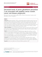
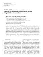
Results of the present study demonstrated the expected
high degree of nanometer surface roughness of silicone
coated with Ti using IPD (Figs. 1, 2). In contrast, uncoated
silicone did not possess a high degree of nanometer surface
roughness.
Decreased Fibroblast Functions on Silicone Coated
with Ti Using IPD
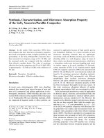
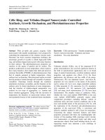
Results of this in vitro study showed for the first time
significantly decreased fibroblast adhesion (Figs. 3, 4),
decreased competitive fibroblast compared to osteoblast
adhesion (Fig. 5), and decreased fibroblast density after 1,
3, and 5 days on silicone coated with Ti using IPD com-
pared to any of the other substrates of interest (Figs. 6, 7).
Half the number of fibroblasts were counted on silicone
coated with Ti compared to uncoated silicone after 4 h. In
addition, four times more osteoblasts competitively ad-
hered to silicone coated with Ti compared to fibroblasts
after 4 h. Of particular interest is that the number of
fibroblasts decreased only on silicone coated with Ti using
IPD from 1 to 3–5 days of culture. Such data provide
strong evidence of the ability of silicone coated with Ti
using IPD to inhibit fibroblast function while promoting
competitive osteoblast function.
Fig. 1 Low magnification scanning electron micrographs of uncoated
and ionic plasma deposited (IPD) Ti on silicone. Numerous
nanometer features were present on IPD coated Ti. Bars = 10 lm
Fig. 2 High magnification scanning electron micrographs of un-
coated and ionic plasma deposited (IPD) Ti on silicone. Numerous
nanometer features were present on IPD coated Ti. Bars = 200 nm for
top and 1 lm for bottom
0
200
400
600
800
Silicone Silicone
Coated with
Titanium
Titanium
)mc erauqs/sllec( ytisneD lleC
*
**
Fig. 3 Decreased fibroblast adhesion on silicone coated with Ti using
ionic plasma deposition after 4 h. Data = mean ± STDEV, n =3;
*p < 0.01 (compared to silicone alone) and **p < 0.01 (compared to
currently-used Ti)
Nanoscale Res Lett (2007) 2:385–390 387
123
Discussion
Ionic plasma deposition is a versatile technique that can be
used to coat different medical devices with diverse chem-
istries. Using conventional deposition methods (such as
plasma-spray deposition), numerous problems exist such as
poor adhesion strength, inability to maintain starting nano-
particle size, change of coating material crystallinity, etc.
[1, 2]. However, in the IPD coating process, ions of the
depositing material are accelerated to ensure that they have
proper energy to coat the specific medical device at room
temperature. As a result, properties of the coatings are
improved and are highly controllable at the nanometer level.
Due to prior studies [4–8], one important property in
material coatings to create to increase osteoblast functions
is nanometer surface features. That is, due to the impor-
tance of nanometer features in promoting bone cell func-
tions and decreasing fibroblast functions, another key
advantage of IPD is that the original particle size, chem-
istry, and crystallinity can be retained due to the low heat
presented during the coating application. Clearly, this
allows IPD to create nanotopographies on conventional
materials to improve their bioactivity properties, as this
study demonstrated. Previous studies have shown that
ceramics and polymers with nanostructured surface fea-
tures decrease fibroblast functions compared to currently
used nanometer smooth implant surfaces [4–8].
Fig. 4 Fluorescent microscopy images of decreased fibroblast
adhesion on silicone coated with Ti using ionic plasma deposition.
Bars = 20 lm
0
500
1000
1500
2000
2500
Silicone Silicone Coated with
Titanium
Titanium
Oste oblasts Fibroblas ts
)mc erauqs/llec( ytisneD lleC
*
**
***
**
***
***
***
Fig. 5 Increased selective osteoblast density on silicone coated with
Ti using ionic plasma deposition after 4 h. Data = mean ± STDEV,
n =3; *p < 0.01 (compared to fibroblast adhesion on respective
sample); **p < 0.01 (compared to respective cell adhesion on silicone
alone); and ***p < 0.01 (compared to respective cell adhesion on Ti)
0
200
400
600
800
1000
1200
1400
1600
1800
2000
Day 1 Day 3 Day 5
)mc erauqs/sl
lec( y
t
isn
eD
lleC
Silicone
Silicone Coated with Titanium
Titanium
*
**
**
*
**
***
*
**
***
**
***
***
Fig. 6 Decreased fibroblast density on silicone coated with Ti using
ionic plasma deposition after 1, 3, and 5 days. Data = mean ± ST-
DEV, n =3;*p < 0.01 (compared to silicone alone at the same time
point); **p < 0.01 (compared to currently-used Ti at the same time
point); and ***p < 0.01 (compared to previous time point on the same
substrate)
388 Nanoscale Res Lett (2007) 2:385–390
123
Such results have consequences not only for orthopedic
applications, in which as discussed the selective promotion
of osteoblast functions are desirable, but also for any im-
plant device in which fibrous tissue encapsulation is
undesirable. For example, for numerous cardiovascular
applications (such as catheters, stents, grafts, etc.), in-
creased fibrous tissue formation decreases the efficacy of a
device. The present results of decreased fibroblast func-
tions on silicone coated with one specific chemistry (Ti)
shows promise for all of these implant applications.
At this time, though, it is unclear what properties of the
coatings enhanced osteoblast adhesion (such as a change in
wettability, chemistry, and/or nanometer surface features).
For example, silicone is a hydrophobic material which may
have been transformed through Ti coatings into hydrophilic
materials to influence cell adhesion. However, as men-
tioned, when compared to traditional Ti (or micron grain
size Ti) which possesses the same chemistry as the poly-
mers coated with Ti, decreased fibroblast adhesion was
measured; this suggests the possibility that nanometer
roughness alone decreased fibroblast adhesion on the
coated samples.
Importantly, a change in nanometer roughness is also
related to changes in wettability since previous studies
have shown lower aqueous contact angles on Ti composed
of nanometer compared to micron grain sizes [11]. Authors
speculated in those studies that hydrophilicity is enhanced
on Ti with nanometer surface features due to the increased
presence of surface defects compared to conventional Ti
[11]. Those studies continued to show greater initial
adsorption of hydrophilic proteins (specifically, vitronec-
tin) and subsequently greater osteoblast functions (from
adhesion to the deposition of calcium containing mineral
on nanograined Ti) and decreased fibroblast functions [12,
13]. More studies, though, are needed for the presently
described IPD process to determine specifically what
properties selectively enhanced osteoblast and decreased
fibroblast adhesion on the coated materials. None-the-less,
this study provides strong evidence for the continued
investigation of IPD for orthopedic applications.
Conclusions
Nanotopography or nanoroughness of an implant surface
is desirable to improve competitive osteoblast functions
while at the same time decrease fibroblast functions
known to contribute to fibrous tissue encapsulation
harmful for orthopedic implant success. With respect to
implant coatings, IPD is an efficient method to deposit
nanostructured coatings onto versatile materials, including
metals and polymers. The current study represents the first
which demonstrated desirable decreased fibroblast
attachment and growth on silicone coated with Ti; this is
in contrast to uncoated silicone or Ti, thus, demonstrating
the strong potential IPD has at increasing conventional
medical device efficacy for a number of biomedical
applications (such as for orthopedic implants, cardiovas-
cular components, etc.).
References
1. = 1405
2. R. Furlong, J.F. Osborn, J. Bone Joint Surg. Br. 73B, 741 (2001)
3. K.C. Baker, M.A. Anderson, S.A. Oehlke, A.I. Astashkina, D.C.
Haikio, J. Drelich and S.W. Donahue, Mater. Sci. Eng.C (in
press)
4. M. Karlsson, E. Palsgard, P.R. Wilshaw, L.D. Silvio, Biomate-
rials 24, 3039 (2003)
5. K.C. Popat, E.E.L. Swan, V. Mukhatyar, K.I. Chatvanichkul,
G.K. Mor, C.A. Grimes, T.A. Desai, Biomaterials 26, 4516
(2005)
6. A.S.G. Curtis, N. Gadegaard, M.J. Dalby, M.O. Riehle, C.D.W.
Wilkinson, G. Aitchison, IEEE Trans. Nanobiosci. 3, 61 (2004)
7. M. Sato, E.B. Slamovich, T.J. Webster, Biomaterials 26(12),
1349 (2005)
8. T.J. Webster, J.U. Ejiofor, Biomaterials 25(19), 4731 (2004)
9. R.Z. Valiev, V.V. Stolyarov, H.J. Rack, T.C. Lowe Medical
Device Materials. (ASM, Materials Park, OH, 2004) p. 362
Fig. 7 Fluorescent microscopy images of decreased fibroblast
density after 5 days on silicone coated with Ti using ionic plasma
deposition. Bars = 20 lm
Nanoscale Res Lett (2007) 2:385–390 389
123
10. C. Yao, V. Perla, J.L. McKenzie, E.B. Slamovich, T.J. Webster,
J. Biomed. Nanotechnol. 1, 68 (2005)
11. T.J. Webster, R.W. Siegel, R. Bizios, Biomaterials 20, 1221
(1999)
12. T.J. Webster, L.S. Schadler, R.W. Siegel, R. Bizios, Tissue Eng.
7, 291 (2001)
13. T.J. Webster, R.W. Siegel, R. Bizios, Biomaterials 21, 1803
(2000)
14. A. Reising, C. Yao, D. Storey, T. J. Webster, J. Biomed. Mater.
(in press, 2007)
15. C. Yao, D. Storey, T. J. Webster, Int. J. Nanomed. (in press, 2007)
390 Nanoscale Res Lett (2007) 2:385–390
123