- Trang chủ >>
- Y - Dược >>
- Y học gia đình
case report thông liên nhĩ asd cia
Bạn đang xem bản rút gọn của tài liệu. Xem và tải ngay bản đầy đủ của tài liệu tại đây (5.01 MB, 41 trang )
<span class="text_page_counter">Trang 1</span><div class="page_container" data-page="1">
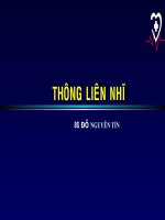
Case Report
Thông Liên Nhĩ(ASD - CIA)
Bs Thiện Châu – Bs Trọng Thịnh
CT MEDIC
</div><span class="text_page_counter">Trang 2</span><div class="page_container" data-page="2">Thông liên nhĩ chiếm khoảng 6% đến 10% các trường hợp mắc bệnh tim bẩm sinh. ASD có thể được phân loại theo vị trí
• Thơng liên nhĩ lỗ thứ phát: vùng lỗ bầu dục: 75%.
• Lỗ nguyên phát : Lỗ thông ở mặt trước dưới của vách liên nhĩ 15%
• Thể xoang tĩnh mạch: Một lỗ thông ở mặt sau của vách ngăn, liền kề với tĩnh mạch chủ trên(10%) hoặc tĩnh mạch chủ dưới và thường liên quan đến quá trình quay trở lại bất thường của tĩnh mạch phổi trên hoặc tĩnh mạch phổi dưới bên phải về tâm nhĩ phải hoặc về tĩnh mạch chủ
• Xoang vành : hiếm gặp
Thơng liên nhĩ :
ASD (ATRIAL SEPTAL DEFECT), CIA (Communication inter auriculaire)
</div><span class="text_page_counter">Trang 3</span><div class="page_container" data-page="3">Bệnh nhân: nữsinh năm 1974Địa chỉ : Q8 thành phố Hồ Chí Minh
Lý do khám bệnh: Mệt, thỉnh thoảng khó thở khi gắng sứcHA = 145/90 , M = 95 , CC = 159cm, CN = 69kg, BMI = 27,3
Bênh nhân đến MEDIC khám bệnh sau được khám và điều trị theo tuyến bảo hiểmy tế, với các kết quả thăm khám siêu âm có biểu hiện nhân giáp , nốt mô vú , đodiện tim và siêu âm tim được chẩn đoán : thiếu máu cơ tim, tăng huyết áp, rối loạntăng đường máu.
Tổng trạng chung thừa cân, không phù, da niêm hồngCảm giác mệt, không ho, không khó thở.
khơng đau bụng.
Xin được khám và kiểm tra lại siêu âm tuyến vú và giáp do bệnh nhân lo lắng nhiềuvề hai bệnh này.
</div><span class="text_page_counter">Trang 32</span><div class="page_container" data-page="32">- Hậu quả sinh lý của mức độ luồng thông qua lỗ thông : Loạn nhịp nhĩ, chẳng hạn nhưnhịp tim nhanh trên thất(SVT),cuồng động nhĩhoặcrung nhĩ, cũng có thể xảy ra.
- Cuối cùng, sự gia tăng áp lực động mạch phổi và sức cản mạch máu phổi có thể dẫn đến sự đổi thành shunt hai chiều với biểu hiện tím khi trưởng thành (Phản ứng
Eisenmengertrong thời gian từ giữa đến cuối giai đoạn tuổi trưởng thành, thường là 40 tuổi).
</div><span class="text_page_counter">Trang 33</span><div class="page_container" data-page="33">Unusual Atrial septal defects :
Sinus venosus type of ASD (<b>SVC TYPE OF ASD)</b>
Atrial septal defects are one of the commonest forms of congenital heart disease.
• The commonest being the ositum secundum ASD ( Which is in fact is a defect in the development of septum primum)
• The next common is ostium primum defect which is a part of AV canal or atrio ventricular septal defect.
Other forms of ASD include
• SVC type /Also called sinus venosus type of ASD .• IVC type
• Coronary sinus defect -Also called partial or complete forms of unroofed coronary sinus
</div><span class="text_page_counter">Trang 34</span><div class="page_container" data-page="34">The exact area of this PV-SVC window occur between anterior surface of right upper lobe PV with postero lateral surface of SVC.
PAPVC* partial anomalous pulmonary venous drainage can be considered an integral part of this defect as RUPV** is linked with SVC.
* partial anomalous pulmonary venous connection (PAPVC)** right upper lobe pulmonary vein (RUPV)
</div><span class="text_page_counter">Trang 35</span><div class="page_container" data-page="35"><b>Can we have a combination of SVC ASD and OS ASD ?</b>
This is possible .But two embryological errors need to occur. This is often seen as a large OS ASD with deficient or absent superior rim. So whenever superior rim of IAS (inter atrial septum) is deficient a PAPVC and a SVC ASD should be looked for.
</div><span class="text_page_counter">Trang 36</span><div class="page_container" data-page="36">- There is a distinct possibility of missing this lesion in routine echo.Minimal RA,RV enlargement may give us a clue.The classical subcostal or 4 chamber view in echocardiography may not visualise these defects.
- So, whenever one encounters mild dilatation of RA and RV and the IAS appears intact, a meticulous search and a focused echo in the superior aspect of IAS is warranted.
Angled superior views may pick up this defect.A transesophageal echocardiogram (TEE) is often required to confirm it.
</div><span class="text_page_counter">Trang 37</span><div class="page_container" data-page="37"><b>Therapeutic issues</b>
• Device closure is not possible
• Surgery involves little more technicality than ASD OS.• Small defects can be patch closed.
• Some times the SVC has to be disconnected from the PV and anastamosed separately on right atrial appendage. SVC resection will aid the surgeon in proper patch closure.
• Post operative follow up is necessary as SVC obstruction or PV obstruction may be a delayed consequence
</div><span class="text_page_counter">Trang 38</span><div class="page_container" data-page="38"><b>SVC ASD – Adult Congenital Heart – Bài học</b>
- Undetected for a lifetime
- Siêu âm khi phát hiện có dãn buồng tim phải thì cần khảo sát kỹ hơn đểtìm nguyên nhân.
- CT giúp chẩn đoán những bất thường trước đây chưa được chẩn đoán. - MRI giúp đánh giá giải phẫu, thể tích tâm thất, Qp:Qs
- MRI & CT hữu ích trong hướng dẫn chẩn đoán, trong can thiệp, theo dõibiến chứng sau phẫu thuật.
</div><span class="text_page_counter">Trang 39</span><div class="page_container" data-page="39"><small>1 - GS Nguyễn Lân Viêt – PGS Pham Manh Hùng , Thực hành chẩn đốn và điều tri bệnh đơng mạchvành , số 5332-Bộ Y Tế 2020 </small>
<small>2 - PGS Phạm Nguyễn Vinh, Siêu âm 2D, Doppler trong bệnh động mach vành, bài giảng siêu âm tim, Viên tim tp HCM </small>
<small>3 - Bs Đỗ thị kim Chi – Thông liên nhĩ, bài giảng siêu âm tim, Viên tim tp HCM </small>
<small>4 -Lee B. Beerman, MD, Thông liên nhĩ (ASD) Children's Hospital of Pittsburgh of the University of Pittsburgh School of Medicine.</small>
<small>5 -DR.S.VENKATESAN MDUnusual Atrial septal defects : Sinus venosus type of ASD. </small>
<small>6 - Valeria Duarte, MD; Keith Ellis, MD: Undetected for a Lifetime: Sinus Venosus Atrial Septal Defect, </small>
<small>Houston Methodist DeBakey CV</small>
<small>7 - Dr Linda Haramati , Atrial septal defect in Adults, Role of CT & MRI in guiding Management (MonteHeart Lectures) </small>