MODERN ARTHROSCOPY doc
Bạn đang xem bản rút gọn của tài liệu. Xem và tải ngay bản đầy đủ của tài liệu tại đây (38.81 MB, 312 trang )
MODERN ARTHROSCOPY
Edited by Jason L. Dragoo
Modern Arthroscopy
Edited by Jason L. Dragoo
Published by InTech
Janeza Trdine 9, 51000 Rijeka, Croatia
Copyright © 2011 InTech
All chapters are Open Access distributed under the Creative Commons Attribution 3.0
license, which allows users to download, copy and build upon published articles even for
commercial purposes, as long as the author and publisher are properly credited, which
ensures maximum dissemination and a wider impact of our publications. After this work
has been published by InTech, authors have the right to republish it, in whole or part, in
any publication of which they are the author, and to make other personal use of the
work. Any republication, referencing or personal use of the work must explicitly identify
the original source.
As for readers, this license allows users to download, copy and build upon published
chapters even for commercial purposes, as long as the author and publisher are properly
credited, which ensures maximum dissemination and a wider impact of our publications.
Notice
Statements and opinions expressed in the chapters are these of the individual contributors
and not necessarily those of the editors or publisher. No responsibility is accepted for the
accuracy of information contained in the published chapters. The publisher assumes no
responsibility for any damage or injury to persons or property arising out of the use of any
materials, instructions, methods or ideas contained in the book.
Publishing Process Manager Ivona Lovrić
Technical Editor Teodora Smiljanic
Cover Designer InTech Design Team
Image Copyright Blank Michael, 2011. Used under license from Shutterstock.com
First published December, 2011
Printed in Croatia
A free online edition of this book is available at www.intechopen.com
Additional hard copies can be obtained from
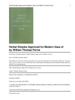
Modern Arthroscopy, Edited by Jason L. Dragoo
p. cm.
ISBN 978-953-307-771-0
free online editions of InTech
Books and Journals can be found at
www.intechopen.com
Contents
Preface IX
Part 1 Arthroscopy of the Temporomandibular Joint 1
Chapter 1 Temporomandibular Joint Arthroscopy 3
Edvitar Leibur, Oksana Jagur and Ülle Voog-Oras
Part 2 Arthroscopy of the Upper Extremity 27
Chapter 2 Arthroscopic Treatment of Recurrent Anterior
Glenohumeral Instability 29
Michael Hantes and Alexandros Tsarouhas
Chapter 3 Anesthesia for Arthroscopic Shoulder Surgery 49
Diego Benítez and Luis M. Torres
Chapter 4 Arthroscopic Treatment of Distal Radius Fractures 65
Yukio Abe and Yasuhiro Tominaga
Part 3 Arthroscopy of the Hip 77
Chapter 5 Arthroscopy after Total Hip Replacement Surgery 79
Cuéllar Ricardo, Ponte Juan, Esnal Edorta and Tey Marc
Part 4 Arthroscopy of the Knee 101
Chapter 6 Management of Knee Articular Cartilage Injuries 103
Joshua D. Harris and David C. Flanigan
Chapter 7 Articular Cartilage Regeneration with Stem Cells 129
Khay-Yong Saw, Adam Anz, Kathryne Stabile, Caroline SY Jee,
Shahrin Merican, Yong-Guan Tay and Kunaseegaran Ragavanaidu
Chapter 8 Traumatic Chondral Lesions of the Knee
Diagnosis and Treatment 179
Masoud Riyami
VI Contents
Chapter 9 Contemporary Anterior
Cruciate Ligament Reconstruction 197
P. Christel and W. Boueri
Chapter 10 The Role of Arthroscopy in
Mini-Invasive Treatment of Tibial Plateau Fractures 225
Şt. Cristea, A. Prundeanu, Fl. Groseanu and D. Gârtonea
Chapter 11 Arthroscopy Following Total Knee Replacement 237
Vaibhav Bagaria, Jami Ilyas, Bhawan Paunipagar,
Darshna Rasalkar and Rohit Lal
Chapter 12 Arthroscopic Soft Tissue Releases of the Knee 257
Michael R. Chen and Jason L. Dragoo
Chapter 13 Extraarticular Arthroscopy of the Knee 273
Shinichi Maeno, Daijo Hashimoto, Toshiro Otani, Ko Masumoto,
Itsuki Yuzawa, Kengo Harato and Seiji Saito
Part 5 Arthroscopy of the Foot and Ankle 285
Chapter 14 Posterior Ankle and Hindfoot Arthroscopy 287
Masato Takao
Preface
Staying current in the rapidly changing field of arthroscopic surgery presents a
challenge for practitioners and students. Books and review articles are often outdated
on the day of release. Recently, rapidly published on-line books have filled an
educational void, assisting millions of practitioners to stay updated on the latest
techniques.
The chapters in this book entitled Modern Arthroscopy were written by a panel of
international experts in the various disciplines of arthroscopy. The goals of this text are
to present the classical techniques and teachings in the fields of Orthopaedics and
Dentistry, but also to include new, cutting-edge applications of arthroscopy, such as
temporomandibular arthroscopy and extra-articular arthroscopy of the knee, just to
name a few. The InTech online publishing format allows rapid publishing of these
new techniques to keep the content accurate and up to date.
The chapters are organized anatomically, with the first chapters introducing basic
arthroscopic techniques, while later chapters describe newer, more advanced
techniques. The surgical technique sections of the chapters are clearly labeled for easy
reference.
It is my hope that this book will either assist readers in learning the current techniques
of arthroscopic surgery, or serve as a springboard to stimulate the creation of new
techniques, which have yet to be described. On behalf of the authors and InTech
Publishing we thank you for your interest in this book, and hope Modern Arthroscopy
becomes a core reference for your arthroscopic surgery practice.
Jason L. Dragoo, MD
Assistant Professor
Department of Orthopaedic Surgery and Sports Medicine
Stanford University,
USA
Part 1
Arthroscopy of the Temporomandibular Joint
1
Temporomandibular Joint Arthroscopy
Edvitar Leibur
1,2,
Oksana Jagur¹ and Ülle Voog-Oras¹
1
Department of Stomatology,
2
Department of Internal Medicine,
Tartu University, University Hospital
Estonia
1. Introduction
Arthroscopy is a technique for direct visual inspection of internal joint structures, including
biopsy and other surgical procedures performed under visual control. In 1918 Takagi first
described arthroscopy of the knee joint examinations using cystoscope (Tag, 1939). Onishi in
1970 was the first to report arthroscopy of the human temporomandibular joint (TMJ) and
the first results were published by him (Onishi, 1975, 1980). The progress in research and
applications of TMJ arthroscopy in joint disease have led to the acceptance of small
operative procedures as a safe, minimally invasive means of effectively treating a number of
intra-articular and degenerative TMJ problems (McCain, 1992; Holmlund &Axelsson, 1996;
Holmlund et al., 2001). Arthroscopic surgery has been an effective treatment for TMJ
disorders refractory to nonsurgical treatments ( Ohnuki et al., 2003; Gonzalez-Garcia et al.,
2008, Leibur et al., 2010). TMJ arthroscopy has been variously reported as successful in up to
80% of cases where outcome of arthroscopic surgery to the TMJ correlates with the stage of
internal derangement (K. Murakami et al., 2000; Sanroman, 2004). Studies have been
variable in their scientific method and some long-term outcomes studies have been
completed where both quality of life and functional outcome have been assessed (Voog et
al., 2003a; Undt et al., 2006; Jagur et al., 2011). For enabling direct comparison of the clinical
results following arthroscopic surgery and open surgery a retrospective study comparing
two centers´ results using the Jaw Pain and Function Questionnaire ( Clark et al., 1989) has
been performed and these treatment results of open surgery were comparable with
arthroscopic treatment results (Undt et al., 2006).
2. Anatomy of the temporomandibular joint
The temporomandibular joint is the articulation between the mandible and the cranium. The
mandibular head (condyle), glenoid (mandibular) fossa, and articular eminence form the
TMJ. These joints serve as one anatomic control for both mandibular movement and the
occlusion, surrounded by a capsule which consists of fibrous material, and a synovial
lining. The capsule is quite thin anteromedially and medially ~ 0,7 mm and thick laterally
and posteriorly ~ 1,8 mm. The inner layer of the capsule or synovial membrane is highly
vascularized layer of endothelial origin cells, producing synovial fluid. The capsule stretches
from the edge of the mandibular fossa to the neck of the mandible, proximal to the
pterygoid fovea, and envelops the articular eminence. TMJ is reinforced by the
temporomandibular and sphenomandibular ligaments. The articular surface of the
Modern Arthroscopy
4
mandible is the upper and anterior surface of the condyle, lined by dense, avascular fibrous
connective tissue. A layer of hyaline cartilage covers the articulating cortical bone. The adult
human condyle is about 15 to 20 mm from side to side and 8 to 10 mm from front to back.
The articular surface is convex when viewed from the side and less when viewed from the
front. Glenoid fossa is the concavity within the temporal bone. The anterior wall is formed
by the articular eminence of the temporal bone and its posterior wall by the tympanic plate,
which also forms the anterior wall of the external auditory meatus. An articular disc is
interposed between the temporal bone and the mandible, dividing the articular space into
upper and lower compartments. The interposed fibrocartilaginous disc has a bow-tie-
shaped biconcave morphology. The anterior and posterior ridges of the disc are termed
anterior and posterior bands and are longer in the mediolateral than in the anteroposterior
dimension. The smaller anterior band attaches to the articular eminence, condylar head, and
joint capsule. The posterior band blends with highly vascularized, loose connective tissue,
the bilaminar zone, and the capsule, the bilaminar zone residing in the retrodiscal space in
the mandibular fossa and attaching to the condyle and temporal bone. Medially and
laterally, the disc is firmly attached to the capsule and the condylar neck. Anteromedially, it
is attached to the superior part of the pterygoid muscle. In a physiologic joint, the disc is
positioned between the mandibular head inferiorly and the articular eminence anteriorly
and superiorly when the jaw is closed. The posterior band of the disc lies within 10° of the
12 o'clock position. The medial and lateral corners of the disc align with the condylar
borders and do not bulge laterally or medially. When the jaw is opened, the disc slides into a
position between the mandibular head and articular eminence. The loose tissue of the
bilaminar zone allows the remarkable range of motion of the disc. The attachments of the
disc prevent luxation during opening. A triangular lateral ligament acts as a strong lateral
stabilizer and inhibits the posterior translation of the mandibular head (Fig. 1).
The muscles of mastication are responsible for the complex movement of the jaw. The
temporal, medial pterygoid, and masseter muscles facilitate jaw closure. Mouth opening is
effected by coordinated action of the lateral digastric, mylohyoid, and suprahyoid muscles.
The lateral pterygoid muscle and part of the fibers of the masseter and medial pterygoid
muscles effect the anterior translation of the mandible. The superior belly of the lateral
pterygoid muscle originates from the greater sphenoid wing and inserts on the disc.
Subsequently, the superior belly plays a key role in upholding the physiologic position of
the disc as it pulls the disc forward when the jaw is opened, in a combined translation and
rotation. The inferior head of the lateral pterygoid muscle stretches from the lateral lamina
of the pterygoid process to the pterygoid fovea. The medial pterygoid muslcle originates
from the pterygoid fossa and inserts near the medial aspect of the mandibular angle
(Sommer et al., 2003). The blood supply to the TMJ, outer and inner ear is provided mainly
by branches from the internal maxillary artery as follows: temporal superficial artery,
superior auricular artery, anterior tympanic artery and pterygoid artery. Innervation is
provided by the auriculotemporal nerve (sensory branch of the mandibular nerve), deep
temporal nerve, masseteric nerve. Sensory cervical sympathetic ramifications are going to
the disc and capsule. The auriculotemporal nerve runs medial to the joint, then runs
laterally, crossing the condylar neck, where it divides into branches to innervate the capsule,
disc attachments, the tympanic membrane, the anterior surface of the cochlea, the upper
part of the auricle, the tragus of the ear, the skin lining the external auditory meatus, the
temporal region,. Nerve receptors as Ruffin receptors, Golgi tendon organs, Vater-Pacini
corpuscules free nerve endings are in the capsule and substance P nerve fibres are also
available in both the auriculotemporal and masseteric nerves, and have been demonstrated
in the capsule, disc attachments but they are not present in the disc (Fig. 2).
Temporomandibular Joint Arthroscopy
5
Fig. 1. A sagittal section through the left temporomandibular joint .
Fig. 2. Branches of trigeminal nerve. Innervation and blood supply of temporomandibular
joint (by R.Schmelzle, 1989).
Modern Arthroscopy
6
3. Classification of temporomandibular joint disorders:
- Arthritis- acute, chronic, infectious (specific, nonspecific)
- Osteoarthritis/arthrosis – most often disorder
- Injuries – luxations, concussion, fracture
- Ankylosis (fibrous, fibro-osseous, osseous)
- Tumours (benign and malignant)
- Congenital disturbances: I & II branchial arch malformations, condylar hypo-,
hyperplasia
- Idiopathic condylar resorption
- Systemic conditions affecting the TMJ (rheumatoid arthritis, psoriasis, pseudogout etc.)
4. Aetiology and pathogenesis of temporomandibular disorders
4.1 Aetiology
Most scientists regard osteoarthritis as an inflammatory process, being most frequent TMJ
disorder, characterised with proliferative changes in the synovia and primary degeneration
of the cartilage and surrounding tissues with destruction of the bone structures. (Holmlund
& Axelsson, 1996; Emshoff , 2005). It is found that 28% of the adult population have signs of
temporomandibular joint disorder. In systemic diseases (rheumatoid arthritis, psoriasis etc.)
involvement of TMJ occurs (Voog et al., 2003b; 2004). Main aetiological factors of TMJ
disorders are as follows: systemic diseases ( rheumatoid arthritis, psoriasis, pseudogout,
ankylosing spondylitis etc.), secondary inflammatory component from the neighbouring
regions (otitis, maxillary sinusitis, tonsillitis ), trauma (chronical), prevalence of dental arch
defects e.g. missing of molar teeth, (Tallents et al. 2002), malocclusion, endocrinological
disturbances, odontogenic infections (third molars). Presence of specific bacterial species as
Staphylococcus aureus, Streptococcus mitis, Mycoplasma fermentas, Actinobacillus
actinomycetemcomitans (Aa) in the synovial fluid have been found (Kim et al., 2003). Serum
antibodies against Chlamydia spp. in patients with monoarthritis of the TMJ have been
occurred. An association may exist between the presence of Chlamydia trachomatis and TMJ
disease (Paegle et al., 2004).
4.2 Pathogenesis
Knowledge about the pathogenesis on a molecular level of disorders of the TMJ has been
improved in recent years giving a possibility to use these data for the evidence based
treatment. Inflammation mainly affects the posterior disc attachement (Holmlund & Axelsson,
1996; Leibur et al., 2010). Several inflammatory mediators play an important role in the
pathogenesis of TMJ diseases as tumor necrosis factor α (TNFα), interleukin-1β (IL-1β),
prostaglandin
E
2
(PGE
2
)
,
leukotrien B
4
(LkB
4 ),
matrix metalloproteinases (MMP
s
), serotonin- 5-
hydroxytryptamine (5-HT), (Alstergren et al., 1999; Voog et al., 2003b). MMP
s
are responsible
for the metabolism of extracellular matrix, being an early marker to determine TMJ arthritis.
High level of MMP-3 has been determined in the synovial fluid in TMJ osteoarthritis patients
(Kamada et al., 2000). Serotonin, mediator of pain and inflammation, is produced in the
enterocromaffin cells of the gastrointestinal mucosa and absorbed by platelets. It is produced
also in the synovial membrane and is present in the synovial fluid and in blood in case of
rheumatoid arthritis and is involved in the mediation of TMJ pain in systemic inflammatory
joint diseases (Alstergren & Kopp, 1997; Voog et al., 2000). It plays a role also in bone
metabolism (Warden & Haney, 2008). Tissue response in case of inflammation is as follows:
Temporomandibular Joint Arthroscopy
7
vasodilatation, extravasation, releasing of mediators, activation of nociceptors, release of
neuropeptides as substance P (SP), neuropeptide Y (NPY), which stimulate releasing of
histamin and serotonin from afferent nerve endings and hyperalgesia in TMJ occurs.
5. Diagnostics of the temporomandibular disorders
5.1 Clinical data
The most frequent complaint is pain and a decrease in the maximal interincisal opening
(MIO), which normal values are between 35 - 50 mm (Fig. 3).
The following symptoms as pain (at rest, during maximum mouth opening and upon
chewing), tenderness to digital palpation of the joint, sounds (clicking, crepitation), restricted
mandibular mobility e.g. difficulty in opening the mouth, intermittent lock, closed lock,
stiffness in the morning are observed. The stages of disease are usually classified according to
Wilkes (1989; Table 1) by reviewing the case histories, clinical data, radiological records
(computerized tomography images, magnetic resonance images, ortopantomography and/or
plain radiographs by Schüller, Parma).
I. Early stage
a. Clinical: No significant mechanical symptoms other than opening reciprocal clicking;
no pain or limitation of motion
b. Radiologic: Slight forward displacement , good anatomic contour of the disc,
negative tomograms, no bone structure changes
c. Pathoanatomy: Excellent anatomic form; slight anterior displacement, passive in-
coordination demonstrable
II. Early intermediate stage
a. Clinical: One or more episodes of pain: beginning major mechanical problems
consisting of mid-to-late opening loud clicking; transient catching and locking
b. Radiologic: Slight forward displacement; beginning disc deformity, slight thickening
of posterior edge; negative tomograms, no bone structure changes
c. Pathoanatomy: Anterior disc displacement; early disc deformity; good central
articulating area
III. Intermediate stage
a. Clinical: Multiple episodes of pain; major mechanical symptoms consisting of locking
( intermittent or fully closed): restriction of motion, function difficulties
b. Radiologic: Anterior disc displacement with significant deformity or prolapse of disc
(increased thickening of posterior edge), negative tomograms, no bone structure
changes
c. Pathoanatomy: Marked anatomic disc deformity with anterior displacement; no hard
tissue changes
IV. Late intermediate stage
a. Clinical: Slight increase in severity over intermediate stage
b. Radiologic: Increase in severity over intermediate stage; positive tomograms
showing early-to-moderate degenerative changes - flattening of eminence,
deformation of condylar head, erosions, sclerosis
c. Pathoanatomy: Increase in severity over intermediate stage; hard tissue degenerative
remodelling of both bearing surfaces (osteophyts), multiple adhesions in anterior and
posterior recesses; no perforation of disk or attachments
Modern Arthroscopy
8
V. Late stage
a. Clinical: Characterized by crepitus; variable and episodic pain; chronic restriction of
motion and difficulty with function
b. Radiologic: Disc or attachment perforation, filling defects, gross anatomic deformity
of disc and hard tissues, positive tomograms with essentially degenerative arthritic
changes
c. Pathoanatomy: Degenerative changes of disc and hard tissues; perforation of
posterior attachement; multiple adhesions, osteophyts, flattening of condyle and
eminence, subcortical cyst formation
Table 1. Classification for internal derangement of the TMJ by Wilkes (1989).
Fig. 3. Maximal interincisal opening (MIO) is 13 mm.
Symptom related factors obtained by questionnaire, the scores pre- and posttreatment
maximal interincisal opening (MIO) and visual analogue scale (VAS) for pain are to be
documentated and compared. Joint pain is assessed with 100 mm visual analogue scale with
end points marked „no pain“ and „worst pain ever experienced“. The absence of pain is
scored as 0. If pain is present the patient is asked to select marked field from 1mm to 100
mm.
It is known that inflammation often is accompanied by pain. Evaluation and estimation of
the impact of pain is a complicated matter, since pain has many different ways to interfere
with everyday life. The impact of pain on the health status and quality of life in patients
with chronic inflammatory joint diseases has been recognized, but there is a lack of
knowledge about the specific impact of TMJ pain on daily activities in patients with clinical
involvement of the TMJ. A scale for measuring the activity of daily living (ADL), (List &
Helkimo, 1995) is a useful tool for assessment of the restriction of activities of patients with
TMJ disorders in their everyday life (Voog et al., 2003a; Kaselo et al., 2007; Jagur et al., 2011).
Temporomandibular Joint Arthroscopy
9
5.2 Radiographic investigations
Radiological changes of the TMJ are evaluated by orthopantomography (OPTG), computed
tomography (CT), magnet resonance imaging (MRI) (Ohnuki et al. 2003; Voog et al., 2003b,
2004; Whyte et al., 2006) as well as ultrasonography (C.A. Landes et al., 2007).
OPTG is mainly used to demonstrate the structural bone changes in the TMJ and it has the
advantage of being easily available but gaves limited information about the above
mentioned joint being an alternative method to other radiological methods. To obtain a
more detailed anatomic picture, CT or MRI are recommended. By evaluating the OPTGs the
following radiographic signs of bone structural changes can be achieved such as presence of
erosions, flattening and osteophytes of the condyle as well as temporal bone (Rohlin et al.,
1986). Erosion in condyles in the radiographs is scored according to Helenius et al. (2004) as
follows: score 1 - very slight erosion; score 2 - erosion on top of the condyle; score 3 - half of
condyle is eroded; score 4 - condyle totally eroded. The first report of TMJ CT was published
by Suarez et al. (1980) and this method is superior to plain transcranial or transmaxillary
imaging for detecting bone changes. CT allows detailed three-dimensional examination of
the TMJ and it is capable to detect even small bone changes not demonstrable by
conventional tomographic procedures (Raustia et al., 1985; Larheim & Kolbensvedt, 1990).
The CT sections are evaluated for presence of radiographic signs of bone changes within
three regions (lateral, central and medial) of the mandibular and temporal part (eminence)
of the TMJ. The recording of the signs is made in the axial, coronal and sagittal views
(Emshoff et al., 2003; Voog et al., 2003). The changes are defined as follows: erosion - a local
area with decreased density of the cortical joint surface including or not including adjacent
subcortical bone (Fig.4), sclerosis - a local area with increased density of the cortical bony
joint surface that may extend into the subcortical bone (Fig. 5), subchondral pseudocyst - a
well defined, local area of bone rarefication underneath an intact cortical outlining of the
joint surface, flattening - a flat bony contour deviating from the convex form (Fig. 6),
osteophyte - a marginal bony outgrowth (Fig. 6). The grade of the total changes of the TMJ
can be evaluated according to the scoring system developed by Rohlin & Petersson (1989) as
well.
Fig. 4. Osteoarthritis of the TMJ. Signs of erosions on the surfaces of the condyles in a
coronal view of the CT. An irregular outline is revealed on the condyles. The bone structure
of the both glenoid fossa is normal.
Modern Arthroscopy
10
Fig. 5. Axial view of the CT. Sign of sclerosis in the medial and central parts of the right
condyle of the mandible ( arrow) . Reduced space is seen.
Fig. 6. Sagittal view of the CT from the left temporomandibular joint. Sign of flattening of
the condyle.
Temporomandibular Joint Arthroscopy
11
MRI has a diagnostic value for internal derangements of the TMJ and rapidly surpassing CT
as the imaging method of choice
.
MRI can detect not only TMJ soft tissue abnormalities like
disc displacements, pathology of synovial membrane or capsule, pathology in the posterior
attachement but also hard tissue morphologic changes can be demonstrated with MRI
(Lieberman et al., 1996; Larheim et al., 1999). Sections in the oblique sagittal plane (i.e.
perpendicular to the horizontal long axis of the mandibular condyle) and oblique coronal
plane (i.e. parallel with the long axis of the condyle), and bilateral temporomandibular base
surface coils are used (Larheim et al., 2001) for obtaining the images (Fig. 8).
The biting device (MEDRAD; Pittsburg) which enables dynamic imaging can be used as bite
blocks during the open jaw phase of the imaging procedure (Gaggl et al.,1999). Dynamic
magnetic resonance imaging is a recent method that investigate directly in vivo articular
function and shows much promise as a noninvasive method of the disc function, however
this limitation should diminish with continuing technological advances in the imaging field.
Ultrasonography has been a helpful diagnostic approach for patients with TMJ disorders,
having a possibility to diagnose with considerable reliability when compared with MRI and
being a sensitive tool for assessing joint function (C. Landes et al., 2000; C.A. Landes et al.,
2006).
Fig. 7. An osteophyte in the medial part of the right mandibular condyle in a sagittal view of
the CT., cortical destruction of the glenoid fossa surface.
Modern Arthroscopy
12
Fig. 8. Sagittal view of the MRI in a patient with internal derangement of the left TMJ.
Anterior disc displacement (arrow), destruction of the disc. Changes of bone structures,
effusion in the anterior recess.
6. Temporomandibular joint arthroscopy
6.1 Indications for arthroscopy
Indications for arthroscopy are radiological bone changes in TMJ characteristic to
osteoarthritis with disc displacement or deformity and non effectiveness of conservative
treatment with NSAIDs, intraoral splints or arthrocentesis. In practice, the decision to
operate and the choice of the method seems to be a matter of the individual surgeon´s
training, experience, and attitude toward the surgical management of TMJ disorders.
Involvement of the TMJ in patients with rheumatoid arthritis or other connective tissue
diseases is rather common and arthroscopy with simultaneous biopsy is indicated in these
situations. Posttraumatic complaints may also be an indication for arthroscopy. Arthroscopy
is contraindicated in case of acute arthritis. In these situations as large medial osteophyts on
the condyle, large central cartilaginous perforations, fibrous, fibro-osseous, osseous
ankylosis are better to handle via open reduction. Arthocentesis is considered as an
intervening treatment modality between nonsurgical treatment and arthroscopic surgery.
All cases for arthroscopy are usually classified as advanced Wilkes (1989) stages IV and V, in
rare cases stage III (Table 1).
6.2 Technique for arthroscopy
Arthroscopy is performed under nasotracheal general anaesthesia which makes possible to
manipulate the mandible during the operation. First the zygomatic arch and the condyle are
palpated. The condyle is then forced in anterior position by the assistant and the
Temporomandibular Joint Arthroscopy
13
preauricular concavity is formed in the skin, marking a point for the injection. Usually
arthroscope KARL STORZ GmbH & Co.KG is used. Although various arthroscopic
approaches to the TMJ have been described, the one most commonly used is the
posterolateral approach to the upper joint space. After the condylar head of the TMJ has
been determined , a marking line and puncture points are made on the skin surface (Fig. 9).
Fig. 9. A marking line and the puncture points on the skin surface for TMJ arthroscopy
The puncture site is located by manipulating the mandible anterio-inferiorly. For distension
of the superior compartement and in order to avoid iatrogenic damage to the cartilaginous
surfaces during introduction of the trocar, 1% lidocain solution 2,0 mL is inserted. The
needle is aimed in a medial and slightly anteriosuperior direction until the contact with the
glenoid fossa is achieved. The posterior recess of the superior joint space is reached when
there is a backflow into the syringe of the solution injected into the joint space (Fig.10).
Fig. 10. Distension of the superior compartment of the right temporomandibular joint with
2% lidocaine solution.
Modern Arthroscopy
14
Through the small skin incision 0,75 – 1,0 cm from the center of the tragus at the injection
site the lateral capsule is punctured with a sharp trocar in an arthroscopic sheath inserted in
the same direction as the previous injection needle. The sharp trocar is exchanged for a blunt
one and the arhroscopic sheath is advanced further into the upper joint space. Puncture
with arthroscope sheath (trocar) with a blunt obturator inserted into upper posterior recess
is performed angling it medially upward ~ 2,5 cm. Another skin incision is made ~ 0,75 cm
from the first skin incision in anterolateral direction for outflow cannula to be inserted into
the upper joint anterior recess.
Following insertion of the trocar (diameter 1,8 mm, length 4 cm) into the joint space, blunt
obturator is removed and forward-oblique telescope 30º (HOPKINS®), diameter 1,9 mm,
length 6,5 cm, fiber optic light transmission incorporated is inserted (Fig. 11).
Fig. 11. Forward - oblique telescope 30° (HOPKINS®) fiber optic light transmission
incorporated and outflow cannula are inserted into the right upper temporomandibular
joint space.
Initial recognition of anatomical structures as the superior surface of the disc, articular fossa,
and internal aspects of the posterior and medial capsule is performed. The fluid level in the
arthroscope sheath should move with the jaw, confirming that the sheath is correctly
positioned in the joint upper space.The upper joint compartment is examined from the
posterior pouch via the intermediate zone to the anterior pouch. Disc may give the
impression of being obstructed against the arthrotic surface of the temporal cartilage. The
anterior part of the disc surface looks usually smooth and collagen fibres could clearly seen.
The condylar cartilage is normally smooth, but in case of pathology e.g. in osteoarthritis
where irregularities of the surface as erosions, osteophyts can be seen. Sever arthrotic
changes of both fossa cartilage and disc may also observed. Adhesions between the disc and
glenoid fossa are quite common. In rare cases the arthrotic or inflammatory changes are
found in the anterior recess. Upper compartment is swept clear under constant irrigation
with isotonic saline solution. This manipulation allow translation of the disc along the
eminence, allowing the condyle to complete its natural path. After the diagnostic
Temporomandibular Joint Arthroscopy
15
arthroscopy has been completed, either forceps, palpation hook or blunt probe are used to
cut fibres, mainly fibers of the pterygoid muscle anterior to the disc, in order to reduce pull
in the anterior direction and facilitate repositioning of the disc. Cutting of adhesions
facilitate repositioning of the disc. During arthroscopy a sweeping procedure between the
disc and fossa release the adhesions and fibrillations increasing the mobility in the joint.
Release of the adhesions and fibrillations of the superior suface of the disc and shaving the
surface of articular fossa in the upper joint compartment are performed with the aid of a
blunt obturator or hook and with grasping forceps, scissors or double-edged knife. Removal
of the superficial layer of cortical bone induces capillar bleeding stimulating formation of
fibrocartilage on bone. Quite often a displaced disc may be found during arthroscopy.
Surgical procedure is completed by irrigating the joint space to remove small tissue
fragments. The outflowing fluid is collected and may be retained for diagnostic purposes.
Arthroscopic lysis and lavage includs also a lateral release of the upper joint compartment
performed with the aid of the blunt obturator or hook.Thus the locked disc could be
mobilized sufficiently. Clinical, radiographic and arthroscopic findings in patients who
underwent arthroscopy are given in Table 2 (Leibur et al. 2010).
Signs and
symptoms
Sum
%
abn
Radiographic findings Sum
%
abn
Arthro-
scopic
findings
Sum
%
abn
Pain 25 86 Flattening 10 34 Adhesions 29 100
Hypomo-
bility
23 79
Bone cyst / Subchondral
pseudocycts
9 31
Chondro-
matosis
5 17
Closed lock 5 17 Erosions 20 69 Fibrillations 22 76
Intermittent
lock
5 17 Reduced space 10 34 Synovitis 9 31
Deviation 4 14 Sclerosis 8 27
Eburneation
of fossa
15 52
Hypomobilityof
condyle Osteophyts
4 5
14
17
Displaced
disc
23 23
Sum = total number of patients with findings; % abn = percentage of individuals with abnormal
findings.
Table 2. Clinical, radiographic and arthroscopic findings in patients who underwent
arthroscopy (N=29).
Arthroscopic findings are as follows: irregularities of joint surfaces, foldings and synovitis –
hyperaemia of the inner wall, localising also in the posterior part of the disc, intra-articular
fibrous adhesions, intracapsular adhesions, fibrillations of superior surface of the disc and
arthrotic lesions of temporal cartilage, pseudowalls, foreign bodies - chondromatosis (Fig.
12, 13, 14, 15).